Introduction
According to recent international recommendations, middle ear effusion (MEE) is defined as the presence of fluid in the middle ear cavity with no associated signs of infection.Reference Rosenfeld, Shin, Schwartz, Coggins, Gagnon and Hackell1–3 MEE is a common childhood condition, with reports showing that it affects over 50 per cent of children under the age of one and 60 per cent under the age of two.Reference Simon, Haggard, Rosenfeld, Jia, Peer and Calmels4 Parallel to that, nearly 90 per cent of children with cleft lip and palate (CLP). have been reported to have a history of non-trivial MEE.Reference Ponduri, Bradley, Ellis, Brookes, Sandy and Ness5
Although the main pathomechanism of MEE remains elusive, Eustachian tube dysfunction (ETD) has been postulated as the main contributing factor.Reference Bruce, Harman, Williamson, Tierney, Callery and Mohiuddin6 Failure of the Eustachian tube to equalise pressure between the middle ear and nasopharynx results in the development of negative pressure and, subsequently, fluid build-up within the middle ear space. Children with CLP are prone to ETD due to dysfunction or deficiency of the palatal muscle responsible for maintaining the Eustachian tube orifice.Reference Maheshwar, Milling, Kumar, Clayton and Thomas7, Reference Civelek, Çelebioğlu, Sağit and Akin8 Nevertheless, the literature has reiterated that children with CLP are susceptible to developing prolonged MEE or recurrent MEE.Reference Bruce, Harman, Williamson, Tierney, Callery and Mohiuddin6
The optimal treatment of MEE remains a debate to date. The current guidelines advocate for watchful waiting for three months and then consideration of a hearing aid or ventilation tube insertion in children with persistent MEE.2 Ventilation tube insertion has been a favoured approach in recurrent MEE in many centres, although it has the risk of persistent perforation and the need for repeated insertion. Eustachian tube balloon dilatation (ETBD) has recently gained attention as an intervention for MEE in children. The procedure is rapid, simple and safe for children. ETBD has also shown an improvement in clinical symptoms in most children.Reference Saniasiaya, Kulasegarah and Narayanan9 However, no studies have investigated the outcome of ETBD in CLP children. We conducted a prospective randomised controlled trial pilot study to determine the efficacy and outcome of ETBD in children with CLP. The rationale for performing this study is to investigate if ETBD can be a safe and effective intervention option in CLP children with MEE.
Methods
Following written informed consent from parents, 14 children were included in the study, among which seven children who underwent ETBD (14 ears). The study period was over one year: August 2021 to July 2022. These children were from our Otorhinolaryngology Clinic at the University Malaya Medical Centre, who fulfilled the inclusion criteria and were randomly assigned to both arms of the study. There were no children who declined to participate after being allocated their randomised arm for the study.
All procedures performed in this study involving human participants were in accordance with the ethical standards of the institutional and/or national research committee and with the 1964 Helsinki Declaration and its later amendments or comparable ethical standards. Parents and the patient were informed regarding the off-label use of the ETBD in the paediatric age group.
Patient selection
ETBD was performed as a second-line treatment in our study. All children included in both arms of the study have undergone prior myringotomy and grommet insertion and present with persistent or recurrent MEE. The allocation into the intervention and control arms were randomised. This study included children under 18 years of age with CLP who fulfilled our inclusion and exclusion criteria.
Inclusion criteria
Children with cleft palate or cleft lip between the ages of 2 to 17 and (1) parents agreeable and able to give informed consent; (2) symptoms of hearing loss supported by PTA findings; (3) supported by clinical findings of (a) tympanic membrane (TM) retraction or middle ear effusion and (b) tympanometric evidence of flat or negative ear pressure (type B or C); and (4) history of previous myringotomy and grommet insertion surgery.
Exclusion criteria
(1) Active chronic or acute otitis media, (2) acute upper respiratory infection, (3) syndromic or other craniofacial anomalies other than cleft lip or palate and (4) enlarged adenoids (adenoids engulfing the opening of Eustachian tube noted during nasal endoscopy).
Flow of research
A single assessor performed complete anamnesis, clinical examination and documented audiometry examinations of the included children with accompanying parent/caretaker in the Otorhinolaryngology Clinic at the University Malaya Medical Centre.
The children in Group 1 underwent ETBD using an ENT Dilation System Entellus XprESS® device (Entellus Medical, Plymouth, MN), followed by the intranasal steroid fluticasone furoate 27.5 mg daily for six weeks. Children in Group 2 (control group) were prescribed only intranasal steroid fluticasone furoate 27.5 mg daily for six weeks. Before the intervention, all children (both groups) underwent nasoendoscopic and otoendoscopic examination in the clinic, audiometry examination with tympanometry and PTA and a questionnaire to quantitatively assess Eustachian tube-related symptoms, the Eustachian Tube Dysfunction Questionnaire (ETDQ-7).Reference McCoul, Anand and Christos10 The ETDQ-7 questionnaire was completed by both the parent and the child to ensure the child understands and answers correctly.
After six weeks, a follow-up assessment was carried out, which included otoendoscopic examination, tympanometry, PTA and the ETDQ-7 questionnaire. Only children who completed the six-week follow-up were included in this study.
Outcome criteria
We aimed to determine two primary outcomes of ETBD in CLP with MEE. The primary outcome is to determine the normalisation of tympanometry and improvement of air conduction thresholds six weeks post-intervention. The secondary outcome of this study is to quantify the subjective improvement six weeks post-intervention using ETDQ-7Reference McCoul, Anand and Christos10 questionnaire.
Sample size derivation
As this is a pilot study, there is no prior information on which to base the sample size calculation. According to a study by S. A. Julious, 12 subjects per group for a pilot interventional study is recommended so that the precision around the estimates can be used to design further studies.Reference Julious11 Hence, 14 ears (7 patients) per group were included.
Interventional procedures
Children in the intervention group underwent ETBD using the ENT Dilation System, Entellus XprESS® device ENT Dilation System (Entellus Medical, Plymouth, MN), which was selected by the surgeons based on their preferences and was used off-label in this age group. Informed consent was obtained from both the children and parents or caretakers prior to intervention. The ETBD was the only procedure performed by two surgeons (J.S. & J.K.). The procedure was performed in the operating theatre in a care setting with the child ventilated with a laryngeal mask airway. The nasal cavity was decongested with diluted adrenaline (1:10) soaked pledgets before the procedure. The nasopharynx was visualised using a 0- and 30-degree Hopkins rod telescope (4 mm or 2.7 mm). The ETBD device was inserted with endoscopic guidance and gently inserted into the Eustachian tube ostia until single or double marker, depending on the age of the patient. In children under the age of 7, the device was inserted until the single marker and in children above 7, the device was inserted at the junction between the first and second marker.
The balloon was inflated for 12 atm and held for two minutes. A similar procedure was identically repeated on the contralateral side. Post-intervention, each child in the intervention group was prescribed fluticasone furoate 27.5 mg nasal spray for six weeks. Patients were given a six-week post-operative appointment following the procedure and were discharged home the same day.
Ethical approval
Ethical approval was obtained from the Medical Research Ethics Committee (MREC) of the University of Malaya Medical Centre, MREC ID No: 202149-10053.
Data analysis
Descriptive statistics were applied to describe the background of the study. Numerical variables were presented by mean plus or minus standard deviation (SD), while categorical variable was presented as frequency (per centage). For continuous data, normality of data distribution was checked initially for mean difference analysis. According to the Shapiro–Wilk test, the data were found to be normally distributed (p > 0.05). Fisher’s exact test was used to determine the pre- and post-tympanometry findings. A paired t-test was performed to determine the mean difference in PTA and ETDQ-7 scores between pre- and post-tests within each group.
Privacy and confidentiality
Subject’s names were kept in a password-protected database and linked only with a study identification number for this research. Other patients’ identifiable data (phone numbers, identity numbers) were collected in a separate patients’ ID list locked away from the rest of the study data. The subject data sheets used the identification number instead of patient identifiers. All data were entered into a computer that is password protected. On completion of the study, the data in the computer were copied to CDs and the data in the computer were erased. CDs and any hard copy data were stored in the investigator’s locked office and maintained for a minimum of three years after the completion of the study. The CDs and data will be destroyed after three years. Subjects cannot view their study data, as it has been consolidated into a database.
Results
Demographic description
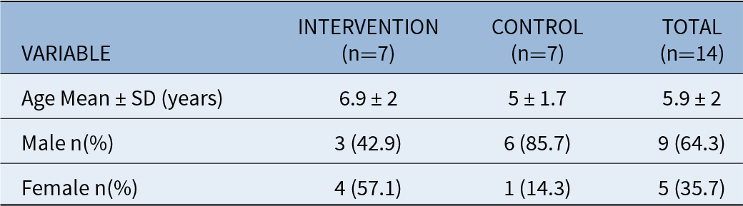
We identified 14 patients who met the inclusion criteria. All patients included in the study participated until the end of the study and attended the six-week follow-up. Their ages ranged from three to eight years (mean 5.93 years). In the interventional group, three (42.9 per cent) were male and four (57.1 per cent) were female. In the conservative group, six (85.7 per cent) were male and one (14.3 per cent) was female (Table 2). The control group was age and gender matched. Table 1 summarises the demographic data of the included children.
Demographic data for age and gender of included children (N = 14)

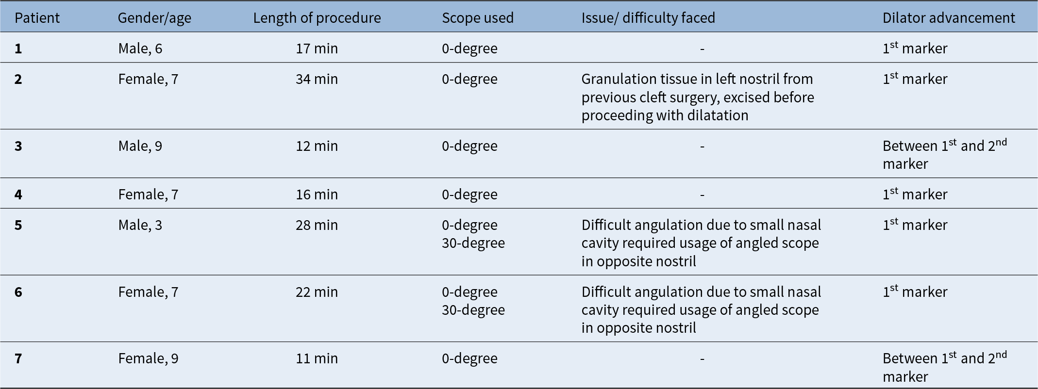
The average time to perform ETBD was 20 minutes, with longer times taken in case 2 due to the presence of granulation tissue and in cases 5 and 6 due to the narrow nasal passage. We utilised the 0-degree scope in all cases except for cases 5 and 6, where the 30-degree scope was utilised via the opposite nostril. In all but two cases, the device was inserted up to the first marker line, 1.75 mm. In older children, the insertion depth was between the first and second maker line, 2.35 mm. For all patients, the balloon was inflated to 12 atm for two minutes. Table 2 provides a summary of duration and challenges for ETBD.
Summary of duration and challenges for ETBD in Group 1

Outcome
For the primary outcome of tympanometry and PTA
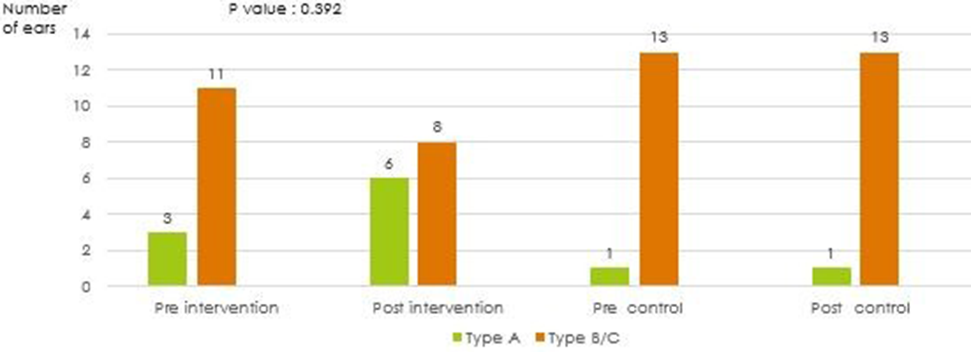
In this study, we found improvement in tympanometry in the intervention group and no improvement in the conservative group, although this difference was not statistically significant (p = 0.392) (Figure 1: Tympanometry pre- and post-intervention in Group 1 and Group 2).
Tympanometry pre- and post-intervention and control.

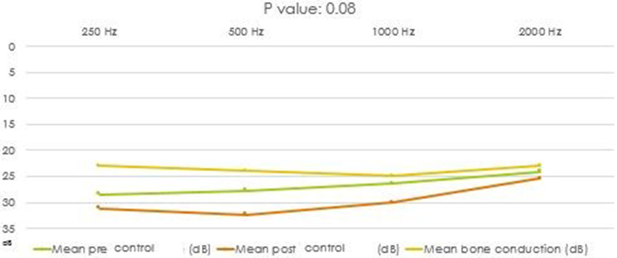
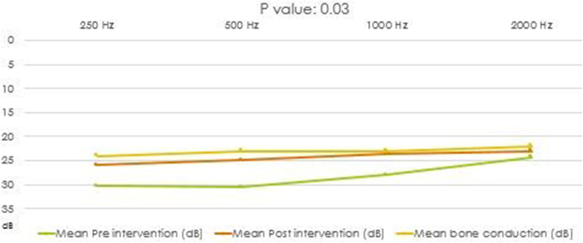
The hearing across 250 to 1 kHz significantly improved post-ETBD intervention compared to the control methods (p = 0.03). We also found that the air-bone gap in the intervention group also reduced significantly (p = 0.04) (Figures 2 and 3: Air-bone gap post-ETBD).
Pure tone audiometry pre and post, control.

Pure tone audiometry pre- and post-intervention.

For the secondary outcome of the ETDQ-7 score
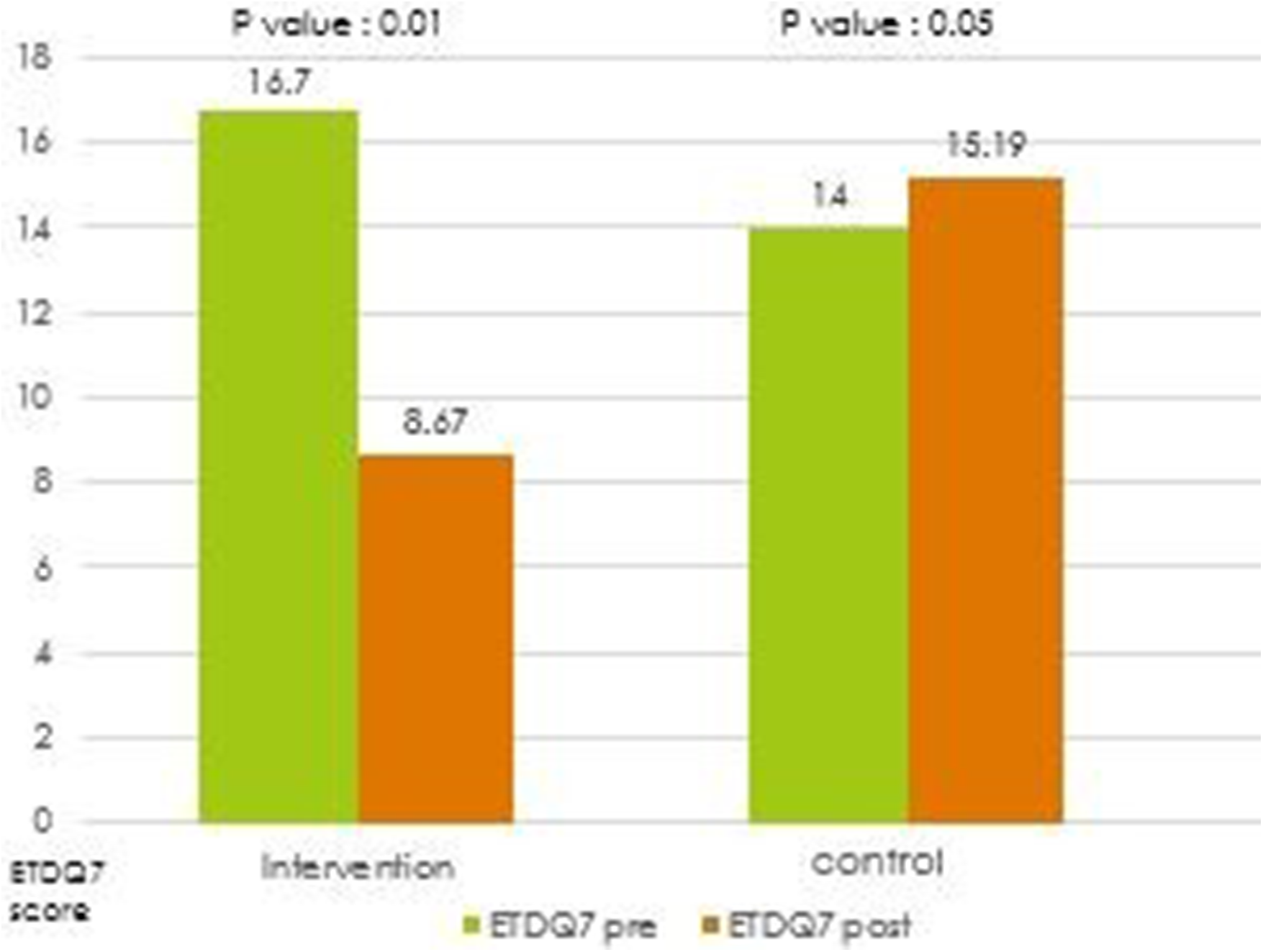
The mean score was significantly reduced in the intervention group compared to the conservative group (p = 0.01) (Figure 4).
Eustachian Tube Dysfunction Questionnaire-7 (ETDQ-7) score pre- and post-intervention and control.

No reported adverse reaction was found among the children in the intervention group besides minimal bleeding (50 per cent), controlled with digital compression over the nasal alar and blood-stained nasal discharge (50 per cent).
Discussion
CLP is a prevalent congenital craniofacial anomaly with a reported incidence of one in every 700 individuals.Reference Flynn, Möller, Jönsson and Lohmander12 The incidence of MEE in children with CLP is as high as 90 per cent,Reference Ponduri, Bradley, Ellis, Brookes, Sandy and Ness5, Reference Flynn, Möller, Jönsson and Lohmander12 with a reported overall incidence between the ages of one and five of 75 per cent, compared to only 19 per cent in non-CLP children.Reference Flynn, Möller, Jönsson and Lohmander12
The National Institute for Health and Care Excellence (NICE) guidelines advocate watchful waiting for three months, followed by intervention with either a ventilation tube or hearing aid.2 Ventilation tube insertion is the most favoured treatment option in most centres. Yet, the devastating long-term consequences of ventilation tube insertion, such as chronic perforation and retraction with cholesteatoma, have led to the exploration of novel techniques.
Children with CLP are prone to multiple ventilation tube insertions and, thus, are susceptible to long-term complications due to recurrent MEE. A study that included 8269 cleft patients revealed that 33.5 per cent of these children would require multiple grommet insertions.Reference Fitzsimons, Copley, Van Der Meulen, Panagamuwa and Deacon13 In another study involving 87 children with MEE who underwent ventilation tube insertion, complications were reported in 63.5 per cent of patients, notably myringosclerosis (34.6 per cent), tympanic membrane atrophy (23.5 per cent) and persistent otorrhea (5.6 per cent).Reference Yaman, Yilmaz, Alkan, Subasi, Guclu and Ozturk14
ETBD is gaining popularity worldwide as an intervention for recurrent or persistent MEE, especially in children. ETBD is found to be a rapid, simple and safe method for treating children with middle ear effusion.Reference Howard, Babu, Haupert and Thottam15 Clinical improvement was reported in over 80 per cent of children, supporting this intervention as a promising avenue. Parallel to that, complications of ETBD are also found to be much less compared to conventional surgical methods (i.e., myringotomy and grommet insertion).Reference Saniasiaya, Kulasegarah and Narayanan9, Reference Howard, Babu, Haupert and Thottam15 The successful outcomes of ETBD in the adult population have been well-established since 2010,Reference Ockermann, Reineke, Upile, Ebmeyer and Sudhoff16, Reference Toivonen, Kawai, Gurberg and Poe17 with emerging reports on favourable outcomes in the paediatric population.Reference Saniasiaya, Kulasegarah and Narayanan9, Reference Maier, Tisch and Maier18–Reference Gürtler, Husner and Flurin20
Our study found a statistically significant improvement in hearing threshold (p = 0.03) and the air-bone gap in audiometry (p = 0.04) among CLP children with MEE post-ETBD. Similarly, 33 per cent of the included children showed improvement in hearing as measured by the average threshold from 18 to 10 dBHL by Maier et al.Reference Maier, Tisch and Maier18 Toivonen et al. reported improvement in hearing threshold from 17. 5 dB HL to 5.7 dB HL at 36 months post-intervention.Reference Toivonen, Kawai, Gurberg and Poe17
Toivonen et al. reported tympanometry normalisation to Type A in 50 per cent at six months, 59 per cent at 12 months and 85 per cent at 36 months.Reference Toivonen, Kawai, Gurberg and Poe17 Although improvement was noted in our study, it was not statistically significant. Leichtle et al. found that improvement in Type A tympanogram was reported from 14 per cent to 50 per cent after one year of follow-up.Reference Leichtle, Hollfelder, Wollenberg and Bruchhage19 All these previous studies had adjunct procedures during the ETBD, which would have improved both objective and subjective parameters.
In the present study, a slight deterioration in hearing thresholds was observed in the control group following treatment with intranasal corticosteroids alone. This finding likely reflects the natural course and fluctuation of MEE commonly seen in children with CLP, who often have persistent ETD despite medical therapy. Over the six-week observation period, some subjects may have experienced residual or recurrent effusion, resulting in a marginal worsening of audiometric thresholds. Additionally, minor variations in middle ear pressure or tympanometric status at the time of testing could have contributed to these threshold changes. In contrast, children who underwent ETBD demonstrated improved Eustachian tube ventilation and middle ear aeration, supporting its potential role as an adjunctive intervention in this population.
Statistically significant subjective improvement was also demonstrated in our cohort following ETBD intervention using ETDQ-7 (p = 0.01). This is in keeping with other studies involving children, whereby Gürtler et al., 2024 reported an 80 per cent improvement in symptoms, mainly in pressure, pain, hearing impairment and difficulties equalising pressure.Reference Gürtler, Husner and Flurin20 Several studies have shown stable long-term results following ETBD in children,Reference Maier, Tisch and Maier18–Reference Gürtler, Husner and Flurin20 with the longest reported at a mean of 2.3 years of follow-up.Reference Toivonen, Kawai, Gurberg and Poe17 Yet, we cannot refute the possibility that the improvement of symptoms could be due to the natural course of the disease: ETD.Reference Gürtler, Husner and Flurin20
ETBD may improve symptoms, although the release of chronic adhesive and non-adhesive retraction and normalisation of tympanometry may not be achieved.Reference Gürtler, Husner and Flurin20
Tips and tricks for performing ETBD in CLP children
The approach for ETBD in children with CLP may be more challenging than for healthy children. Among the challenges faced is a significant narrow nasal passage in CLP children, which may vary from the nostril to the nasopharynx. It is worth noting that nasal distortions post-primary CLP operative manoeuvres have been reported widely reported. We summarise tips and tricks for ETBD in CLP children in Figure 4.
Our study has its limitations. We acknowledge that the small sample size limits the study. The study period is relatively short (six weeks), which may not represent the long-term outcome. The ETDQ-7 questionnaire was completed by parents or caretakers, which could have introduced parental bias and may need to be more representative. We relied on the parents’ answers to the questionnaire, interpreting them to the best of their ability from their children. Having said that, our study is the first to assess the efficacy and outcome of ETBD in CLP with MEE. Owing to its beneficial effect on both symptom and hearing outcomes, we advocate further analysis to investigate its long-term impact on MEE among children with various craniofacial anomalies apart from CLP. Age differences between groups may have influenced the outcomes observed. The control group was significantly younger than the intervention group, which could partially explain the persistence or recurrence of MEE, given that younger children typically have more immature Eustachian tube function. However, as this was a pilot study with a small sample size, the age imbalance was unintentional and highlights the need for larger randomised studies with age-matched cohorts to further validate these findings.
• Children with cleft lip and palate are prone to develop refractory middle ear effusion due to Eustachian tube dysfunction
• Eustachian tube balloon dilatation was well tolerated and showed improvement in hearing and symptoms among children with cleft lip and palate with middle ear effusion
• Eustachian tube balloon dilatation appears safe and may serve as a useful adjunctive intervention pending validation in larger studies
• Hearing assessment and Eustachian Tube Dysfunction Questionnaire show significant improvement following Eustachian tube balloon dilatation in cleft lip and palate children with middle ear effusion
• Long-term outcomes in cleft lip and palate children with middle ear effusion post-Eustachian tube balloon dilatation remain unexplored
Conclusion
We observed significant improvement in hearing thresholds on audiometry, as well as favourable changes in tympanometry and symptom scores (ETDQ-7) among CLP children with MEE following ETBD. While the tympanometric changes did not reach statistical significance, the overall clinical outcomes suggest that ETBD may offer symptomatic and functional benefits in this population. The procedure was well tolerated, with no major complications reported. These findings support ETBD as a safe and potentially useful adjunctive intervention in CLP children with MEE, warranting further validation through larger, controlled studies.
Acknowledgements
All parents and children involved
Funding
None.
Competing interests
All authors have no conflict of interest