INTRODUCTION
Unicellular eukaryotic organisms exist in morphologically and biologically distinct forms during their life cycle. Some stage-specific proteins could play important roles in both infection and cell development therefore providing potential targets for drug and vaccine design. Previous efforts to separate and purify different cell stages have significantly facilitated some large-scale studies on the stage-specific expression of proteins in these parasites. In Plasmodium falciparum, a high-throughput proteomic analysis of 4 isolated stages (sporozoites, merozoites, trophozoites, and gametocytes) led to the functional profiling of 2415 proteins, responsible for approximately 46% of all gene products predicted for the malaria agent (Florens et al. Reference Florens, Washburn, Raine, Anthony, Grainger, Haynes, Moch, Muster, Sacci, Tabb, Witney, Wolters, Wu, Gardner, Holder, Sinden, Yates and Carucci2002). Another example is provided by a combination of 2-DE-MS and 2-DE-free MS techniques that revealed 177 unique proteins expressed in late sporogonial stages of Encephalitozoon cuniculi (Brosson et al. Reference Brosson, Kuhn, Delbac, Garin, Vivares and Texier2006).
Microsporidia are highly specialized obligate intracellular eukaryotes with more than 1200 individual species capable of parasitizing a wide range of vertebrate and invertebrate hosts, including humans (Sprague et al. Reference Sprague, Becnel and Hazard1992). Nosema bombycis is the first described microsporidium (Naegli, Reference Naegli1857) recognized as the causative agent of silkworm pebrine which prevailed in Europe, America and Asia during the mid-19th century (Becnel and Andreadis, Reference Becnel, Andreadis, Wittner and Weiss1999) and is still epizootic and causing heavy economic losses in silk-producing countries such as China. The general life cycle of microsporidia can be divided into 3 phases: the infective phase (the extracellular phase of the cycle), the proliferative phase, and the sporogony phase. The proliferative phase begins when the sporoplasm (infectious content of the spore) travels into a new host cell through the everted polar tube. The morphologically simple cells called the meronts repeat their division one or several times and then differentiate into sporonts characterized by the deposition of a dense cell coat, initiating the sporogony phase. Subsequently, sporonts undergo the last division producing sporoblasts. Sporoblasts continue their morphogenesis process and finally develop into mature spores. N. bombycis is in direct contact with the host cell cytoplasm, and develops asynchronously. Due to its small size (mean spore size: 3·1–4·26×1·9–2·6 mm) (Mei and Jin, Reference Mei and Jin1989) and fragility, large-scale separation of different N. bombycis cell stages from heavily infected silkworm larvae is considered to be a difficult task. Here we present a density gradient-based method to purify the intracellular life-cycle stages of N. bombycis from infected tissues of the host, as a first ongoing effort to analyse differentiation stages of these parasites.
In addition, we used 2D gel electrophoresis (2D-PAGE) to evaluate the fractionation method for comparison of protein expressions between late sporoblasts and mature spores. By MALDI-TOF/TOF MS analysis, we identified an exosporal protein NbSWP5 which could only be found in mature spores. Subsequently, gene cloning, protein expression, immunoelectron microscopy analysis and spore phagocytosis assays were carried out to further address the location and function of this protein.
MATERIALS AND METHODS
Microsporidia
N. bombycis spores originally isolated from infected silkworms in Zhejiang, China (Mei and Jin, Reference Mei and Jin1989) were produced and harvested from laboratory-reared silkworm larvae as previously described (Zhang et al. Reference Zhang, Lu, Kumar, Zhu, Chen, Chen and Hong2007).
Percoll-gradient separation of microsporidian life-cycle stages
N. bombycis cell stages were isolated and purified as previously described (Seleznev et al. Reference Seleznev, Issi, Dolgikh, Belostotskaya, Antonova and Sokolova1995; Taupin et al. Reference Taupin, Metenier, Vivares and Prensier2006) with some modifications. Briefly, the fifth instar moulted silkworm larvae were challenged by feeding on mulberry leaves artificially polluted by N. bombycis spores (107 spores/100 larvae). The midguts of the infected larvae were dissected at day 5 of the fifth instar, and gently homogenized in 0·1 m PBS, pH 7·4. Large silkworm debris was removed by filtration on 4 layers of cheesecloth. After centrifugation (20 000 g, 5 min), the sediment was re-suspended in 15 ml of a solution containing 0·05% saponin and 0·05% (v/v) Triton X-100 in PBS. The cell suspension was passed through a plastic syringe with an inserted glass wool wad. Successive fractions of the filtrate were collected and thoroughly checked by light microscopy. Spores were concentrated by centrifugation (20 000 g, 5 min) after most of the contaminant was removed. The resulting pellet was dispersed with PBS and loaded onto a polycarbonate tube containing 12 ml of 70% Percoll (Pharmacia Fine Chemicals) in 0·25 m sucrose. Centrifugation was performed in a S58A angle rotor of a Himac CS150GXII ultracentrifuge (Hitachi Koki Co., Ltd) at 45 000 g for 30 min at 4°C. N. bombycis cells at different developmental stages were distributed to 3 major layers according to their respective densities. The top (A) and the middle (B) layers were collected and subjected to further centrifugation on 30% Percoll–0·25 m sucrose gradient and 50% Percoll–0·25 m sucrose gradient, respectively, generating empty germinated spore husks (A2), early sporoblasts (B1) and late sporoblasts (B2). All the fractions were washed and stored for further use.
Transmission electron microscopy (TEM)
Pellets derived from all PBS-washed gradient fractions were fixed in 2% glutaraldehyde, 0·05% ruthenium red, and 0·07 m cacodylate buffer (pH 7·4) for 1 h at room temperature. After washing for 30 min in 0·1 m cacodylate buffer (pH 7·4), cells were post-fixed for 1 h in 1% OsO4, dehydrated through a graded ethanol series, infiltrated in propylene oxide and embedded in Epikote resin (Agar Scientific). Ultrathin sections obtained with an UltracutSLeica ultramicrotome were placed on 300-mesh nickel grids coated with Formvar and carbon, then double-stained with uranyl acetate and lead citrate. Grids with sections were examined using a JEM-1230 transmission electron microscope.
Protein extraction and 2D gel electrophoresis
Late sporoblasts and mature spores were separately disrupted in a lysis buffer (109 cells/ml) containing 0·1 m DTT, 4% CHAPS and 0·2% SDS using acid-washed glass beads (Sigma, diameter: 425 μm-600 μm) in a FastPrep-24 (MP BIO). Proteins from broken cells were extracted with a solution containing 7 m urea, 2 m thiourea, 100 mm DTT, 4% CHAPS and 0·2% SDS for 6 h at room temperature. Insoluble material was removed by centrifugation for 10 min at 20 000 g. After quantification using Plus-One 2D Quant kit (Amersham), solubilized extracts (450 μg) were subjected to 24 cm IPG gel strips with a non-linear separation range of pH 3–10 (Amersham Biosciences) by in-gel sample rehydration. IEF was carried out at 20°C using an IPGphor unit (Amersham Biosciences) with the following conditions: 500 V for 1 h, 1000 V for 1 h, 5000 V for 1 h and 8000 V for 9 h. After equilibration with 50 mm Tris–HCl (pH 8·8), 6 m urea, 30% glycerol, 2% SDS, 100 mm DTT, and then 135 mm iodoacetamide, strips were applied on top of a 12·5% SDS-PAGE 20×24 cm2 format on a vertical Ettan-Dalt six unit (Amersham Biosciences). Gels were stained using CBB (Bio-Rad) then scanned with an ImageScanner (Amersham Biosciences) and image analysis was performed with the ImageMaster 2D Platinum 6.0 software (Amersham Biosciences).
MALDI-TOF/TOF MS analysis and database search
Protein spots were excised from the preparative gels, destained with 100 mm NH4HCO3 in 30% ACN. After removing the destaining buffer, gel pieces were lyophilized and rehydrated in 30 μl of 50 mm NH4HCO3 containing 50 ng trypsin (sequencing grade; Promega, Madison, WI, USA). After an overnight digestion at 3°C, the peptides were extracted 3 times with 0·1% TFA in 60% ACN. Extracts were pooled together and lyophilized. The resulting lyophilized tryptic peptides were kept at −80°C until mass spectrometric analysis. A protein-free gel piece was treated as above and used as a control to identify autoproteolysis products derived from trypsin. MS and MS/MS spectra were obtained using ABI 4800 Proteomics Analyzer MALDI-TOF/TOF (Applied Biosystems, Foster City) operating in a result-dependent acquisition mode. Peptide mass maps were acquired in positive ion reflector mode (20 kV accelerating voltage) with 1000 laser shots per spectrum. Monoisotopic peak masses were automatically determined within the mass range 800–4000 Da with a signal to noise ratio minimum set to 10 and a local noise window width of m/z 250. Up to 5 of the most intense ions with minimum signal to noise ratio: 50 were selected as precursors for MS/MS acquisition, excluding general trypsin autolysis peaks and matrix ion signals. In MS/MS-positive ion mode, spectra were averaged, collision energy was 2 kV, and default calibration was set. Monoisotopic peak masses were automatically determined with a signal to noise ratio minimum set to 5 and a local noise window width of m/z 250. The MS together with MS/MS spectra were searched against the UniprotKB/SwissProt database using the software GPS Explorer, version 3.6 (Applied Biosystems) and MASCOT version 2.1 (Matrix Science) with the following parameter settings: trypsin cleavage, 1 missed cleavage allowed, carbamidomethylation set as fixed modification, oxidation of methionines allowed as variable modification, peptide mass tolerance set to 100 ppm, fragment tolerance set to ±0·3 Da, and minimum ion score confidence interval for MS/MS data set to 95%.
Sequence analysis and recombinant protein expression
The gene encoding HSWP5 was amplified from N. bombycis genomic DNA using primers 5′-CGGAATTCAAAGAAAATAAGAATGTGCCG-3′ (forward) and 5′-CCGCTCGAGTTATTTATCCGAAGGTGC-3′ (reverse) containing an EcoR I and an Xho I restriction site (underlined), respectively. The amplified fragment was 495-bp corresponding to the amino acid regions 23–186 of the 186-amino acid protein. The PCR product was digested with EcoR I and Xho I then ligated into pET-30a (Novagen). After DNA sequence verification, the recombinant plasmid was transformed into Escherichia coli BL21 cells and expression of recombinant HSWP5 was induced for 6 h at 37°C in the presence of 1 mm isopropyl-b-thio-galactopyranoside (IPTG). The expressed fusion protein was then purified with His-Bind Purification Kit (Invitrogen).
Polyclonal antibody production and immunoblotting
Polyclonal antiserum against purified recombinant HSWP5 was produced by immunization of 2 native rabbits using the 56-day antibody production protocol. Eighty μg recombinant protein homogenized with Freund's complete adjuvant was subcutaneously injected for the first time and Freund's incomplete adjuvant for subsequent injections (at days +21, +35 and +49). Sera were collected 1 week after the last injection and stored at −20°C.
For immunoblot analysis, total proteins of N. bombycis were transferred to a polyvinylidene fluoride membrane after SDS-PAGE. Membranes were blocked for 1 h in Tween-Tris buffered saline (TTBS, 0·05% (v/v) Tween-20 in TBS) with 5% (w/v) skim milk and then incubated with either 1:1000 diluted anti-Hswp5 polyclonal antibody or control sera. After washing, membranes were reacted with peroxidase-labelled goat anti-mouse IgG antiserum (Sigma), at 1:1000 dilution. Bands were detected by addition of the substrate tetrahydrochloride (DAB).
Immunoelectron microscopy (IEM) analysis
N. bombycis spores were fixed with 3·0% paraformaldehyde and 1% glutaraldehyde in 0·1 m sodium cacodylate buffer, pH 7·2, for 1 h. After rinsing several times in buffer, the fixed spores were dehydrated with graded ethanols and sequentially permeabilized in low temperature embedding medium, Lowicryl K4M at −20°C. The resin was exposed to a 360-nm UV light source at room temperature until it was totally polymerized. Ultra-thin sections were placed on 300-mesh nickel grids coated with Formvar and carbon. For immunostaining, these grids were incubated in a blocking solution (1% bovine serum albumin (Sigma), 0·02% PEG 20000 (Promega), 0·1 m NaCl, 1% NaN3 in 50 mm PBS, pH 7·2) for 30 min, followed by incubation with a 1:200 dilution of primary antiserum (anti-SWP sera or negative control sera) for 1 h at 25°C, rinsed in PBS, and then incubated with a 1:200 dilution of 10-nm colloidal gold-conjugated goat anti-rabbit IgG at 25°C for 1 h. Grids were rinsed 5 times in PBS, stained with uranyl acetate and lead citrate, and then examined and photographed by a JEM-1230 transmission electron microscope.
Spore phagocytosis assays
For phagocytosis experiments, BmN cells from ovaries of Bombyx mori were used as host cells and seeded in glass-bottomed culture dishes (GBD-35-20, Nest Biotechnology Co., Ltd, China) at approximately 1×10⁶ with 2 ml of growth medium and allowed to settle to the bottom of the dishes for a minimum of 16 h. In order to measure the rate of phagocytosis, 2·5×107 freshly recovered mature spores, late sporoblasts and late sporoblasts incubated at 28°Cfor 4 h with NbSWP5 recombinant proteins (0·1–5 μg/ml), and late sporoblasts incubated with control sample (non-transformed total E. coli protein) were added to each dish with 2 ml of fresh medium respectively. The phagocytosis process was visually monitored by an inverted fluorescent microscope (Nikon Eclipse Ti-S). The numbers of free spores per field were counted using a 100× oil immersion objective. For each dish, the result was presented as a mean±standard deviation of 20 fields of magnification at 1000× assessed in a blinded fashion. Statistical significance among the control and experimental assays was determined by Student's t-test, values were considered significantly different if P was <0·001.
RESULTS
Separation and purification of N. bombycis life-cycle stages
The first centrifugation generated a reproducible pattern of 3 layers in the following order of increasing density. Transmission electron microscopy showed the first layer (fraction A) was a mixture of early sporonts, empty germinated spore husks and cellular debris derived both from host and parasite. The middle layer (fraction B) contained a population of sporoblasts with different degrees of maturity. The lower white layer (fraction C) contained mostly electron-dense mature spores and was virtually free of host contaminants.
In order to reduce the heterogeneity of fractions A and B, cells of corresponding bands were individually collected, PBS-washed, then loaded onto a 30% Percoll–0·25 m sucrose gradient and a 50% Percoll–0·25 m sucrose gradient respectively. Three separated fractions derived from fraction A were taken and examined by transmission electron microscopy. Cellular debris (A1) concentrated on the top layer was removed and discarded. Fraction A2 mainly consisted of empty spores. Further centrifugation of fraction B gave 2 major bands on the Percoll gradient. TEM analysis indicated that the light band B1 was rich in early sporoblasts while the heavier band B2 consisted of late sporoblasts.
In summary, 4 cell fractions representative of different life stages of N. bombycis were successfully isolated and purified: A2 for empty germinated spore husks, B1 for early sporoblasts, B2 for late sporoblasts, and C for mature spores (Fig. 1).
TEM micrographs of Nosema bombycis collected from different Percoll gradient fractions. Fraction A (A) is a mixture of sporonts (St) and empty spores (ES). Fraction A2 (B) contains only empty germinated spore husks. Fraction B1 (C) is rich in early sporoblasts marked by darkening sporoplasm. Fraction B2 (D) contains late sporoblasts with corrugated developing wall (arrow). Fraction C (E) contains mature spores with a typical thick wall.

Comparison of 2D protein profiles of isolated sporoblasts and mature spores
To evaluate the fractionation method for analysing protein expression, fractions B2 (late sporoblasts) and C (mature spores) were chosen for protein extraction followed by 2D-PAGE. A wide pH gradient (pH 3–10) and large-sized (24 cm) gels revealed high spot resolution. As shown in Fig. 2, major proteins were located at highly similar isoelectric point (pI) and molecular mass (MM) intervals for both fractions. However, several spot pattern differences were also observed. Spot 1, one of the most abundant protein spots, seemed to be specific in mature spores.
A comparison of 2DE maps of proteins from isolated late sporoblasts (A) and mature spores (B). Spot 1 is an abundant protein found only in mature spores. Rectangle regions are known to contain polar tube proteins.

MALDI-TOF/TOF MS and sequence analysis
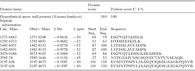
One hundred abundant protein spots of mature spores were excised and analysed by MALDI-TOF/TOF MS. However, due to the lack of N. bombycis genome and proteome databases, only several conservative proteins were identified (e.g. spot 2 and spot 3 were identified to be actins). Protein spot 1, the most abundant protein expressed in mature spores but absent in late sporoblasts, matched a 186-amino acid protein (Table 1) which was previously described as NbHSWP5 under GenBank Accession number EF683105 (Wu et al. Reference Wu, Li, Pan, Tan, Hu, Zhou and Xiang2008). By in silico analysis, HSWP5 possesses a 20-amino acid signal peptide (www.cbs.dtu.dk/services/SignalP/) and is predicted to be an extracellular (e.g. cell wall) protein (http://www.psort.org/). By NetNglyc and NetOglyc (http://www.cbs.dtu.dk), no N-glycosylation sites are predicted in HSWP5; however, 8 O-glycosylation sites are predicted. Neither protein homologies nor protein domains are predicted by using the BLAST program (www.ncbi.nlm.nih.gov) and the Pfam database (www.sanger.ac.uk/Software/Pfam/search.shtml).
MALDI-TOF/TOF MS analysis of protein spot 1

Expression of HSWP5 fusion protein
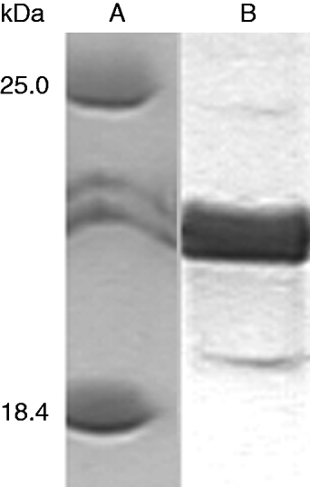
To further characterize HSWP5, we produced a recombinant protein in E. coli. The recombinant protein was expressed in E. coli and purified by affinity chromatography using His-Bind Purification Kit (Novagen). HSWP5-specific polyclonal antibody generated by immunizing rabbits with purified protein was used in Western blot analysis. A single ∼20 kDa band was detected from the N. bombycis spore protein lysate (Fig. 3).
Immunoblotting with antiserum of spore protein NbSWP5 of Nosema bombycis. (A) Protein marker. (B) A single 20 kDa N. bombycis protein recognized with the anti-NbSWP5 antibody.

Ultrastructural localization of HSWP5 in the N. bombycis spores
The specific antibody was used for immunoelectron microscopy to confirm the cellular location of HSWP5 in spores. As shown in Fig. 4, many gold particles were well distributed around the surface coat of mature N. bombycis spores, indicating that HSWP5 was a highly expressed exosporal protein. This result is consistent with the result of 2D-PAGE.
Immunoelectron microscopy of Nosema bombycis spores using anti-NbSWP5 antibody reveals labelling in the exospore. Arrows indicate numerous gold particles abutted round the thick dense surface spore coat.

Exogenous NbSWP5 protein protects N. bombycis from phagocytosis by host cells
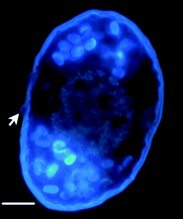
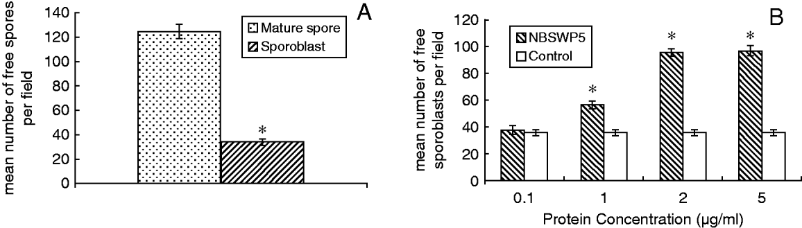
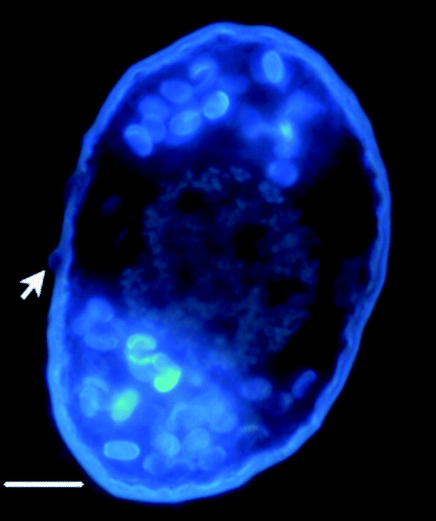
A series of assays was conducted to investigate the role of NbSWP5 in cell invasion. During the investigation of the interaction between N. bombycis and its host cells in vitro, we observed that spore precursor cells lacking NbSWP5 can be easily taken up by host cells (Fig. 5 and Fig. 6A) while freshly recovered mature spores with an intact spore wall barrier exhibited resistance. Additionally, phagocytosis was reduced by rNbSWP5 in a in a dose-dependent manner. Compared with the control, maximum inhibition of phagocytosis of spore precursor cells occurred when 2 μg/ml rNbSWP5 was used (Fig. 6B).
Nosema bombycis late sporoblasts phagocytosed by BmN cells are visualized by DAPI staining. Arrow indicates a bubble–like claw on the host cell surface showing a N. bombycis sporoblast being phagocytosed. Scale bar=10 μm.
NbSWP5 protects Nosema bombycis from being phagocytosed by BmN cells. (A) Sporoblasts can be more easily internalized by host cells. (B) Exogenous recombinant NbSWP5 inhibited sporoblast phagocytosis. The asterisks indicate significant differences (P<0·001).

DISCUSSION
While various proteomic studies have been performed on some unicellular parasites, very little has been done for N. bombycis. This is partly due to problems purifying the early stages of developing N. bombycis from their host cells. With its complete genome sequence project (running at Chongqing, China) nearing completion, more and more important information is provided (Xu et al. Reference Xu, Pan, Fang, Li, Tian, Li, Zhou and Xiang2006a; Wu et al. Reference Wu, Li, Pan, Tan, Hu, Zhou and Xiang2008; Xiang et al. Reference Xiang, Pan, Zhang, Xu, Li, Li, Zhou and Xiang2010). With that motivation, we developed a Percoll gradient-based procedure to separate the stages of N. bombycis life cycle from its natural host. Gentle detergent pre-treatment and glass-wool filtration efficiently reduced contaminating material. Utilization of a continuous gradient consisting of a Percoll–sucrose mixture provides each spore fraction with satisfying degrees of purity. Careful performance of the procedure yields sufficient cells of each stage for further biochemical study. A considerable quantity of sporonts was observed in the silkworm mid-gut; however, most of them were disrupted during the procedure, leaving a few of them distributed among the empty spore husks centrifuged in Fraction A. More work is needed to develop more gentle strategies for the isolation of those highly fragile earlier stages.
Sporoblasts are products of the final division of the entire intracellular development and the cells that undergo morphogenesis into spores marked by the biogenesis of a thick cell wall (Becnel and Andreadis, Reference Becnel, Andreadis, Wittner and Weiss1999). Thus, late sporoblasts and mature spores were chosen for protein extraction followed by 2D-PAGE to assess the fractionation method by analysing differential protein expression and identifying spore wall proteins. Differences in spot number and intensity between 2D protein profiles of spore precursor cells and mature spores were observed. Some spots were present in the spore profile but absent in the late sporoblast profile.
The study of spore wall proteins is a promising route for the development of a prospective target in diagnostic research and the treatment of microsporidiosis. So far, 7 spore wall proteins have been identified in the Encephalitozoonidae family (Bohne et al. Reference Bohne, Ferguson, Kohler and Gross2000; Hayman et al. Reference Hayman, Hayes, Amon and Nash2001; Brosson et al. Reference Brosson, Kuhn, Prensier, Vivares and Texier2005; Peuvel-Fanget et al. Reference Peuvel-Fanget, Polonais, Brosson, Texier, Kuhn, Peyret, Vivares and Delbac2006; Xu et al. Reference Xu, Takvorian, Cali, Wang, Zhang, Orr and Weiss2006b; Southern et al. Reference Southern, Jolly, Lester and Hayman2007). In N. bombycis, 2 exosporal proteins (SWP26 and SWP32) and 2 endosporal proteins (SWP25 and SWP30) have been reported (Wu et al. Reference Wu, Li, Pan, Tan, Hu, Zhou and Xiang2008, Reference Li, Wu, Pan, He, Zhang, Hu and Zhou2009; Li et al. Reference Li, Wu, Pan, He, Zhang, Hu and Zhou2009). There are reports suggesting that some microsporidian spore wall proteins are involved in spore adherence and host invasion processes (Southern et al. Reference Southern, Jolly, Lester and Hayman2007; Li et al. Reference Li, Wu, Pan, He, Zhang, Hu and Zhou2009). However, our preliminary investigation showed that KOH-treated spores and cold-stored spores with spore wall defects can be more easily internalized by host cells than freshly recovered intact spores. Indeed, freshly recovered spores seemed to be resistant in host cell phagocytosis. In the current study, we further demonstrated that spore precursor cells lacking spore wall protein 5 is more likely to be phagocytosed by host cells than mature spores. Moreover, we found that exogenous recombinant NbSWP5 could reduce spore precursor cell phagocytosis in a dose-dependent manner.
Given the evidence from recent phylogenetic analyses and genome data, it is now widely acknowledged that microsporidia are closely related to fungi (Katinka et al. Reference Katinka, Duprat, Cornillot, Metenier, Thomarat, Prensier, Barbe, Peyretaillade, Brottier, Wincker, Delbac, El Alaoui, Peyret, Saurin, Gouy, Weissenbach and Vivares2001; Keeling, Reference Keeling2003). Pathogenic fungi have evolved efficient strategies such as physical barriers (chitin and proteins) to avoid innate immune recognition (Seider et al. Reference Seider, Heyken, Luttich, Miramon and Hube2010). A recent study, working with A. fumigatus conidia, confirmed a 19 kDa protein RodAp on the cell wall surface, masking the immunogenicity of the dormant conidia while hydrofluoric acid-treated dormant wild-type conidia or germinating conidia lacking the hydrophobin layer could be easily recognized by host cells (Aimanianda et al. Reference Aimanianda, Bayry, Bozza, Kniemeyer, Perruccio, Elluru, Clavaud, Paris, Brakhage, Kaveri, Romani and Latge2009). Microsporidia have evolved a sophisticated mechanism to infect host cells. Their unique infection apparatus, consisting of a coiled polar tube and the infective sporoplasm, are wrapped in a thick rigid spore wall. Staying out of a host cell is beneficial for these microbes to trigger the germination and transportation of the sporoplasm. We therefore infer that NbSWP5 is a candidate protective protein like RodAp to avoid spore pathogen-associated molecular patterns being recognized by responsive cells. Further studies to investigate the molecular function of NbSWP5 on parasite–host interactions are in progress.
ACKNOWLEDGEMENTS
This research was supported by the Natural Science Foundation of China (Project no.: 30270898).