INTRODUCTION
In the minds of most people the term parasite suggests adverse effects on the host such as disease and pathogenicity. But the reverse of this image is that parasites can sometimes be of benefit to their hosts. For example, recent studies such as those of Galli et al. (Reference Galli, Crosa and Occhipinti-Ambrogi1998), Siddall and Sures (Reference Siddall and Sures1998), Sures and Siddall (Reference Sures and Siddall1999), Sures and Taraschewski (1999, Reference Sures and Taraschewski2001), Sures et al. (Reference Sures, Siddall and Taraschewski1999), Sures (Reference Sures2001, Reference Sures2004) and Williams and MacKenzie (Reference Williams and MacKenzie2003) have shown that metal concentrations in some helminth parasites are very much higher than those in their hosts. In this way the parasites can reduce heavy metal concentrations in the tissues of their hosts. A classic example of this effect is the acanthocephalan parasite Pomphorhynchus laevis (Acanthocephala) in chub Leuciscus cephalus. In this host/parasite system the lead and cadmium concentrations in parasite tissues are 2700 and 400 times higher respectively than their concentrations in host muscle (Sures and Taraschewski, Reference Sures and Taraschewski1999).
According to MacKenzie (Reference MacKenzie1999), there are 3 good reasons for selecting parasites for use as bioindicators. First, there are more parasitic than free-living species. Secondly, in parasites with complex life-cycles, the numbers of potential indicators increase with the number of different developmental stages. Thirdly, some parasites are highly sensitive to changes in their environment while others are much more tolerant. Methodologically, parasites provide a quick and sensitive method for estimating the pollution level, especially in remote areas where pollution levels are low (Sures, Reference Sures2003).
In the present study, the lead and cadmium concentrations in the tissues of the shark Carcharhinus dussumieri (Müller and Henle, 1839) and its cestode parasites, Anthobothrium sp. and Paraorygmatobothrium sp., were measured and compared.
MATERIALS AND METHODS
Sixteen specimens of the shark C. dussumieri were collected from Hurmozgan province in the Persian Gulf by Ferdows fishing trawler. Fish were dissected, sexed and examined for helminth parasites. Tissue samples from the muscle, liver, intestine and gonad as well as specimens of adult cestodes from the spiral intestine were collected. Samples, varying in weight from 0·0005 g (in some parasites) to 3 g were digested in a mixture of 3 ml of nitric acid (65%) and 3 ml of perchloric acid (75%) for 24 h. To speed digestion, specimens were heated to 50°C. The organic parts present in the solutions were extracted by 5 ml of chloroform. Finally, the aqueous layer was diluted up to 10 ml with distilled water and analysed in an Inductively Coupled Plasma-OES Vista MPX (Varian, Inc.). Samples of standard reference materials (ICP multi-element standard solution IV Certipur Lot. no. OC317366) were used to check the accuracy of the analysis. Detection limits of the measurements were 0·2 μg/l and 1·5 μg/l for cadmium and lead, respectively. Data were analysed using the SPSS 13 package.
RESULTS
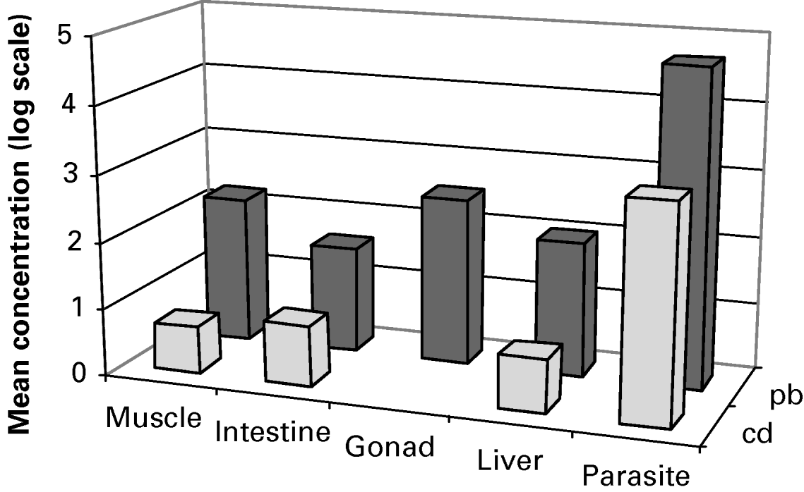
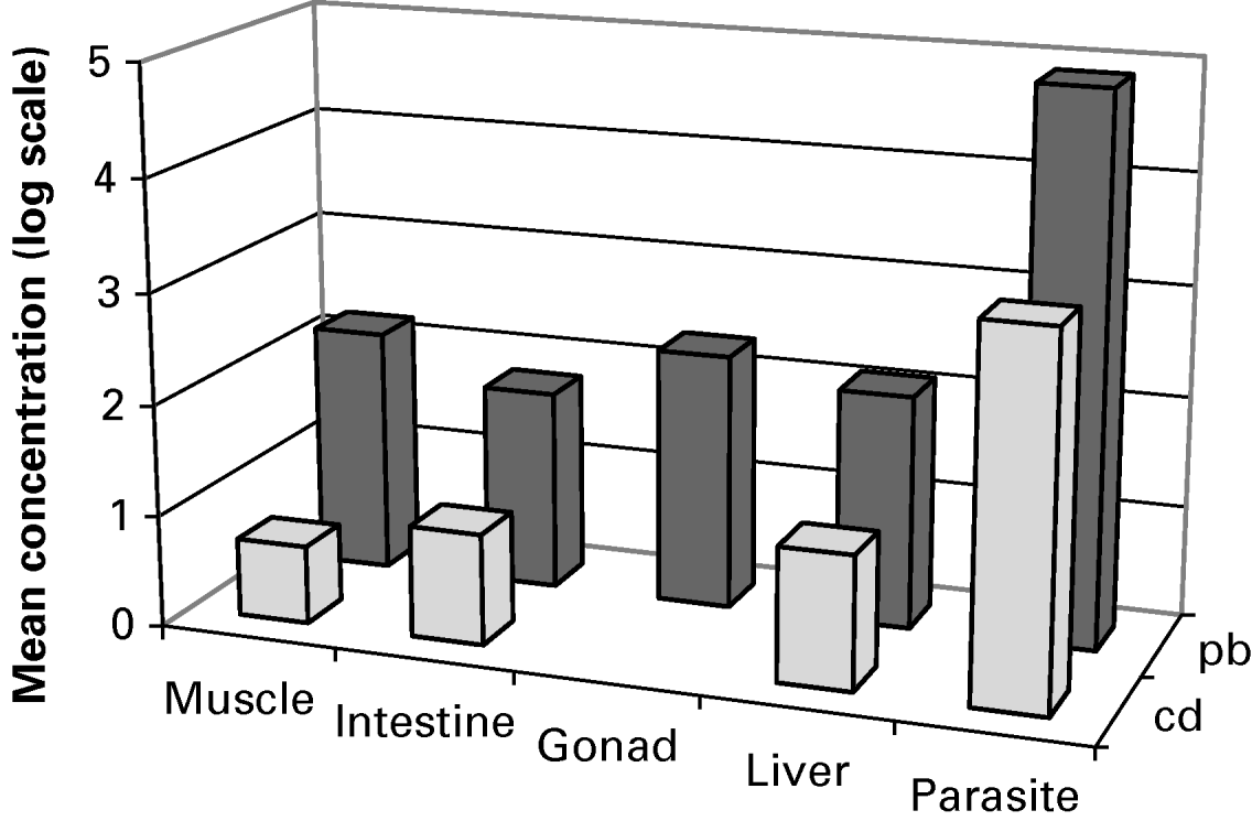
Cadmium and lead concentrations were much higher in the 2 cestode parasites than in the host tissues (Table 1, Figs 1 and 2), with levels 278 and 332 times higher in Anthobothrium sp., and 455 and 438 times higher in Paraorygmatobothrium sp., than in the muscle of the host. In Anthobothrium sp. the mean cadmium and lead concentrations (±s.e.) were 0·018 (±0·008) and 0·47 (±0·086) μg g−1 wet weight, respectively, while in Paraorygmatobothrium sp. the mean cadmium and lead concentrations (±s.e.) were 0·02 (±0·016) and 0·77 (±0·26) μg g−1 wet weight, respectively. The concentrations in Paraorygmatobothrium sp. were higher than in Anthobothrium sp., but the Mann-Whitney U-test showed that the differences were not statistically significant.
Relative concentrations, converted to logarithmic scale, of cadmium and lead in Paraorygmatobothrium sp. and its host tissues.

Relative concentrations, converted to logarithmic scale, of cadmium and lead in Anthobothrium sp. and its host tissues.

Mean cadmium and lead concentrations in cestode parasites and host tissues

* ND=No data (in some gonad samples the cadmium concentrations were very low and were considered unreliable).
The results of Wilcoxon tests showed that the lead and cadmium concentrations in Paraorygmatobothrium sp. were very significantly higher than those in the host muscle, liver and intestine (0·002<P<0·043). In Anthobothrium sp. the cadmium concentration was also significantly higher than in host muscle (P=0·043), and the lead concentrations were very significantly higher than in the host muscle, liver, intestine and gonad tissues (0·005<P<0·043).
DISCUSSION
Sures and Siddall (Reference Sures and Siddall1999) pointed out that, as acanthocephalans are not able to synthesize their own cholesterol and fatty acid, they have become extremely efficient at taking them up from the intestinal lumen. Organometallic complexes in the host bile pass down the bile duct into the small intestine where they are taken up by acanthocephalans concurrently with bile salts. The same might also happen in cestode parasites (Sures, personal communication 2006). Consequently, this will reduce the amount of metal uptake by the host's intestinal wall. So, by acting as heavy metal filters or sinks, these parasites may actually have a beneficial effect on the health of their hosts.
Marine fish lose water through their gills as a result of osmoregulation. To compensate for this, they drink seawater, so that their intestinal helminths are exposed mainly to inorganic metal ions via oral intake rather than organometallic complexes in the bile. One would therefore expect lower heavy metal levels in marine fish cestodes than in their freshwater counterparts (Sures and Siddall, Reference Sures and Siddall1999). In the present study, 2 shark parasites, like parasites of freshwater fish, had much higher heavy metal concentrations than the host tissues. This unexpected result could be related to the salt gland and high urea level in shark blood. Marine elasmobranchs maintain an osmotic pressure that is slightly higher than that of sea water by retaining urea in the blood (Naylor, 2006, personal communication). Therefore, their intestinal helminth parasites might be expected to show a similar pattern of heavy metal accumulation to those of freshwater teleosts.
Most studies of fish parasites as accumulators of heavy metals have so far focused on acanthocephalans (Sures et al. Reference Sures, Taraschewski and Jackwerth1994a, Reference Sures, Taraschewski and Jackwerthb, Reference Sures, Taraschewski and Jackwerthc; Sures and Taraschewski, Reference Sures and Taraschewski1995; Galli et al. Reference Galli, Crosa and Occhipinti-Ambrogi1998; Siddall and Sures, Reference Siddall and Sures1998; Sures and Siddall, Reference Sures and Siddall1999; Sures and Reimann, Reference Sures and Reimann2003; Thielen et al. Reference Thielen, Zimmermann, Baska, Taraschewski and Sures2004), but cestodes have also been shown to accumulate heavy metals at rates many times higher than their fish hosts (Sures et al. Reference Sures, Taraschewski and Rokicki1997; Tenora et al. Reference Tenora, Baruš, Kracmar and Dvoracek2000; Turčekova et al. Reference Turčekova, Hanzelova and Spakulova2002). The present study confirms the value of cestodes as sensitive bioindicators in environmental monitoring and is also the first such study in a marine elasmobranch. The results strongly support the view that helminth parasites are extremely sensitive early warning bioindicators of heavy metal pollution, particularly in sensitive environments and remote areas such as the Antarctic, as suggested by Sures (Reference Sures2003). We further suggest that cestode parasites of elasmobranchs could be useful indicators of heavy metal pollution in another remote environment – the ocean depths – where elasmobranch fish are a major component of the fauna.
The project was financed by the Research Affairs Department of The University of Tehran. We are indebted to Professor Janine Caira for the confirmation of the parasites' identification. We are grateful to Dr Valinasab for providing the fishing facilities.