Over 16 000 missense mutations are linked to human diseases (Refs Reference Sanders and Myers1, Reference Stenson2). A missense mutation – also known as a nonsynonymous single-nucleotide polymorphism – is a heritable change of a single base in a coding region of a gene that causes an amino acid change in the corresponding protein. These mutations are responsible for about half of the genetic changes known to cause disease (Ref. Reference Ng and Henikoff3). A common theme emerging from detailed studies of mutant proteins linked to disease is that point mutations have deleterious effects on folding and trafficking of proteins that are synthesised in the endoplasmic reticulum (ER) (Ref. Reference Sanders and Myers1). The term trafficking is used to describe the delivery of proteins from the ER to other parts of the cell. We refer to mutants synthesised in the ER that are defective in protein folding or trafficking as processing mutants.
The underlying defects in diseases caused by processing mutants can be divided into two broad categories: reduced or absence of functional protein at a required location; and defective protein folding leading to the accumulation of toxic aggregates. In the first case, misfolded proteins are recognised by the cell's quality-control mechanism as being defective, are usually retained in the ER and degraded by the ER-associated degradation (ERAD) system (Refs Reference Hebert, Garman and Molinari4, Reference Kopito5), although in some instances (e.g. in Charcot–Marie–Tooth disease) they may be degraded in the lysosome (Ref. Reference Pedrola6). In the second case, in some tissues mutant proteins that are prone to aggregation appear to avoid ERAD, and accumulate in post-ER sites (Ref. Reference Sekijima7). Cells respond to the accumulation of misfolded protein in the ER or to protein aggregation by undergoing an unfolded-protein response (UPR) (Ref. Reference Schroder and Kaufman8) that tries to reduce ER stress through increased expression of housekeeping proteins, increased degradation of misfolded proteins and suppression of translation initiation (Ref. Reference Pearse and Hebert9).
The misfolding of a protein leading to its aggregation and accumulation as protein deposits is a characteristic of several neurodegenerative diseases, including Alzheimer disease, Creutzfeldt–Jacob disease, Parkinson disease, Huntington disease, serpin-deficiency disorders, amyotrophic lateral sclerosis and over a dozen other rare diseases (reviewed by Refs Reference Horwich10, Reference Stefani11, Reference Soto, Estrada and Castilla12). For example, in Alzheimer disease, accumulation of protofibrillar protein aggregates in the brain leads to neurodegeneration and dementia. Although the proteins involved in these diseases do not share sequence or structural identity, the formation of protein aggregates shows some similarities. The amyloid-forming proteins usually contain stacks of β-sheets (Ref. Reference Soto, Estrada and Castilla12); these have a high tendency to form large polymers because β-sheets are stabilised by intermolecular interactions. The mechanisms involved in protein aggregation in disease are reviewed in detail in Refs Reference Horwich10, Reference Soto, Estrada and Castilla12 and Reference Rochet13.
A classic example of a disease arising from reduction or absence of functional protein at a required location as a result of defective protein folding is cystic fibrosis. About 90% of cystic fibrosis patients express a mutant of the cystic fibrosis transmembrane conductance regulator (CFTR) that is missing one amino acid (ΔF508 CFTR). The mutant is defective in folding and delivery to the cell surface. Other examples of diseases resulting from defective folding and/or trafficking of proteins (reviewed in Refs Reference Stenson2, Reference Aridor and Hannan14 and Reference Aridor and Balch15) are familial hypercholesterolaemia, long QT syndrome (cardiac arrhythmias), diabetes insipidus, persistent hyperinsulinaemic hypoglycaemia of infancy, retinitis pigmentosa, Charcot–Marie–Tooth disease, a type of autism (Ref. Reference De Jaco16), and hereditary hearing loss. This review focuses on methods to improve the folding and trafficking defects in this class of mutant proteins.
Synthesis of membrane proteins in the ER
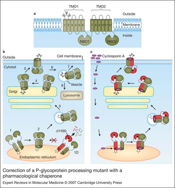
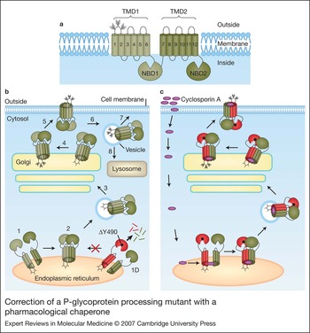
Synthesis of proteins destined for the plasma membrane, organelles (e.g. lysosomes) or for export is a complex process. Figure 1b is a schematic representation of synthesis of a polytopic membrane protein.
Correction of a P-glycoprotein processing mutant with a pharmacological chaperone (a) The four domains of human multidrug resistance P-glycoprotein (P-gp) are shown: two transmembrane domains (TMDs) and two nucleotide-binding domains (NBDs). Each TMD consists of six transmembrane (TM) segments (numbered cylinders). The branched lines represent glycosylated sites. (b) Synthesis of ΔY490 mutant (red and green model) and wild-type (green model) P-gps. In the first stages of synthesis and folding of the polypeptide chain, wild-type P-gp undergoes some cotranslational folding to yield a biosynthetic intermediate where the four domains (NBD1, NBD2, TMD1, and TMD2) are loosely associated and packing of the TM segments is incomplete (step 1). The protein then matures as the four domains form tight interactions (step 2). Complete folding to yield a compact structure enables the protein to leave the endoplasmic reticulum (ER) and enter the Golgi complex (step 3). In the Golgi, the high-mannose carbohydrate groups (three lines joined to the first loop) are modified to yield complex carbohydrate (multibranched lines) (step 4). The mature, fully glycosylated protein is delivered to the cell surface in a functional form (step 5). Proteins at the cell surface can undergo endocytosis to be retrieved from the plasma membrane (step 6). The retrieved protein can be recycled to the plasma membrane (step 7) or targeted for degradation in the lysosome (step 8). If P-gp contains a processing mutation such as ΔY490, which is analogous to the ΔF508 mutation of the cystic fibrosis transmembrane conductance regulator (CFTR), then the presence of the mutation alters the NBD1 surface (ragged edge) that normally makes contact with TMD1, and introduces a thermodynamic hurdle that interferes in the ability of the protein to complete the folding process. This defective protein is retained in the ER (step 1D) and is targeted for degradation by cytosolic proteasomes that cleave the protein into fragments (short lines). (c) Expression of ΔY490 P-gp in the presence of a pharmacological chaperone. When the ΔY490 mutant is expressed in the presence of cyclosporin A (purple ovals), the drug diffuses into the cell and acts as a pharmacological chaperone by binding at the interface between the two TMDs to promote packing of the TM segments, resulting in a tightly folded compact structure. The rescued P-gp can then be delivered to the cell surface in a functional form.
Synthesis of membrane proteins is not only complex but can also be inefficient. The efficiency of synthesis of functional protein at the proper destination is less than 50% for many wild-type membrane proteins, including the CFTR (Ref. Reference Seibert17), delta opioid receptor (Ref. Reference Petaja-Repo18), nicotinic acetylcholine receptor (Ref. Reference Paulson19), AE1 anion exchanger (Ref. Reference Quilty, Li and Reithmeier20), and EnaC sodium channel (Ref. Reference Valentijn, Fyfe and Canessa21). The low folding efficiency of many membrane proteins suggests that the energetic balance between complete and incomplete folding must be quite delicate (Ref. Reference Sanders and Myers1). It has been estimated that a 1.5 kcal/mole change in free energy associated with a single amino acid change would reduce the efficiency of a folding process from 50% at body temperature to less than 10% (Ref. Reference Sanders and Myers1). Therefore, the delicate folding process of many membrane proteins makes them particularly sensitive to single amino acid changes.
Approaches to correct protein-folding and -trafficking defects
Chemical chaperones
Studies on transporters such as the human multidrug resistance P-glycoprotein (P-gp) (Ref. Reference Loo, Bartlett and Clarke22) and CFTR (Ref. Reference Chen23) suggest that processing mutations interfere in folding by trapping the protein in a partially folded structure. The structure of the mutant biosynthetic intermediate is similar to a biosynthetic intermediate of the wild-type protein but the presence of the point mutation creates a thermodynamic hurdle for completion of the folding process. The biosynthetic intermediate is unstable and is targeted for degradation. Therefore, one approach to promote folding of processing mutants would be to stabilise the mutant protein during the folding process through the use of chemical chaperones. Chemical chaperones refer to small molecular weight compounds that promote folding by stabilising proteins without actually binding to them (see Refs 24 and 25 for reviews). The most common types of chemical chaperones used to promote folding are osmolytes such as glycerol and trimethylamine-N-oxide (TMAO).
Osmolytes stabilise proteins by increasing the hydration of proteins (Ref. Reference Gekko and Timasheff26) and/or reducing the mobility of individual protein domains (Ref. Reference Sampedro and Uribe27). Increased hydration of proteins promotes protein stabilisation by increasing the free-energy difference between a partially folded protein (which has increased surface area) and its more compact native structure (Ref. Reference Arakawa24): the native structure therefore becomes relatively more stable. In addition, osmolytes reduce the free movement of proteins to prevent aggregation of partially folded molecules. Deuterated water might function by a similar mechanism to promote protein folding (Ref. Reference Papp and Csermely25).
Osmolytes appear to be nonspecific in their ability to promote folding. Processing mutants of cystic fibrosis (CFTR) (Ref. Reference Sato28), liver disease (α-antitrypsin) (Ref. Reference Burrows, Willis and Perlmutter29), Menke disease (copper transporter), nephrogenic diabetes insipidus [aquaporin-2 (Ref. Reference Tamarappoo and Verkman30) or vasopressin V2 receptor (V2R) (Ref. Reference Tan, Nickols and Limbird31)] and maple syrup urine disease (branched chain α-ketoacid dehydrogenase) (Ref. Reference Song and Chuang32) could be rescued with osmolytes.
Not all chemical chaperones function as osmolytes. Compounds such as dimethyl sulphoxide, 4-phenyl butyrate and lipids/detergents are not osmolytes but can promote folding of mutant proteins (reviewed in Ref. Reference Papp and Csermely25).
It might not be practical to use chemical chaperones in clinical studies. The body could metabolise osmolytes such as glycerol, and the high concentrations of osmolytes required might also be too toxic; for example, treatment of mice expressing the ΔF508 CFTR gene with sufficient TMAO to rescue the protein also killed half of the mice (Ref. Reference Fischer33). Probably the biggest problem with osmolytes is that they are not specific in their action and they could potentially affect multiple metabolic pathways. The compound 4-phenylbutyrate is an exception, however. The US Food and Drug Administration has approved 4-phenylbutyrate for use in treating urea-cycle disorders, where it acts as an ammonia scavenger (Ref. Reference Brusilow and Maestri34), and for clinical trials in the treatment of diseases such as cystic fibrosis (Ref. Reference Rubenstein, Egan and Zeitlin35) and thalassaemia (Ref. Reference Collins36). In addition to acting as a chemical chaperone to promote folding of proteins, 4-phenylbutyrate has also been reported to suppress general ER stress (Refs Reference Kubota37, Reference Ozcan38). Suppression of ER stress by 4-phenylbutyrate could be a potential form of treatment for type 2 diabetes, since ER stress is postulated to be a key link between obesity and insulin resistance (Ref. Reference Ozcan38).
Pharmacological chaperones
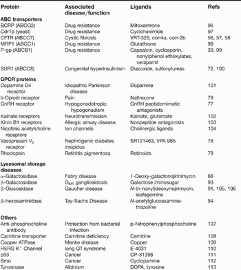
Another strategy to overcome folding defects in processing mutants is to stabilise folding intermediates in the ER with specific hydrophobic ligands that can diffuse into the cell. The feasibility of using such ‘pharmacological chaperones’ or ‘pharmacochaperones’ for a membrane protein was first demonstrated with rescue of processing mutants of P-gp (Ref. Reference Loo and Clarke39) by carrying out expression in the presence of drug substrates such as capsaicin, cyclosporin A, verapamil or vinblastine. The P-gp mutants were delivered to the cell surface in a functional form. Processing mutants of many other proteins can also be rescued with pharmacological chaperones (Table 1).
Misfolded proteins rescued by pharmacological chaperones

Key abbreviations: ABC, ATP-binding casette; GPCR, G-protein-coupled receptor.
The advantages of pharmacological chaperones are that they can be specific and nontoxic, and can be used at much lower concentrations than chemical chaperones. Examples of processing mutants rescued by pharmacological chaperones are discussed below.
Examples of proteins rescued with pharmacological chaperones
ABC transporters
Humans express 48 different members of the ATP-binding cassette (ABC) family of transporters. These proteins are characterised by the presence of four domains: two cytoplasmic nucleotide-binding domains (NBDs); and two transmembrane domains (TMDs) each of which has six transmembrane (TM) segments. Defects in 14 of these transporters have been linked to diseases such as cystic fibrosis, familial persistent hyperinsulinaemic hypoglycaemia of infancy, insulin-dependent diabetes mellitus, progressive familial intrahepatic cholestasis, Tangier disease, Stargardt disease, age-related macular degeneration, immune deficiency, Dubin– Johnson syndrome, adrenoleukodystrophy and sitosterolaemia. Many of the problems are caused by the presence of processing mutations in an ABC transporter (Ref. Reference Dean and Annilo40).
P-glycoprotein
The human multidrug resistance P-gp (ABCB1) has been a useful model system for studying the mechanism of rescue with pharmacological chaperones because truncation mutants as well as processing mutants (mutations scattered throughout the molecule) can be corrected. P-gp functions as a drug-efflux pump, and contributes to the phenomenon of multidrug resistance during cancer chemotherapy (reviewed in Ref. Reference Loo and Clarke41). In P-gp, tyrosine 490 (Y490) in NBD1 occupies the equivalent position to F508 in CFTR, such that deletion of Y490 (ΔY490 P-gp) inhibits maturation and delivery of P-gp to the cell surface (Ref. Reference Hoof42). Expression of ΔY490 P-gp in the presence of drug substrates and modulators was found to promote maturation of the mutant protein to yield a functional transporter at the cell surface (Ref. Reference Loo and Clarke39).
A model of P-gp folding and the effect of processing mutations and pharmacological chaperones are shown in Figure 1. During protein synthesis, the protein undergoes some cotranslational folding to yield the four domains of the molecule arranged in a loosely folded conformation (Fig. 1b). The protein then undergoes superfolding (Ref. Reference Loo and Clarke43) as contacts are established between the four domains and packing of the TM segments is completed. The molecule is now in a tightly folded compact structure that can leave the ER and be delivered to the cell surface in a functional form. Processing mutations act as thermodynamic hurdles in the folding process (Fig. 1b). The molecule does not complete the folding process. Folding intermediates exist transiently before being degraded. Expression of a processing mutant in the presence of a pharmacological chaperone such as a drug substrate promotes folding of the partially folded structure to yield a functional protein at the cell surface (Fig. 1c). The drug appears to induce folding by promoting interactions between the TMDs since truncation mutants lacking both NBDs can still be rescued with drug substrates (Ref. Reference Loo and Clarke44). Both TMDs, however, are required for drug rescue (Ref. Reference Loo and Clarke43). The effects of substrates or modulators on P-gp appear to be mimicked by the presence of suppressor mutations in the drug-binding pocket of the protein (Ref. Reference Loo, Bartlett and Clarke45).
CFTR
Cystic fibrosis is an autosomal recessive disease and affects 1 in 2500 live births (Ref. Reference Rowe, Miller and Sorscher46). In the Caucasian population, one in 25 persons are carriers of the defective gene. Although more than a thousand disease-associated mutations have been identified in the CFTR gene, one particular mutation – deletion of phenylalanine 508 (ΔF508 CFTR) – has been found in at least 90% of patients. The deletion of F508 in CFTR causes a defect in protein folding.
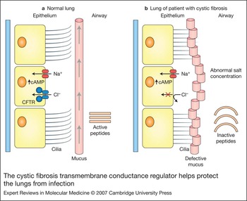
CFTR (ABCC7) is a cAMP-activated chloride channel located at the cell surface of a variety of cells (Ref. Reference Riordan47). The lack of chloride channel activity in cystic fibrosis patients leads to mucosal obstruction of a variety of ducts within organs such as the pancreas, liver, sweat glands, salivary glands and lungs. The main cause of morbidity in cystic fibrosis patients is the presence of thick, tenacious secretions that obstruct distal airways and submucosal glands (Fig. 2). These patients have chronic lung infections that cause a decline in respiratory function and eventual lung failure.
The cystic fibrosis transmembrane conductance regulator helps protect the lungs from infection (a) The normal lung is protected from bacterial infections by the presence of antimicrobial peptides and a mucus layer that lines the airways. Bacteria inhaled into the lung are caught in the mucus layer and their growth is inhibited by the presence of the microbial peptides. The action of cilia moves the mucus along with the bacteria out of the lung. The cystic fibrosis transmembrane conductance regulator (CFTR) contributes to these protective measures by maintaining normal salt levels in conjunction with sodium channels. Normal physiological salt levels are required for the antimicrobial peptides to function and to maintain hydration of the mucus layer. (b) In the cystic fibrosis lung, an inactive or absent CFTR inhibits chloride absorption and alters the salt balance. Perturbation of ion levels reduces the effectiveness of antimicrobial peptides and causes the mucus to become dehydrated and viscous. Bacterial infections are not effectively eradicated and persist in the lung. Chronic inflammation leads to lung failure.

When ΔF508 CFTR is expressed in cultured cells, the protein accumulates in the ER and is rapidly degraded (Ref. Reference Cheng48). Retention of ΔF508 CFTR in the ER has also been observed in primary cultures of airway epithelial cells from cystic fibrosis patients homozygous for ΔF508 CFTR (Ref. Reference Yang49) and in immunocytochemical studies of epithelial cells of sweat glands from cystic fibrosis patients (Ref. Reference Kartner50). The ΔF508 mutation causes a temperature-sensitive defect in folding of CFTR because expression of the mutant at a reduced temperature (27°C) increased CFTR expression and cAMP-activated chloride channel activity at the cell surface (Refs Reference Drumm51, Reference Denning52). Single-channel studies of the rescued ΔF508 CFTR at the cell surface showed that it had reduced activity compared with wild-type CFTR (Ref. Reference Dalemans53). Therefore, it appeared that ΔF508 CFTR could still function if it could be coaxed to the cell surface.
An important goal in developing a treatment for patients with cystic fibrosis is to improve the maturation of ΔF508 CFTR. The low efficiency of ΔF508 CFTR maturation appears to be a problem of incomplete folding rather than misfolding. In the early stages of biosynthesis both wild-type and ΔF508 CFTR appear to be very similar in that both are hypersensitive to digestion by proteases (Refs Reference Zhang, Kartner and Lukacs54, Reference Chen, Bartlett and Clarke55). The proteolytic cleavage patterns of ΔF508 CFTR were indistinguishable from those of the early folding intermediate of the wild-type CFTR. Wild-type CFTR, however, becomes relatively protease-resistant in the later stages of biosynthesis, whereas ΔF508 CFTR appears to be trapped in an early folding step. Therefore, it appears that the ΔF508 mutation does not cause misfolding but causes the protein to accumulate in a form that resembles a folding intermediate of the wild-type CFTR.
In the past few years, high-throughput screening of chemical libraries has been used to identify correctors of ΔF508 CFTR folding (Refs Reference Pedemonte56, Reference Van Goor57). Several classes of correctors were identified that increased the yield of active CFTR protein on the cell surface of cells expressing ΔF508 CFTR. One corrector, VRT-325, could promote folding of misfolded mutants of both CFTR and P-gp (Ref. Reference Loo, Bartlett and Clarke58). It appeared to function by promoting interactions between the TMDs and inducing proper packing of the TM segments into a native conformation (Refs Reference Loo59, Reference Wang60, Reference Wang61). Specific rescue of misfolded CFTR mutants was also observed with compounds such as a bisaminomethyl bithiazole derivative (corr-4a), a thiazole derivative (corr-2b) or a pyrazol derivative (VRT-532) (Ref. Reference Wang60). These new corrector compounds such as VRT-325 and corr-4a were more efficient in promoting maturation of ΔF508 CFTR (Ref. Reference Wang61) than methods that employed curcumin (Ref. Reference Egan62), miglustat (Ref. Reference Norez63), thapsigargin (Ref. Reference Egan64) or transcomplementation (rescue of a mutant protein by coexpression with a fragment of the wild-type protein) (Ref. Reference Cormet-Boyaka65).
The mechanism that compounds such as VRT-325, corr-2b, corr-4a and VRT-532 use to promote maturation of ΔF508 CFTR may be different from that used by curcumin, miglustat, thapsigargin or transcomplementation. Curcumin, thapsigargin and miglustat have been postulated to promote ΔF508 CFTR maturation by disrupting interactions between the protein and molecular chaperones in the ER (Ref. Reference Norez63). Trans complementation has been suggested to rescue ΔF508 CFTR through interaction of the mutant protein with a wild-type N-terminal half-molecule form of CFTR (Ref. Reference Cormet-Boyaka65). Correctors such as corr-2b and VRT-532, however, appear to promote rescue of ΔF508 CFTR through direct interactions with the protein in the ER (Ref. Reference Wang61).
Although pharmacological chaperones such as corr-4a can promote maturation of ΔF508 CFTR with some specificity, the level of correction is still quite small. In studies on airway epithelial cells from a patient homozygous for ΔF508 CFTR, treatment with corr-4a increased cAMP-activated chloride secretion from undetectable levels to about 8% of levels observed in normal epithelia (Ref. Reference Pedemonte56). This level of correction, however, might be adequate as it has been estimated that 6–10% of normal CFTR activity might be enough to prevent or reduce lung pathology in CF patients (Ref. Reference Johnson66). Another type of pharmacological chaperone that might be potentially useful in restoring ΔF508 CFTR function is one that could both promote maturation and increase the channel activity. A problem with the ΔF508 CFTR protein even when it is folded properly at the cell surface is that its open probability (the probability that the channel is open) is only about 30% of that of wild-type CFTR. Compounds that can increase the channel activity of CFTR are called potentiators. One such compound, VRT-532 (Ref. Reference Van Goor57), has also been shown to promote maturation of ΔF508 CFTR (Ref. Reference Wang60). However, its ability to rescue ΔF508 CFTR was less efficient than that of correctors such as VRT-325 or corr-4a. An additional issue to address is the more rapid degradation of ΔF508 CFTR than wild-type CFTR at the cell surface (Refs Reference Sharma67, Reference Gentzsch68). The possible effect of correctors on the half-life of ΔF508 CFTR at the cell surface should be assessed; also, inhibition of endocytosis after rescue with pharmacological chaperones could be explored as a method to increase ΔF508 CFTR activity at the cell surface.
It is possible that the pharmacological chaperones and potentiators identified by high-throughput screening could serve as lead compounds for synthesis of more-potent or more-specific compounds that could further increase the efficiency of ΔF508 CFTR maturation and/or increase channel activity. Further in vivo studies will also be needed to test the pharmacology and toxicity of these compounds in preparation for clinical trials.
Sulphonylurea receptor protein
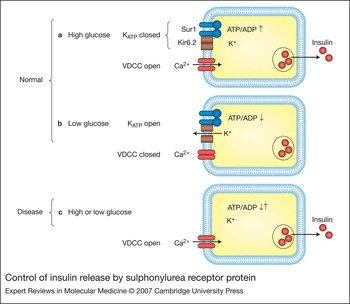
Sulphonylurea receptor protein (SUR1; ABCC8) interacts with the inward rectifier potassium channel Kir6.2 in the ER and is delivered to the cell surface of β-cells to form an ATP-sensitive (KATP) channel complex (Ref. Reference Clement69). The KATP channel complex regulates insulin secretion in response to blood glucose levels. At high glucose levels the ratio of ATP/ADP is high and the binding of ATP to KATP at the β-cell membrane in the pancreas inhibits channel activity (Fig. 3). In the absence of KATP activity, voltage-dependent calcium channels (VDCCs) are open and the calcium influx acts as a signal for insulin release. The ATP/ADP ratio falls in the presence of low glucose and increased binding of ADP to KATP opens the channel. The release of potassium alters the membrane potential, closes the VDCCs and inhibits insulin release.
Control of insulin release by sulphonylurea receptor protein (a) Sulphonylurea receptor protein SUR1 and potassium channel Kir6.2 subunits form an ATP-sensitive potassium channel complex (KATP) at the cell surface. High glucose levels in the bloodstream increase the ATP/ADP ratio in the β-cells of the pancreas. Binding of ATP to KATP inhibits potassium channel activity, which in turn promotes opening of voltage-dependent calcium channels (VDCCs). Enhanced intracellular calcium levels induce release of insulin by promoting fusion of vesicles loaded with insulin with the plasma membrane. (b) As blood glucose levels fall, the ATP/ADP ratio decreases. Interaction of ADP with KATP opens the potassium channels and the resulting alteration in membrane potential closes the VDCCs and inhibits insulin release. (c) In the disease state, the absence of the KATP complex at the cell surface causes the β-cells to release insulin even when the ATP/ADP ratio is low. Persistent insulin release leads to severe hypoglycaemia.

Mutations in SUR1 that interfere with folding inhibit delivery of both SUR1 and Kir6.2 to the cell surface (Refs Reference Sharma70, Reference Cartier71, Reference Yan72), and cause congenital hyperinsulinism. The lack of KATP channels at the cell surface results in the calcium channels remaining open and insulin being released even when glucose levels fall: the resulting hypoglycaemia leads to brain damage.
Since sulphonylurea drugs such as glibenclamide can inhibit the KATP channels by binding to SUR1, they were tested for pharmacological chaperone activity on misfolded SUR1 mutants (Ref. Reference Yan, Casey and Shyng73). It was found that expression of two different SUR1 processing mutants in the presence of glibenclamide promoted maturation of SUR1 and resulted in a functional KATP complex at the cell surface. Rescue of the SUR1 mutants likely involved binding of the sulphonylurea to the drug-binding site in a folding intermediate of SUR1 in the ER, because the presence of a mutation that abolished sulphonylurea binding also diminished drug rescue of the protein (Ref. Reference Yan, Casey and Shyng73).
G-protein-coupled receptors
G-protein-coupled receptors (GPCRs) are a large class of plasma membrane proteins that have seven TM helices, a ligand-binding site on their extracellular surface, and a G-protein-binding site on their cytoplasmic surface. They transduce signals from the outside of the cell to the intracellular environment by coupling ligand binding to activation of the bound G protein.
At least ten different diseases have been linked to GPCR mutants that are defective in folding and delivery to the cell surface (reviewed in Refs Reference Bernier, Bichet and Bouvier74, Reference Ulloa-Aguirre75). The mutant proteins are degraded soon after synthesis in the ER. Examples of diseases associated with GCPR processing mutants include familial adrenocorticotropic hormone resistance, familial hypocalciuric hypercalcaemia, Hirschsprung disease, hypogonadotropic hypogonadism, obesity, retinitis pigmentosa, and nephrogenic diabetes insipidus. The first demonstration that ligands could act as pharmacological chaperones to promote folding of GCPR processing mutants to yield active receptors at the cell surface was a study on mutants of the vasopressin receptor V2R (which utilised the cell-permeable antagonists SR121463 or VPA-985) (Ref. Reference Morello76). Pharmacological chaperones have also been demonstrated to promote folding of other GPCR processing mutants including those of the gonadotropin-releasing hormone receptor (which causes hypogonadotropic hypogonadism) (Ref. Reference Janovick, Maya-Nunez and Conn77), rhodopsin (which causes retinitis pigmentosa) (Ref. Reference Noorwez78) and the δ-opioid receptor (involved in pain) (Ref. Reference Petaja-Repo79).
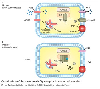
V2R plays an important role in water reabsorption in the inner and medullary collecting ducts of the kidney to concentrate urine (Fig. 4). Mutations in V2R that inhibit delivery of the receptor to the cell surface cause nephrogenic diabetes insipidus (reviewed in Refs Reference Sands and Bichet80, Reference Morello and Bichet81). Patients with diabetes insipidus produce large quantities of dilute urine (>10 l per day); in infants, large water losses cause growth and brain defects due to severe episodes of dehydration. Although mutations that inhibit hormone binding or G-protein coupling would also inhibit water reabsorption, the majority of mutant V2Rs from patients with diabetes insipidus appear to be defective in folding (Ref. Reference Bernier, Bichet and Bouvier74).
Contribution of the vasopressin V2 receptor to water reabsorption. (a) The vasopressin V2 receptor (V2R) is synthesised in the ER, folds into a native structure and is delivered to the cell surface. The receptor binds arginine vasopressin (AVP) on an extracellular site, resulting in activation of G protein (G) in the cytoplasm. The dissociated G protein increases cAMP levels by activating adenyl cyclase (AC). Protein kinase A (PKA), which is activated by cAMP, initiates fusion of cytoplasmic vesicles carrying aquaporin 2 (AQ2) water channels to the lumenal membrane. The urine can now be concentrated as water enters the AQ2 channels on the lumenal surface and exits through aquaporin 3 (AQ3) water channels on the basolateral membrane. (b) A mutant V2 receptor (MV2R) containing a processing mutation (blue dot) does not fold correctly and is degraded. The cells lack the ability to insert AQ2 channels into the lumenal membrane in response to AVP. The urine is not concentrated and the patient produces large quantities (>10 l per day) of dilute urine.

It has been proposed that the pharmacological chaperones SR121463 or VPA-985 enhance cell-surface expression of V2R processing mutants by binding to the mutants post-translationally to increase conformational stability (Ref. Reference Wuller82). In addition, binding of ligands to sites within the TM segments may cause a rearrangement of the TM helices to correct altered packing. Another possibility is that packing of the TM segments is a slow process that takes place post-translationally. Processing mutations might trap the protein in a pre-folded state that can be overcome by binding of ligand.
Lysosomal storage diseases
Lysosomal storage diseases are a class of about 40 genetic diseases caused by deficient activity of a lysosomal hydrolase (reviewed in Refs Reference Futerman and van Meer83, Reference Sawkar, D'Haeze and Kelly84). The decreased hydrolase activity causes accumulation of substrates such as sphingolipids, mucopolysaccharides and oligosaccharides. The intralysosomal accumulation of undegraded metabolites eventually leads to variable clinical symptoms such as bone and skeletal changes, enlarged liver, respiratory or cardiac problems and brain dysfunction. Characterisation of the mutant hydrolases suggests that many have processing defects that could be corrected with pharmacological chaperones (Ref. Reference Sawkar, D'Haeze and Kelly84).
Fabry disease
Fabry disease is characterised by the accumulation of glycosphingolipids such as globotriosylceramide (Gb3) in the lysosome. Many patients with Fabry disease express processing mutants of α-galactosidase A (Refs Reference Ishii85, Reference Okumiya86). The mutants are defective in folding, so little functional protein is delivered to the lysosome. For example, mutant Q279E was found to be thermolabile at the neutral pH levels found in the ER (Ref. Reference Kase87), but could be stabilised by carrying out expression in the presence of galactose. Galactose could also be used as a pharmacological chaperone to stabilise other α-galactosidase A processing mutants to yield functional enzyme in the lysosome.
The iminosugar 1-deoxygalactonorijymicin (DGJ) is another promising pharmacological chaperone for treating Fabry disease. It is a nontoxic competitive inhibitor of α-galactosidase A (Refs Reference Fan88, Reference Asano89). In cell culture studies using lymphoblasts from a patient expressing the R301Q α-galactosidase A processing mutant, addition of 1–10 µm DGJ significantly increased its enzyme activity. Treatment of fibroblasts from patients with other types of processing mutations in α-galactosidase A also increased delivery of active enzyme to the lysosome (Ref. Reference Yam, Zuber and Roth90). The lymphoblasts showed a near-normal lysosomal phenotype and successful clearance of Gb3. Therefore, DGJ might be a more effective pharmacological chaperone for Fabry patients expressing α-galactosidase A processing mutants.
Gaucher disease
Gaucher disease is the most common lysosomal storage disease (Ref. Reference Sawkar, D'Haeze and Kelly84). It is characterised by the accumulation of glycosphingolipid glucosylceramide in the lysosome. Another iminosugar, N-(n-nonyl)deoxynojirimycin (N-nonyl-DNJ), was shown to act as a pharmacological chaperone for lysosomal glucocerebrosidase (also called β-glucosidase) processing mutants (N370S and G202R) associated with Gaucher disease (Ref. Reference Sawkar91). N-nonyl-DNJ is thought to act as a pharmacological chaperone by binding to the active site of glucocerebrosidase processing mutants to stabilise the proteins in the ER to prevent their degradation (Ref. Reference Sawkar92). It appears that glucocerebrosidase processing mutants such as N370S and G202R are less stable in the neutral pH of the ER compared with the acidic pH of the lysosome (Ref. Reference Sawkar, D'Haeze and Kelly84).
GM1 gangliosidosis
Defects in lysosomal β-galactosidase cause accumulation of GM1 glycosphingolipids in lysosomes of the brain and other tissues (GM1 gangliosidosis). A potent iminosugar inhibitor of lysosomal β-galactosidase has been found to act as a pharmacological chaperone for processing mutants in fibroblasts from patients with GM1 gangliosidosis (Ref. Reference Matsuda93). In an in vivo mouse model of GM1 gangliosidosis, administration of a galactose iminosugar inhibitor increased the lysosomal β-galactosidase activity in the brain and other tissues. Immunohistochemical staining of neuronal cells from the treated mice showed a decrease in the amount of GM1 glycosphingolipids. This study was important as it showed that pharmacological chaperones could be used to promote folding of processing mutants expressed in the brain.
Tay–Sachs and Sandhoff diseases
Processing mutants of β-hexosaminidase A and B result in disorders such as Tay–Sachs and Sandhoff disease. Inhibitors of hexosaminidase can act as pharmacological chaperones in fibroblasts from patients with Tay–Sachs or Sandhoff diseases (Ref. Reference Tropak94): the presence of the inhibitor was found to increase the intralysosomal hexosaminidase activity.
Clinical implications
Pharmacological chaperones have been used in two clinical studies. In one study, five patients who had X-linked nephrogenic diabetes insipidus as a result of expression of V2R processing mutants were treated with the pharmacological chaperone SR49059 (Ref. Reference Bernier, Bichet and Bouvier74). The pharmacological chaperone was administered orally for 7 days and water intake and urine output were monitored. It was found that urine volume and water intake decreased by about a third. Therapy with SR49059 had to be halted because of its adverse effects on the cytochrome P450 pathway. Future therapies with better V2R pharmacological chaperones such as YM087 might begin after Phase I trials.
In the second study, an adult male with Fabry disease expressing a G328R α-galactosidase A processing mutant was treated with the pharmacological chaperone galactose. Infusions of 1 g galactose per kg body weight every other day for over 2 years resulted in significant improvement such that the patient no longer required cardiac transplantation (Ref. Reference Frustaci95).
Limitations of pharmacological chaperones
There are limitations to the use of pharmacological chaperones. They might not be able to rescue processing mutants where the mutation has affected the active site or the folding defect is too great to be reversed. The compound must be hydrophobic or be able to enter the cells through an alternative pathway and reach the ER at concentrations sufficient to promote folding. The concentration of pharmacological chaperone required for rescue might be too high and produce side effects. The short half-life of the rescued protein or pharmacological chaperone might require continuous infusion of the compound and result in adverse side effects. A way to overcome some of these problems is to screen for stable high-affinity compounds. Despite these potential problems, the initial cell culture and clinical studies suggest that pharmacological chaperones offer an exciting new therapeutic strategy to treat genetic diseases.
Research in progress and outstanding research questions
It is important to determine which genetic diseases will respond to treatment with pharmacological chaperones. Only a small percentage of disease-associated proteins containing point mutations have been characterised in detail. The processing defects must be identified and tested to determine whether they can be rescued with pharmacological chaperones. Pharmacological chaperones have to be identified that are nontoxic, show specificity and do not inhibit function of the protein once it has been rescued.
Many other questions remain unanswered. Could pharmacological chaperones be used over a long period of time? Could pharmacological chaperones be identified that are effective at lower concentrations to reduce adverse side effects? Are the effects of multiple pharmacological chaperones additive or synergistic? Is there ‘plasticity’ in the folding process upon interaction of the misfolded protein with a pharmacological chaperone? Answers to these questions may enhance the therapeutic value of pharmacological chaperones.
Acknowledgements and funding
D.M.C. is funded by the Canadian Institutes of Health Research and is the recipient of the Canada Research Chair in Membrane Biology. Comments from the peer reviewers are appreciated.