Cognitive decline and brain atrophy are often considered common characteristics of the ageing process (Erickson, Leckie, & Weinstein, Reference Erickson, Leckie and Weinstein2014). However, not all cognitive processes or brain regions are equally affected. Indeed, the frontal brain regions that support the cognitive domain of executive functions (EFs) appear to be particularly vulnerable to the effects of advancing age (Fjell & Walhovd, Reference Fjell and Walhovd2010). EFs comprise higher order cognitive functions such as Shifting, Updating/Working Memory, Inhibition, Verbal Generativity, and Non-verbal Reasoning (see Fisk & Sharp, Reference Fisk and Sharp2004; Frost et al., Reference Frost, Weinborn, Gignac, Markovic, Rainey-Smith, Sohrabi and Brown2019; Lezak, Reference Lezak2012; Miyake & Friedman, Reference Miyake and Friedman2012 for detailed descriptions of these EF subdomains). It is unsurprising, then, that age-associated EF declines can affect quality of life and functional independence (Davis, Liu-Ambrose, Marra, & Najafzadeh, Reference Davis, Liu-Ambrose, Marra and Najafzadeh2010). Promisingly, physical activity, and aerobic exercise in particular, has been found to be important in maintaining neurocognitive health (Hertzog, Kramer, Wilson, & Lindenberger, Reference Hertzog, Kramer, Wilson and Lindenberger2008; Stillman, Esteban-Cornejo, Brown, Bender, & Erickson, Reference Stillman, Esteban-Cornejo, Brown, Bender and Erickson2020). Moreover, it appears that the most age-vulnerable frontal brain regions are most amenable to exercise-related neuroprotective factors and plasticity (the ability of the brain to undergo biological changes as a result of intrinsic or extrinsic stimuli; Colcombe & Kramer, Reference Colcombe and Kramer2003).
Exercise, cardiorespiratory fitness, and frontal brain health
Although interventional research examining the effect of exercise on neuroimaging-derived brain parameters is relatively sparse, meta-analytic and intervention studies have demonstrated exercise-induced frontal volume benefits, disproportionate to other brain regions (Colcombe & Kramer, Reference Colcombe and Kramer2003; Reiter et al., Reference Reiter, Nielson, Smith, Weiss, Alfini and Smith2015). Colcombe and colleagues (Reference Colcombe, Erickson, Scalf, Kim, Prakash, McAuley and Kramer2006) demonstrated increased prefrontal grey matter (GM) volume in older adults (60–79 years) following an aerobic walking exercise intervention, whereas the control group (stretching and toning) showed decline in the same brain region. Congruently, Ruscheweyh and colleagues (Reference Ruscheweyh, Willemer, Krüger, Duning, Warnecke, Sommer and Flöel2011) reported increased prefrontal GM volume in those in an aerobic walking intervention, compared to control, in a slightly younger population (50–78 years).
Cardiorespiratory fitness is an objective measure of aerobic capacity, modifiable by participation in regular aerobic exercise. However, it is not in and of itself a measure of exercise levels, and is considered functionally separate when it comes to neurocognitive benefits (Erickson et al., Reference Erickson, Leckie and Weinstein2014). However, similar to the aerobic exercise research, cardiorespiratory fitness shows a robust association with frontal brain volume. Since the seminal Colcombe et al. study (Reference Colcombe, Erickson, Raz, Webb, Cohen, McAuley and Kramer2003) that reported the first empirical confirmation of the relationship between cardiorespiratory fitness and frontal brain volume, many others have corroborated this finding (Alosco et al., Reference Alosco, Brickman, Spitznagel, Griffith, Narkhede, Raz and Gunstad2013; Bugg & Head, Reference Bugg and Head2011; Flöel et al., Reference Flöel, Ruscheweyh, Krüger, Willemer, Winter, Völker and Knecht2010). While both cardiorespiratory fitness and general participation in exercise have been found to be predictive of greater frontal GM volume, the effect of exercise intensity as it relates specially to frontal lobe GM volume remains relatively underexplored. However, those studies that have examined the effect of intensity on brain volume more generally found that vigorous-intensity aerobic exercise, as compared with moderate- or low-intensity exercise, led to increased brain volume (Best, Chiu, Liang Hsu, Nagamatsu, & Liu-Ambrose, Reference Best, Chiu, Liang Hsu, Nagamatsu and Liu-Ambrose2015; Cabral et al., Reference Cabral, Rice, Morris, Rundek, Pascual-Leone and Gomes-Osman2019; Colcombe et al., Reference Colcombe, Erickson, Scalf, Kim, Prakash, McAuley and Kramer2006; Erickson et al., Reference Erickson, Voss, Prakash, Basak, Szabo, Chaddock and Kramer2011; Niemann, Godde, & Voelcker-Rehage, Reference Niemann, Godde and Voelcker-Rehage2014).
In summary, there is clear evidence to suggest that exercise and cardiorespiratory fitness are associated with frontal neuroprotective benefits. However, the role of exercise intensity in this relationship remains underexplored. Thus, to extend knowledge on the nature of the relationship between exercise and frontal brain GM volume in older adults, we have examined the effect of a systematically manipulated exercise intervention (self-paced high-intensity vs. moderate-intensity vs. control) on structures within the frontal lobe. We also examined the longevity of any observed effects at 12-month post-intervention follow-up. We hypothesised that there would be an increase in frontal GM volume in the high- and moderate-intensity aerobic exercise groups, but not for the control group. We also hypothesised that this effect would be more pronounced for the high-intensity group as compared with the moderate intensity exercise group. Also, based on cognitive findings from our previous study (Frost et al., Reference Frost, Weinborn, Gignac, Rainey-Smith, Markovic, Gordon and Brown2020) that reported an association between cardiorespiratory fitness and some EF cognitive subdomains, we aimed to assess the relationship between change in post-intervention cardiorespiratory fitness and change in frontal brain volume. These analyses were approached in an exploratory manner.
METHODS
Standard protocol approvals, registrations, and patient consents
The current study drew data from the single-blind randomised controlled trial (RCT), the Intense Physical Activity and Cognition (IPAC) study (Australian New Zealand Clinical Trials Registry number: ACTRN12617000643370). The study was approved by the Human Research Ethics Committees of Edith Cowan University and Murdoch University. All participants provided signed informed consent before enrolment into the study. Procedures of this RCT followed the principles of the Declaration of Helsinki for Human Rights.
Participants and design
Comprehensive information about participants, recruitment, inclusion/exclusion criteria, intervention, and outcome measures for the IPAC study have been detailed previously (Brown et al., Reference Brown, Rainey-Smith, Castalanelli, Gordon, Markovic, Sohrabi and Peiffer2017). Briefly, participants underwent cardiorespiratory fitness assessment and Magnetic Resonance brain imaging at three time points: Baseline (pre-intervention), 6-months (directly post-intervention) and 18-months (12-months post-intervention). Supplementary Table 1 provides information related to the timeline of all measurements, outcome measures, and interpretation of scores. As outlined in the CONSORT diagram (Figure 1), 228 potential participants were screened to exclude those with significant cognitive impairment (i.e., <26 on the Montreal Cognitive Assessment and results of the baseline cognitive assessment; Nasreddine et al., Reference Nasreddine, Phillips, Bédirian, Charbonneau, Whitehead, Collin and Chertkow2005). Additionally, participants were between 60 and 80 years, did not have any uncontrolled medical conditions known to influence cognitive function, and did not regularly engage in high-intensity exercise, as evaluated by an Exercise and Sports Science Australia accredited exercise physiologist.
Flowchart of participants in high-intensity exercise intervention, moderate-intensity exercise intervention, and control groups from screening to 12-month post-intervention follow-up.

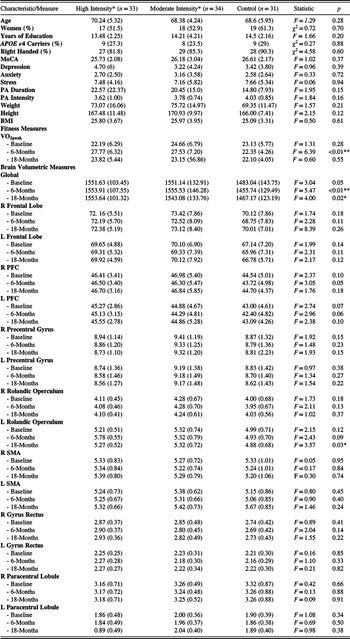
One-hundred and eight participants were successfully recruited and attended at least one baseline assessment appointment. Based on power calculations, a sample size of 30 per group was required to detect (with 80% power; α = .05; 2-sided; Cohen’s d: 0.25; small effect size) variance between the intervention and control groups. Our recruitment sample (N = 108) allowed for a ˜15% attrition rate from baseline to post-intervention follow-up. Nine participants were withdrawn prior to baseline completion due to incomplete cognitive data or a baseline cognitive assessment suggestive of mild cognitive impairment. One participant was withdrawn from the study due to missing imaging data. Participants who completed baseline (N = 98) were randomised, via computer-generated block randomisation, into either a self-paced high- or moderate-intensity exercise, or control (no exercise) group. Table 1 summarises the baseline (and relevant 6- and 18-month) demographic, clinical, cardiorespiratory fitness and brain volumetric characteristics of participants by group.
Baseline (and relevant 6- and 18-month) demographics, clinical characteristics, cardiorespiratory fitness, and frontal brain regions of interest volumetrics

Note. *Self-paced intensity levels based on the Borg Scale of Perceived Exertion (ranging between 0 and 20); moderate-intensity (continuous cycling at 13 Borg scale); high-intensity (intervals of 18 Borg scale, interspersed with active recovery at 12 Borg scale). Data are expressed as mean (± SD), except where otherwise stated. χ2 is Pearson Chi-squared statistic, degrees of freedom (2); F: Analysis of variance statistic, degrees of freedom (2, 95). p = probability value. Handedness: Right, left, ambidextrous, or corrected right. MoCA, score ranges between 0 and 30. Depression, anxiety and stress were measured by the Depression, Anxiety, and Stress Scale – 21, highest possible score for each subscale was 42. Duration and Intensity reflect self-report PA levels derived from the International Physical Activity Questionnaire. PA duration is hours/week, PA intensity is metabolic equivalents/duration. Weight is measured in kilograms, height is measured in centimetres. BMI, a score less than 18.5 falls within the underweight range, 18.5 to <25, falls within the normal range, 25 to <30 falls within the overweight range and 30.0 or higher falls within the obese range. PA VO2peak is measured in ml/kg/min. Brain ROI volume measures in cm3; Global is the total volume of all brain regions. Abbreviations. APOE ϵ4, apolipoprotein E ϵ4 allele; MoCA, Montreal Cognitive Assessment; PA, physical activity; BMI, body mass index; VO2peak, peak volume of oxygen uptake in ml/kg/min; ROI, region of interest; R, right; L, left; PFC, prefrontal cortex; SMA, supplementary motor area. p < 0.05*; p < 0.01**.
Intervention
Participants were randomised to either a self-paced high- or moderate-intensity exercise intervention group, or no-exercise control. Those study participants randomised to an intervention group completed a six-month cycling program consisting of two sessions per week at 50-minutes per session (under the supervision of an Accredited Exercise Physiologist). Exercise was completed on a cycle ergometer (WattbikePro; Wattbike, Australia) allowing accurate assessment of intensity level (Wattage). Target intensity was set using the 6 to 20 Borg Scale of Perceived Exertion (Borg, Reference Borg1998). Additionally, radiotelemetric heart rate monitors (Garmin HRM1G, Garmin, USA) were used to assess physiological intensity (Brown et al., Reference Brown, Rainey-Smith, Castalanelli, Gordon, Markovic, Sohrabi and Peiffer2017).
Participants in the moderate-intensity condition were encouraged to maintain a continuous moderate pace of cycling at a rating of perceived exertion of 13.0 Borg Scale (50%–60% aerobic capacity) for 50 minutes. Those participants in the high-intensity condition undertook high intensity interval training (HIIT), bookended with a warm up and cool down period. Specifically, high-intensity participants were encouraged to work at a vigorous pace (>80% aerobic capacity; rating of perceived exertion of 18.0 Borg Scale) during the 11 cycling-based intervals of 1-minute of high exertion, and a moderate pace during the 2-minutes of active recovery (30%–40% aerobic capacity; rating of perceived exertion of 12.0 Borg Scale).
Both high- and moderate-intensity intervention protocols were work-matched based on an 80 kg person with a maximal aerobic capacity of 27 ml.kg−1.min−1 to yield approximately 386 Met.min−1 for a moderate-intensity session and 380 Met.min−1 for a high-intensity session (Brown et al., Reference Brown, Rainey-Smith, Castalanelli, Gordon, Markovic, Sohrabi and Peiffer2017). We observed the high-intensity group cycled at a higher percentage peak aerobic power in the active condition (120.6 ± 26.3%), compared with the moderate-intensity group (70.2 ± 18.0; p<0.001), indicating we successfully delivered the exercise interventions at different intensities (Brown et al., Reference Brown, Frost, Rainey-Smith, Doecke, Markovic, Gordon and Peiffer2021).
Participants in the control group were invited to an information session on the benefits of diet and exercise in relation to neurocognitive ageing; however, these individuals did not receive any other exercise-related instruction or intervention (Brown et al., Reference Brown, Rainey-Smith, Castalanelli, Gordon, Markovic, Sohrabi and Peiffer2017).
Cardiorespiratory fitness measurement
As detailed previously (Brown et al., Reference Brown, Rainey-Smith, Castalanelli, Gordon, Markovic, Sohrabi and Peiffer2017; Frost et al., Reference Frost, Weinborn, Gignac, Rainey-Smith, Markovic, Gordon and Brown2020), the measure for cardiorespiratory fitness was VO2peak. Cycle-based assessment required participants to pedal against increasing resistance until volitional fatigue. Heart rate was recorded continuously, and expired ventilation was assessed at 15-second mean values, using a Parvo TrueOne (ParvoMedics, USA) metabolic cart, for the measurement of rate of oxygen consumption (VO2) and carbon dioxide production (VCO2). VO2peak was classified as the highest 15 second mean VO2 value obtained during the last two minutes of the test. Additionally, participants must have reached a maximal heart rate greater than 85% of their age predicted maximum (i.e., (220 – age) * 0.85) and a respiratory exchange ratio (VCO2/VO2) greater than 1.15 (Brown et al., Reference Brown, Rainey-Smith, Castalanelli, Gordon, Markovic, Sohrabi and Peiffer2017).
Structural magnetic resonance imaging (MRI) segmentation and frontal lobe region of interest (ROI) GM volume measurement
MRI acquisition was performed at all three time points using two 3T scanners (Magnetom Verio and Vida, Siemens, Erlangen, Germany). Only a subset of participants at 18-month follow-up were scanned using the 3T Vida scanner. In order to estimate the scanner effect on the volumetric measurement, repeated MRI scans were acquired from 18 participants using both scanners. T1-weighted structural brain images were acquired with the magnetisation-prepared rapid gradient-echo (MPRAGE) sequence (TE/TR =2.52/1900 ms, flip angle = 9°, FoV = 250 mm, 1.00 mm slice thickness, 176 slices).
The 3D T1-weighted MPRAGE images were first segmented into GM, white matter (WM), and cerebrospinal fluid using an in-house implementation of the expectation maximisation algorithm (Van Leemput, Maes, Vandermeulen, & Suetens, Reference Van Leemput, Maes, Vandermeulen and Suetens1999). Topological constraints were applied to force GM to be a continuous layer covering the WM, and GM segmentation was also topologically corrected in deep sulci (Rueda et al., Reference Rueda, Acosta, Couprie, Bourgeat, Fripp, Dowson and Salvado2010).
The brain parcellation was performed on cortical and subcortical regions based on the Automated Anatomic Labelling (AAL) parcellation atlas using Learning Embeddings for Atlas Propagation following the work of Wolz et al. (Wolz, Aljabar, Hajnal, Hammers, & Rueckert, Reference Wolz, Aljabar, Hajnal, Hammers and Rueckert2010). This method relies on the construction of an affinity matrix that captures the similarity between each pair of images in the atlas database (M = 20 atlases used here), and therefore, minimises the amount of deformation required for segmentation propagation. The AAL brain parcellation provided the ROI for quantifying regional GM volumes (in centimetres cubed; Boccardi et al., Reference Boccardi, Bocchetta, Morency, Collins, Nishikawa, Ganzola and Frisoni2015), with CurAIBL (https://milxcloud.csiro.au/) providing the MRI pipeline used for the analysis.
The ROI volumetric measures from 18-month follow-up image data were adjusted for a scanner effect using the repeated MRI scans (n = 18) from both scanners. For each ROI, a linear regression model was fitted to volumetric data in this subset in order to estimate the scanner effect. The estimated model was then used to correct volumetric measures from MR images acquired from the 3T Vida scanner for the remainder of participants. Good agreement was observed for volumetric measures of all the selected ROIs from the two different scanners (Pearson’s r > 0.95, p < 0.001). Frontal ROIs within this study were bilateral measurements of the total frontal lobe, prefrontal cortex (PFC; a composite of superior, middle, and inferior frontal gyri; Harms et al., Reference Harms, Wang, Campanella, Aldridge, Moffitt, Kuelper and Csernansky2010), precentral gyrus, rolandic operculum, supplementary motor area (SMA), gyrus rectus, and paracentral lobule.
Demographic and covariates measurement
Through interview, participants completed questionnaires to attain demographic data, medical history, medications, alcohol consumption, smoking status, mood, and exercise characteristics (all standardised measures fully described in our protocol paper; Brown et al., Reference Brown, Rainey-Smith, Castalanelli, Gordon, Markovic, Sohrabi and Peiffer2017). To determine Apolipoprotein E genotype (i.e., ϵ4 allele carriage), blood was drawn at baseline and analysed using standard procedures previously described (Brown et al., Reference Brown, Rainey-Smith, Castalanelli, Gordon, Markovic, Sohrabi and Peiffer2017). Additionally, global brain size was used as a covariate for analyses of regional GM volumes.
Statistical analyses
All brain ROI volume and fitness (VO2peak) measurements were considered sufficiently normally distributed (skew <|2.0|) for the purposes of parametric analyses (Mallery & George, Reference Mallery and George2016). Descriptive statistics were used to summarise data. Analysis of variance and χ2 statistics were used to compare baseline characteristics of participants randomly assigned to the three study groups (high-intensity, moderate-intensity, control).
Group effect on cardiorespiratory fitness and frontal ROI GM volume
An intention-to-treat analysis was conducted to examine the effect of the intervention on VO2peak and frontal brain ROI volumes (Mohammady, Sadeghi, & Janani, Reference Mohammady, Sadeghi and Janani2017), using linear mixed models (group*time). Participants were treated as a random effect. Secondary (per-protocol) analyses of the outcome measures included only participants who had adhered to the intervention as per protocol (i.e., those participants in the high- and medium-intensity intervention groups who attended ≥75% of supervised exercise sessions) and control participants. Global brain volume, age, gender, and education level were entered into all linear mixed models as covariates. No adjustment was made for multiple comparisons as the dependent variables were highly intercorrelated: average r = 0.60. Consequently, the familywise error rate could not be considered 0.05*14 given that analyses were not independent tests. Accordingly, using standard adjustments (e.g., Bonferroni) would be considered too conservative, increasing the risk of a Type 2 error (Banerjee, Chitnis, Jadhav, Bhawalkar, & Chaudhury, Reference Banerjee, Chitnis, Jadhav, Bhawalkar and Chaudhury2009).
Changes in cardiorespiratory fitness and frontal ROI GM volume
Post hoc, exploratory analyses examined the associations between change in pre-to-post-intervention fitness (VO2peak) and change in pre-to-post-intervention brain ROI volume. These analyses were conducted separately for control group and intervention group participants for which there were valid baseline and 6-month follow-up data (intervention group participants: n = 57; control group participants: n = 24). We analysed intervention and control groups separately in order to assess individual differences in responsiveness to cardiorespiratory fitness training and how that related to frontal GM volume.
Change in VO2peak and frontal brain ROI volume was assessed through residual scores estimated from linear regression analysis that predicted post-intervention VO2peak/brain ROI volume from pre-intervention VO2peak/brain ROI volume. Outliers (n = 1 for VO2peak in the control group) were identified based on criteria outlined by Hoaglin and Iglewicz (Reference Hoaglin and Iglewicz1987) and subsequently Winsorised (Reifman & Keyton, Reference Reifman and Keyton2010) by way of next highest/lowest data point +/- one increment (i.e., 1 or 0.1 as appropriate). Exploratory analyses were performed by way of linear regression models. Age, gender, education level, and baseline global brain volume were entered as covariates. All statistical analyses were performed using SPSS (version 26; SPSS Inc., Chicago, IL, 2019). Alpha was set at 0.05 and all reported results are two-tailed.
RESULTS
The CONSORT diagram (Figure 1) summarises the flow of study participants from screening through to 18-month follow-up. By the end of the 6-month intervention period (follow-up directly post-intervention) seven participants had withdrawn from the study. At the 18-month follow-up (12-months post-intervention) assessment a further six participants were withdrawn. Participants withdrawn from the study post-randomisation did not differ significantly across any baseline characteristics as compared with those participants that remained in the study to completion. Table 1 summarises baseline demographic, clinical, fitness, and brain ROI volume characteristics across groups for all study participants according to group allocation at randomisation.
An intervention adherence assessment study showed that participants randomised to the high-intensity exercise group adhered to the exercise sessions as outlined in the protocol (Brown et al., Reference Brown, Rainey-Smith, Castalanelli, Gordon, Markovic, Sohrabi and Peiffer2017) at a mean of 85.5% (SD = 12.45; range = 50%–100%), while those randomised to the moderate-intensity exercise group adhered to the exercise sessions as outlined in the protocol at a mean of 86.29% (SD = 9.82; range = 59.62%–100%). Intervention adherence as per protocol did not differ between the two groups (F (1, 60) =0.08, p = 0.778).
Effect of intervention group on cardiorespiratory fitness and frontal ROI GM volume
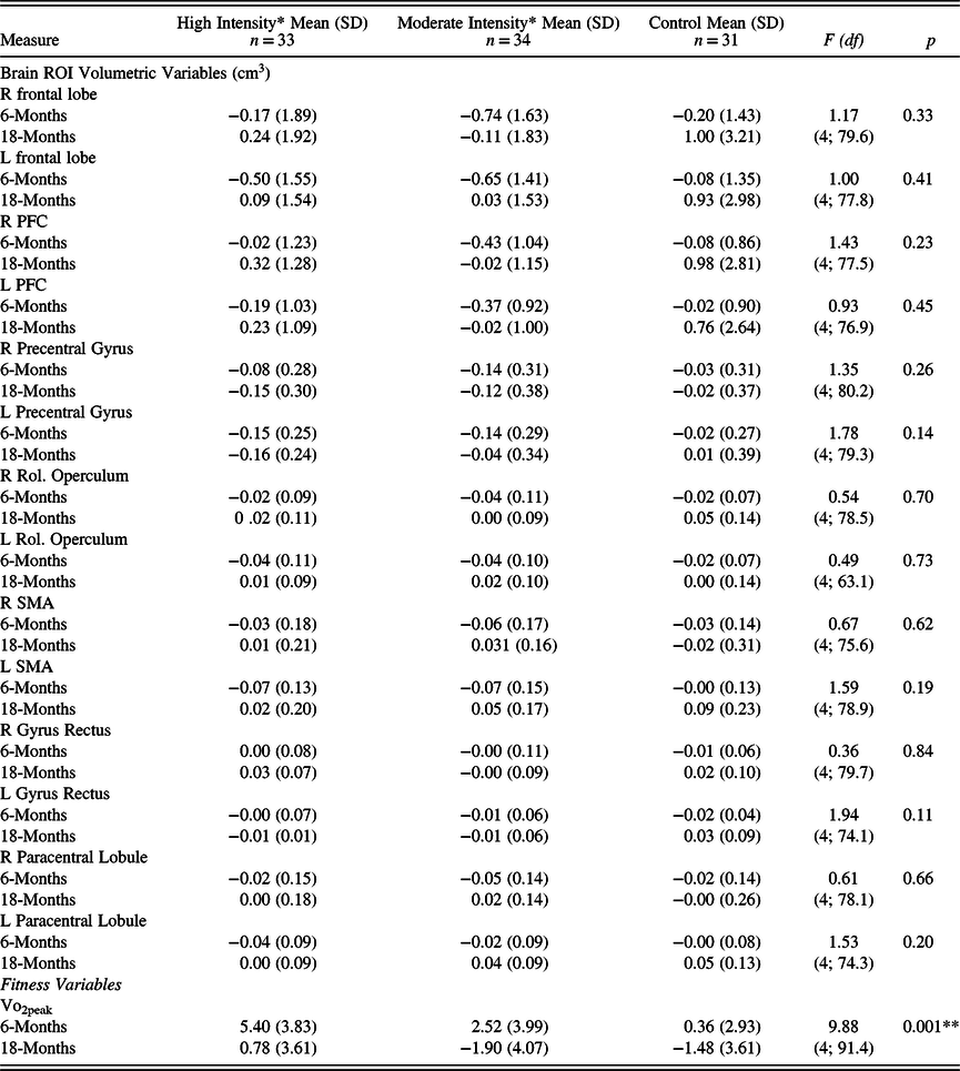
Change in scores from pre- to post-intervention (6-months post baseline), and again at 12-months post-intervention (18-months post baseline) measurement are presented in Table 2.
Observed raw mean difference compared with baseline of brain regions of interest volumetric measures and cardiorespiratory fitness over 18-months for high-intensity exercise intervention participants, moderate-intensity exercise intervention participants, and control participants

Note. *Self-paced intensity levels based on the Borg Scale of Perceived Exertion (ranging between 0 and 20); moderate-intensity (continuous cycling at 13 Borg scale); high-intensity (intervals of 18 Borg scale, interspersed with active recovery at 12 Borg scale). Raw data are expressed as mean (± SD), except where otherwise stated. F: Linear Mixed Model statistic; df is degrees of freedom (numerator degrees of freedom; denominator degrees of freedom). Model adjusted for age, gender, education level, and global brain volume. Abbreviations: SD, standard deviation; ROI, region of interest; R, right; L, left; PFC, prefrontal cortex; Rol, Rolandic; SMA, supplementary motor area; VO2peak, peak volume of oxygen uptake in ml/kg/min. **p < 0.01.
Pre- to post-intervention, there was a significant increase in VO2peak in the high-intensity group as follows: t = 7.87(30), p = <0.001, mean 5.40 (± 3.83). In the moderate-intensity group there was also a significant increase in VO2peak: t = 3.40 (28), p = 0.002, mean 2.52 (± 3.99). Across groups, cardiorespiratory fitness levels differed: high-intensity and moderate intensity were higher than control from pre- to post-intervention follow-up (see Table 2). There were no significant group differences from pre-intervention to 12-month post-intervention follow-up, or group*time effects for any of the frontal ROIs (see Table 2).
Secondary analyses (i.e., per protocol analyses) again revealed that cardiorespiratory fitness differed at a group level (F (4, 76.3) = 10.93, p = < 0.001) with both high-intensity and moderate intensity greater than control from pre- to post-intervention. Again, there were no group*time effects for any of the brain ROI measures (Right Frontal Lobe: F (4, 70.8) = 1.27, p = 0.29; Left Frontal Lobe: F (4; 69.2) =0.97, p = 0.43; Right PFC: F (4; 69.7) = 1.80, p = 0.14; Left PFC: F (4; 69.3) = 1.18, p = 0.33; Right precentral gyrus: F (4; 73.3) = 0.99, p = 0.42; Left precentral gyrus: F (4; 72.4) = 1.34, p = 0.27; Right Rolandic Operculum: F (4; 71.5) = 0.57, p = 0.69; Left Rolandic Operculum: F (4; 54.9) = 0.35, p = 0.84; Right SMA: F (4; 68.1) = 0.45, p = 0.70; Left SMA: F (4; 72.4) = 1.04, p = 0.40; Right Gyrus Rectus: F (4;83.3) = 1.00, p = 0.41; Left Gyrus Rectus: F (4; 66.0) = 2.09, p = 0.09; Right Paracentral Lobule: F (4; 71.4) = 0.32, p = 0.86; Left Paracentral Lobule: F (4; 67.6) = 0.87, p = 0.49).
Change in cardiorespiratory fitness and frontal ROI GM volume
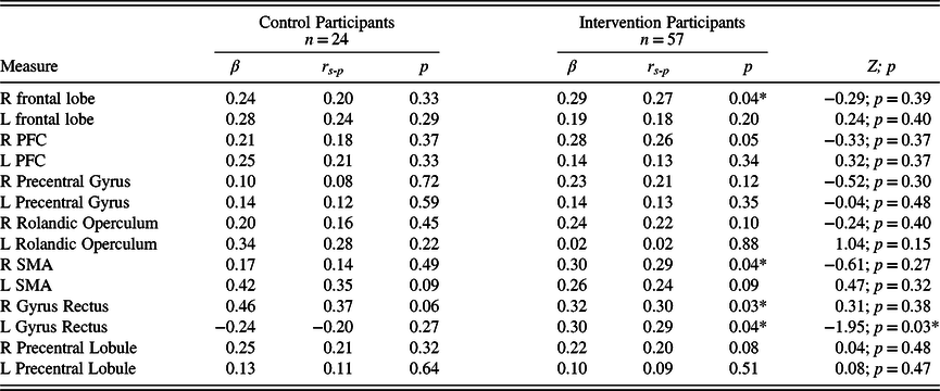
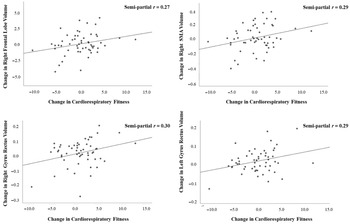
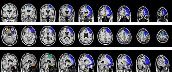
Results from post hoc, exploratory analyses on the association between change in pre-to-post-intervention (6-months) cardiorespiratory fitness and ROI volume are presented in Table 3. There were no associations observed within the control group. Conversely, significant positive associations were observed between change in cardiorespiratory fitness and right frontal lobe volume, right SMA volume, right gyrus rectus volume, and left gyrus rectus volume for intervention group participants (Figures 2 and 3) Specifically, increases in fitness were positively associated with increases in frontal lobe volumes..
Associations between change in pre-to-post-6-month exercise intervention cardiorespiratory fitness and frontal brain region of interest volume assessed separately for intervention and control group participants after controlling for age, gender, education level, and baseline global brain volume

Note. Change assessed through residual scores estimated from linear regression analysis that predicted post-intervention VO2peak/ROI volume from pre-intervention VO2peak/ROI volume. Analyses conducted by way of linear regression modelling. β = Standardised Beta Weight; r s-p = semi-partial correlation. Z = Fishers-z statistic. Degrees of freedom for the control group analyses: (4, 19); Degrees of freedom for the intervention groups analyses (4, 52). Model adjusted for age, gender, education level, and global (whole) brain volume; cardiorespiratory fitness measured as VO2peak, peak volume of oxygen uptake measured in ml/kg/min ; Frontal Brain volume measured in cm3. Abbreviations: R, right; L, left; PFC, prefrontal cortex; SMA, supplementary motor area. *p < 0.05.
Change in cardiorespiratory fitness residual scores was positively correlated with change in Right frontal lobe, Right supplementary motor area (SMA), Right gyrus rectus, and Left gyrus rectus residual scores from pre- to post-6-month exercise intervention in exercise intervention group participants after controlling for age, gender, education, and baseline global brain volume measurement.

Frontal brain areas positively correlated with change in cardiorespiratory fitness post 6-month aerobic exercise intervention in coronal, axial, and sagittal view, respectively (blue: Right frontal lobe, green: Right SMA, yellow: Right gyrus rectus, orange: Left gyrus rectus).

DISCUSSION
In the current study, we examined the effect of a systematically manipulated exercise intervention (self-paced high-intensity vs. self-paced moderate-intensity vs. control) on frontal lobe GM volume in a group of cognitively normal older adults. We did not observe group-level differences in frontal brain volume after intervention. This finding is in contrast with previous works (Colcombe et al., Reference Colcombe, Erickson, Scalf, Kim, Prakash, McAuley and Kramer2006; Colcombe & Kramer, Reference Colcombe and Kramer2003). Our null group findings may be explained by a number of factors. Our intervention was based on public health recommendations of 90–100 minutes per week of vigorous exercise for physical health benefits, and was duration matched between groups to evaluate the consequence of exercise intensity (Füzéki & Banzer, Reference Füzéki and Banzer2018). However, these health recommendations were primarily based on continuous bouts of exercise, rather than interval exercise as examined within the current study. Thus, due to the HIIT methodology, perhaps our intervention fell short of the recommended 90–100 minutes required for physical health benefits. Indeed, while Colcombe et al.’s (Reference Colcombe, Erickson, Scalf, Kim, Prakash, McAuley and Kramer2006) intervention study that reported structural frontal brain benefits examined six-months of 180-weekly minutes of moderate-intensity aerobic exercise, perhaps our intervention’s weekly duration of 100-minutes of HIIT fell under the threshold required to induce structural change at an aggregate level. Correspondingly, other studies reporting benefits of aerobic exercise on non-frontal brain GM in older adults assessed longer overall exercise interventions, compared with ours (i.e., one-year vs. six-months; Erickson et al., Reference Erickson, Voss, Prakash, Basak, Szabo, Chaddock and Kramer2011; Niemann et al., Reference Niemann, Godde and Voelcker-Rehage2014).
Additionally, the participants in our study were both cognitively and physically high-functioning. Mean education level for our cohort was 14.05 years, which is greater than ˜50% of all adults in Australia (ABS, 2016). Accordingly, GM volume in our participants may have already been augmented to a degree, beyond which the additive benefits of our intervention could not be detected (Arenaza-Urquijo et al., Reference Arenaza-Urquijo, Landeau, La Joie, Mevel, Mézenge, Perrotin and Chételat2013). Similarly, while we excluded participants who were above a certain threshold of pre-intervention exercise, mean baseline body mass index (BMI; a measure used to determine whether an individual is in a healthy weight range for their height) for our participants was 25.6. Although this mean falls just within the ‘overweight category’, research conducted by Winter et al., argues that BMI thresholds for the ‘healthy range’ used for young and middle-aged adults are set too low to measure a healthy BMI in older adults (Winter, MacInnis, Wattanapenpaiboon, & Nowson, Reference Winter, MacInnis, Wattanapenpaiboon and Nowson2014). This is particularly relevant given the results reported by Hamer and Batty (Reference Hamer and Batty2019) whereby individuals with higher BMI had lower GM volume in the brain, as compared to those with lower BMIs. Thus, it is conceivable that we may have seen clearer neurostructural group benefits of our exercise intervention in a ‘less healthy’ or a more ‘cognitively vulnerable’ population.
Although we did not observe differences in GM frontal lobe volumes between our study groups, we cautiously reported positive associations between improved cardiorespiratory fitness and increased volume of the right frontal lobe, right SMA, and bilateral gyrus rectus from pre- to post-intervention for participants that completed the intervention. These potential findings complement existing research on the benefits of cardiorespiratory fitness on brain structure (Colcombe et al., Reference Colcombe, Erickson, Scalf, Kim, Prakash, McAuley and Kramer2006; Flöel et al., Reference Flöel, Ruscheweyh, Krüger, Willemer, Winter, Völker and Knecht2010; Weinstein et al., Reference Weinstein, Voss, Prakash, Chaddock, Szabo, White and Erickson2012). Prior research has found the right frontal lobe to be implicated in the EF abilities of shifting and verbal-generativity (Davidson, Gao, Mason, Winocur, & Anderson, Reference Davidson, Gao, Mason, Winocur and Anderson2008), as well as inhibition, impulsivity, emotional lability, and social cognition (Chow, Reference Chow2000). Additionally, the SMA has been associated with working memory (Cañas, Juncadella, Lau, Gabarrós, & Hernández, Reference Cañas, Juncadella, Lau, Gabarrós and Hernández2018) and auditory processing (Lima, Krishnan, & Scott, Reference Lima, Krishnan and Scott2016). The neurocognitive functions mediated by the gyrus rectus remain relatively unclear; however, there is emerging evidence for its involvement in the EF subdomain of inhibition (Knutson et al., Reference Knutson, Dal Monte, Schintu, Wassermann, Raymont, Grafman and Krueger2015), as well as language and memory (Joo et al., Reference Joo, Park, Moon, Chun, Song and Roh2016).
Human and animal studies suggest that exercise-induced fitness contributes to neuron proliferation, growth of capillary beds, and restoration of dendritic spines. Indeed, rodent studies have found that voluntary wheel-running increases dendritic spine density in frontal brain structures as well as expression of synaptic plasticity markers, (e.g., synaptophysin and postsynaptic density protein 95; Brockett, LaMarca, & Gould, Reference Brockett, LaMarca and Gould2015), brain-derived neurotrophic factor (BDNF; Hopkins, Nitecki, & Bucci, Reference Hopkins, Nitecki and Bucci2011), and gliogenesis (Mandyam, Wee, Eisch, Richardson, & Koob, Reference Mandyam, Wee, Eisch, Richardson and Koob2007). Similarly, human studies have shown aerobic exercise to induce increased circulating growth factors such as BDNF (Cotman & Berchtold, Reference Cotman and Berchtold2002), insulin-like growth factor-1 (Stein et al., Reference Stein, Silva, Coelho, Arantes, Costa, Teodoro and Santos-Galduróz2018), and vascular endothelial growth factor (Izzicupo et al., Reference Izzicupo, apos, Amico, Di Blasio, Napolitano, Nakamura and Ghinassi2017). Importantly, neurotrophic factors have properties that could underlie the beneficial effect of exercised-induced fitness on brain health. These growth factors have been found to support the survival and growth of many neuronal subtypes (Barde, Reference Barde1994), and facilitate synaptic efficacy, neuronal connectivity, and neurocognitive plasticity (McAllister, Katz, & Lo, Reference McAllister, Katz and Lo1999; Schinder & Poo, Reference Schinder and Poo2000). Thus, our current findings highlight the potential importance of exercise-induced fitness in maintaining neurocognitive health in older adults.
Limitations
The results reported here should be interpreted in the context of some potential limitations. First, as is the case for many RCTs, our study cohort was fairly homogenous in sociodemographic features: most participants were Caucasian, of high socioeconomic status, cognitively high-functioning and well-educated. However, in an effort to counter the effects of studying such a homogenous group, we controlled for global brain volume and education level. Additionally, our intervention was largely based on public health recommendations of 90–100 minutes per week of vigorous exercise. However, due to the non-continuous, HIIT nature of our self-paced high-intensity exercise protocol, it is possible that our intervention may have fallen marginally short the duration required for physical health benefits. Finally, given the high intercorrelations of the dependent variables, analyses within this study were not adjusted for multiple comparisons in order to maintain the familywise error rate at 0.05. Additionally, we reported associations between change in cardiorespiratory fitness and GM volume of a number of frontal brain regions for intervention but not control participants; however, Fisher’s-z showed was significant the left gyrus rectus only. Therefore, there is a chance that some results reported here may represent a Type 1 error. Ultimately, we encourage further replication of these results in similar populations.
SUMMARY, CONCLUSIONS, AND FUTURE DIRECTIONS
Here, we report associations between increased individual cardiorespiratory fitness and the frontal lobe volumes of the right frontal lobe, right SMA, and bilateral gyrus rectus from pre- to post-intervention. These frontal lobe ROIs have been associated with a broad range of EFs and other cognitive functions: shifting, verbal-generativity, inhibition, impulsivity, emotional lability, social cognition, working memory, auditory processing, language, and memory. Previous work suggests that the mechanism underlying neurocognitive benefits of cardiorespiratory fitness is based in stimulation of human neurotrophic factors, which in turn induce neural protection and regeneration. Thus, our current findings highlight the potential importance of exercise-induced fitness in maintaining neurocognitive health in older adults.
We did not observe group-level differences in brain volume after intervention. The previous studies reporting such benefits typically use greater intervention duration (minutes per week) or length (weeks per intervention) or found the association to be dependent on other variables such as BMI. Future studies that aim to differentiate the effects of exercise intensity on brain volume may wish to consider an intervention of >100 minutes per week duration and/or >6-months in length, and perhaps include investigation of the moderating effect of BMI. Finally, given that our exercise intervention showed limited effects in our generally high sociodemographic status and well-educated sample, it may be prudent to conduct similar trials in sociodemographically diverse or clinically ‘at-risk’ populations.
SUPPLEMENTARY MATERIAL
To view supplementary material for this article, please visit https://doi.org/10.1017/S1355617721001132
ACKNOWLEDGMENTS
We would like to acknowledge The Australian Alzheimer’s Research Foundation who contributed ‘in kind’ support to the study. We would also like to acknowledge the participants of this study and our research volunteers for their time and commitment in helping to further knowledge on factors that may protect against age related cognitive decline.
FINANCIAL SUPPORT
This work was supported by a National Health and Medical Research Council Dementia Research Development Fellowship awarded to BMB (grant number: GNT1097105).
CONFLICTS OF INTEREST
NJF, MW, GG, YX, VD, SRS, SM, NG, SML, and RNM report nothing to disclose. HRS reports grants and non-financial support from The Australian Alzheimer’s Research Foundation, outside the submitted work, JP reports grants from National Health and Medical Research Council (NHMRC), during the conduct of the study, and BMB reports grants from NHMRC, during the conduct of the study.
Appendix 1.