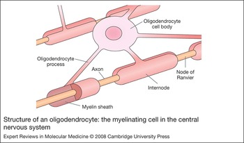
Pelizaeus–Merzbacher disease (PMD; OMIM 312080) is an X-linked leukodystrophy characterised by dysmyelination of the central nervous system (CNS). In PMD, myelin is not formed properly during development and is distinct from demyelination – when myelin forms and is later broken down. PMD is a rare disorder with an estimated incidence of 1 case per 770 000 live births in Germany (Ref. Reference Heim1). Patients have reduced white matter because of the failure of oligodendrocytes to myelinate axons (Fig. 1). Affected individuals are typically males, with females being unaffected carriers. The disease has been recognised for over 100 years, with the first clinical and neuropathological descriptions by Pelizaeus (Ref. Reference Pelizaeus2) and Merzbacher (Ref. Reference Merzbacher3), respectively. Pelizaeus reported a family with males affected by early nystagmus (rapid, involuntary, jerky eye movements), ataxia (failure of muscular coordination), spasticity and developmental delay. Merzbacher investigated the same family 25 years later in 1910 and described the diffuse hypomyelination of the CNS, associated with a reduced number of mature oligodendrocytes.
Structure of an oligodendrocyte: the myelinating cell in the central nervous system. Oligodendrocyte processes spiral around axons during myelination in the CNS.

Discovery of the molecular basis of PMD was significantly aided by studies of allelic animal models, including mice (Refs Reference Dautigny4, Reference Nave5, Reference Hudson6) and rats (Ref. Reference Boison and Stoffel7). In 1986, the gene encoding myelin proteolipid protein (PLP1) was shown to be mutated in the dysmyelinating jimpy mutant mouse (Ref. Reference Dautigny4). Further studies revealed that the molecular defect was a deletion in PLP1 mRNA, leading to aberrant splicing of the protein (Refs Reference Nave5, Reference Hudson6). In 1989, the genetic cause of PMD was recognised when mutations in PLP1, which is located on chromosome Xq22.3 (Ref. Reference Mattei8), were identified in several patients (Refs Reference Hudson9, Reference Gencic10, Reference Trofatter11). Mutations in PLP1 have also been identified in patients with spastic paraplegia type 2 (SPG2) – a clinically distinct, but allelic disorder to PMD (Ref. Reference Saugier-Veber12). However, sequence variations in PLP1 could only account for 15–20% of cases, including some with clear X-linked pedigrees (Ref. Reference Boespflug-Tanguy13). Analogies were made between PMD and the peripheral nervous system (PNS) disorder Charcot–Marie–Tooth disease type 1 (CMT1A), which is caused by duplications of the peripheral myelin protein 22 gene (PMP22) (Ref. Reference Lupski and Garcia14), and this led to the identification of PLP1 duplications in PMD patients (Ref. Reference Ellis and Malcolm15). Increased dosage of PLP1 caused by a duplication of chromosome Xq22 is now recognised to be the major cause of PMD (Refs Reference Sistermans16, Reference Inoue17, Reference Wang18). Deletions of PLP1 can also give rise to PMD or SPG2 but account for very few cases (Refs Reference Raskind19, Reference Inoue20). General correlations between the type of mutation and the clinical phenotype are emerging, with deletions resulting in mild PMD or SPG2, duplications giving rise to moderate PMD and missense mutations leading to severe disease (see below).
Many animal models of PMD with spontaneous point mutations have been characterised, and several mirror the genetic defects found in human disease (Refs Reference Lazzarini21, Reference Griffiths22, Reference Yool23). Transgenic mouse models with additional copies of PLP1 have been generated (Ref Reference Readhead24, Reference Kagawa25, Reference Inoue26), as well as mice with targeted mutations and functional null alleles (Refs Reference Boison and Stoffel27, Reference Klugmann28, Reference Stecca29). Analyses of mouse models and cell culture studies are providing valuable insights into the aetiologies of PMD and opportunities to develop therapeutic approaches.
Clinical description of PMD and SPG2
Together, PMD and SPG2 represent a broad- spectrum CNS myelin disorder, which is reflected by the disease terminology PMD/SPG2. PMD/SPG2 phenotypes range from severe connatal PMD through an intermediate classical form with a slowly progressive course, to mild PMD/SPG2, and generally, the level of dysmyelination correlates well with clinical severity (Refs Reference Steitelberger30, Reference Seitelberger31, Reference Cailloux32). Although the clinical features of PMD are variable, there are common characteristics, which include nystagmus, ataxia, psychomotor developmental delay, spasticity and onset within the first year of life (Refs Reference Boulloche and Aicardi33, Reference Hodes, Pratt and Dlouhy34, Reference Inoue35, Reference Garbern36).
Clinical features of connatal PMD present shortly after birth: patients have severely delayed motor and intellectual development, no head control and die in childhood or within the third decade of life. Additional signs such as seizures, stridor and severe dysphagia may be present (Ref. Reference Boulloche and Aicardi33, Reference Scheffer37), but the seizures are rarely epileptic. Classical PMD is the predominant form of the disease, with symptoms usually presenting in the first year of life, and patients often survive into their sixth decade. Patients have nystagmus, muscular hypotonia evolving to spastic paraparesis, movement disorder, including ataxia and dystonia, and mild cognitive delay. SPG2 has a later onset of 1–5 years, and patients have a milder phenotype and are often able to walk, talk and have a normal lifespan. Symptoms of SPG2 include progressive weakness and spasticity of the lower extremities with or without CNS involvement. Patients are classified as having either ‘complicated’ or ‘pure’ SPG2 accordingly. The clinical presentation of complicated SPG2 is very similar to mild PMD, and patient phenotypes are almost indistinguishable.
A clinical diagnosis of PMD relies on the neurological signs of disease, an X-linked pattern of inheritance and diffusely abnormal myelinating white matter upon cerebral magnetic resonance imaging (MRI). Molecular diagnosis involves screening for dosage alterations and mutations in PLP1, and is essential for confirmation of the clinical findings because there is no biochemical functional assay for PLP1.
Proteolipid protein and PLP1
The PLP1 gene is composed of seven exons extending over approximately 17 kb of genomic sequence on chromosome Xq22 (Ref. Reference Diehl38). An internal splice donor site within exon 3 creates the smaller alternatively spliced transcript DM20 (Ref. Reference Nave39), which lacks exon 3B. PLP1 encodes a 277 amino acid, 30 kDa protein known as proteolipid protein 1 (PLP1) and its smaller isoform DM20, which is a 242 amino acid, 25 kDa protein. PLP1 and DM20 are hydrophobic integral membrane proteins and are the major myelin components in the CNS, constituting approximately 50% of total protein (Ref. Reference Eng40, Reference Lees, Brostoff and Morell41). Myelin basic protein (MBP) is also found in compact myelin and accounts for approximately 30% of total protein. PLP1 and DM20 are highly conserved across species and they are identical in man, mouse and rat at the amino acid level.
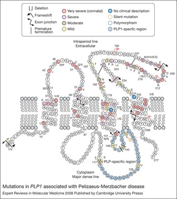
A four-transmembrane domain structure linked by one intracellular and two extracellular loop regions has been predicted for PLP1 and DM20 (Refs Reference Popot, Pham Dinh and Dautigny42, Reference Weimbs and Stoffel43, Reference Gow44) (Fig. 2). The N- and C-termini of both proteins are located in the cytoplasm. The 35 amino acids (residues 117–151) missing from DM20 are within the intracellular loop, which is known as the PLP1-specific region. Post-translational modification of the mature proteins includes removal of the first methionine (Ref Reference Milner45), formation of two disulphide bridges between two pairs of cysteine residues in the second extracellular loop region and acylation of cysteine residues (Ref. Reference Weimbs and Stoffel43).
Mutations in PLP1 associated with Pelizaeus-Merzbacher disease. Putative PLP1 topology includes four transmembrane domains, two extracellular loops and one cytoplasmic loop. The alternatively spliced region present in PLP1 and absent in DM20 is coloured blue (position 117–151) within the cytoplasmic loop. Known PLP1 mutations are shown with the most severe phenotype represented if more than one mutation is found at a single amino acid position. Clinical details and references for the mutations are available from http://www.geneclinics.org/img/table3-PLP1.pdf. Acylated cysteine residues are coloured black. The two disulfide bridges between two pairs of cysteine residues in the second extracellular loop region are shown as black connecting lines (C184–C228 and C201–C220). These cysteine-mediated crosslinks in PLP1 have an important role in correct protein folding. Mutations that lie within this second extracellular loop can lead to abnormal crosslinks that result in retention in the endoplasmic reticulum (ER) and activation of the unfolded-protein response (Ref. Reference Dhaunchak and Nave121). Figure adapted from Ref. Reference Garbern36, with permission from Springer © 2007.

Expression of PLP1 and DM20 is spatially and temporally regulated during development (Ref. Reference Campagnoni and Skoff46). PLP1 is predominantly expressed in oligodendrocytes and is the major isoform expressed during myelination and adulthood. By contrast, DM20 is expressed early in embryonic development, prior to myelination, and has a more ubiquitious expression profile in Schwann cells – the myelinating cells of the PNS – and in nonmyelinating cells, including those of the heart, spleen, thymus and lymph nodes (Refs Reference Campagnoni and Skoff46, Reference Campagnoni47, Reference Pribyl48).
PLP1 is synthesised at the endoplasmic reticulum (ER) within the oligodendrocyte and then transported through the Golgi to the cell surface for incorporation in the myelin membrane (Ref. Reference Nussbaum and Roussel49). PLP1 is associated with cholesterol and galactocerebroside-enriched membrane domains in myelin rafts during transport (Ref. Reference Simons50). The myelin lipid and protein components are assembled in the Golgi, and the formation of rafts may be important in the regulation of intracellular membrane transport and assembly of myelin (Refs Reference Simons50, Reference Simons51). A single oligodendrocyte can myelinate up to 50 axons (Fig. 1), and synthesis of the myelin sheath that envelops each axon occurs during a few days of myelinogenesis. Metabolic activity throughout this period is extremely high, with ~105 myelin-specific protein molecules synthesised every minute.
The abundance and location of PLP1 in compact myelin suggests a structural role in the formation and/or maintenance of the myelin sheath by adhesion and stabilisation of the extracellular surfaces (Refs Reference Klugmann52, Reference Bizzozero53). However, the exact physiological functions of PLP1 and DM20 are still uncertain, and other proposed functions for PLP1 include an ion channel (Refs Reference Knapp54, Reference Kitagawa55), an adhesion molecule (Ref. Reference Klugmann52) and a signalling molecule in oligodendrocyte interactions (Ref. Reference Gudz56). Although PLP1 and DM20 are structurally very similar, there are contradictory data on whether they are functionally equivalent. It has been shown that DM20 cannot replace PLP1 in compact CNS myelin (Ref. Reference Stecca29). Knock-in mice that lack PLP1 but have DM20 expression levels comparable with that of wild-type PLP1, developed progressive neurodegenerative changes similar to those seen in PLP1-knockout mice (Ref. Reference Stecca29). This finding suggests that the 35 amino acid section comprising the PLP1-specific region must have unique properties that are absent in DM20 and essential for a stable compact myelin sheath. However, a transgenic mouse expressing only the DM20 isoform has been created, and the protein was able to assemble into a functionally ordered compact myelin sheath (Ref. Reference Spörkel57). The mice showed no evidence of neurodegeneration or axonal swellings, only late-onset oligodendrocyte death and slow disintegration of the compact myelin sheath (Ref. Reference Spörkel57).
Genetic mechanisms for PMD/SPG2
PLP1 duplication
Duplications of chromosome Xq22, including the entire dosage-sensitive PLP1 gene, account for 60–70% of patients (Refs Reference Ellis and Malcolm15, Reference Sistermans16, Reference Inoue17, Reference Wang18). Duplications are heterogeneous, ranging in size from 100 kb to 4.6 Mb, with great variability in the position of proximal and distal breakpoints (Refs Reference Woodward58, Reference Inoue59, Reference Woodward60). The duplications cannot usually be seen by standard chromosome analysis using the light microscope, and therefore they are termed submicroscopic. The majority of duplications have a tandem head-to-tail orientation within Xq22.2, and predominantly arise from an intrachromosomal event in an ancestral male during spermatogenesis (Refs Reference Woodward58, Reference Inoue59, Reference Mimault61). However, there are exceptions to simple tandem events, because several patients have complex rearrangements, which may include a second region of duplication within Xq22 (Ref. Reference Woodward60). Furthermore, PLP1 triplications, or even a quintuplication, with five copies of PLP1, have been described (Ref. Reference Wolf62).
Other atypical cases with increased dosage of the PLP1 gene include patients with noncontiguous duplications. In these patients, the additional copy of PLP1 has integrated into different regions of the X chromosome (Xp22 and Xq26) (Refs Reference Hodes63, Reference Woodward64), or the Y chromosome (Ref. Reference Woodward60). These unusual duplications are termed submicroscopic transposons. In addition to these cases, there has been at least one individual reported with an atypical PLP1 duplication that did not result in clinical features of the disease (Ref. Reference Inoue20). An unaffected male with two copies of PLP1, one at Xq22, and an additional copy translocated to the subtelomeric region of chromosome 19q, has also been described. The translocated copy is probably either not expressed or not functional owing to a position effect or sequence changes associated with the rearrangement. His mother was a carrier of the translocation and also had a PLP1 deletion on one of her X chromosomes. The rearrangement was discovered because the mother had another son who inherited the X chromosome with the PLP1 deletion and was affected with PMD (Ref. Reference Inoue20).
Genomic duplications
As PMD is caused by alterations in gene dosage, it is referred to as a genomic disorder, but unlike many other genomic disorders such as CMT1A (Refs Reference Lupski65, Reference Inoue and Lupski66), the PLP1 duplications are not common recurrent rearrangements. The largest study of 59 PMD patients with duplications showed that each genomic structure was unique, and sequencing the breakpoint junctions in 13 patients revealed that the tandem duplications are formed by a coupled homologous and nonhomologous recombination mechanism (Ref. Reference Woodward60). The model suggests that duplications are initiated by a double-strand break on one side of PLP1, which is followed by strand invasion of a sister chromatid by one side of the break, and DNA synthesis. The event is completed by a nonhomologous end joining (NHEJ) repair mechanism at the other end of the break (Refs Reference Woodward60, Reference Lee67, Reference Lee, Carvalho and Lupski68).
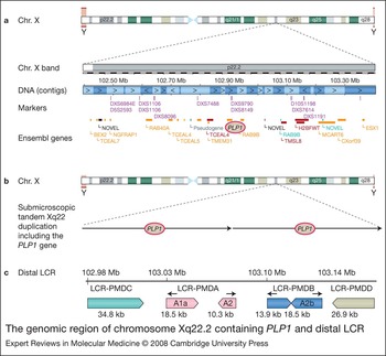
The molecular mechanism for formation of PLP1 duplications therefore differs mechanistically from many other genomic diseases because it does not involve nonallelic homologous recombination (NAHR) between flanking low-copy repeats (LCRs) (Refs Reference Lupski65, Reference Lee67, Reference Shaw and Lupski69). However, there is evidence at the nucleotide sequence level for the presence of structural features near the PLP1 gene, which might have a role in mediating the duplications. Clustering of the distal breakpoints near a large region of LCR has been reported, unlike the less-clustered distribution of proximal breakpoints (Refs Reference Woodward58, Reference Inoue59, Reference Woodward60). The distal LCR has a complicated structure containing inverted and direct repeats termed LCR-PMDA, LCR-PMDB, LCR-PMDC and LCR-PMDD (Refs Reference Inoue59, Reference Woodward60, Reference Lee67) (Fig. 3) and may have a role in increasing susceptibility to DNA double-strand breaks that initiate formation of the PLP1 duplications and deletions. Further LCRs have been identified proximal to the PLP1 gene (Ref. Reference Lee67); many are more than 1 Mb proximal to PLP1 – beyond the region of DNA breakpoints in patients – but it has been suggested that this novel genomic sequence arrangement could be involved in stimulating the DNA rearrangements within Xq22 (Ref. Reference Lee67).
The genomic region of chromosome Xq22.2 containing PLP1 and distal LCR.PLP1and distal LCR. a. Genes and markers within a 1 Mb region surrounding the PLP1 gene. PLP1 lies at position 102918410–102934201 (102.9 Mb) (Ensembl 49, NCBI build 36) on the X chromosome and is indicated with a red circle. b. Illustration of a tandem submicroscopic duplication of chromosome Xq22 including the PLP1 gene. c. Organisation of low-copy repeats (LCRs) distal to PLP1. The position of the start of each LCR on chromosome X and the size of each LCR is shown (Ensembl 49, NCBI build 36). Arrows represent the direction of each repeat.

Detection of PLP1 duplications
Since duplications are the most frequent cause of PMD, molecular diagnosis usually commences with PLP1 dosage analysis and is followed by PLP1 sequencing if no duplication is detected. Many methods have been described to detect PLP1 duplications. Traditionally, techniques such as interphase fluorescence in situ hybridisation (FISH) (Refs Reference Woodward58, Reference Inoue59) and quantitative multiplex PCR (Refs Reference Inoue17, Reference Woodward58) were used, but now there is a move towards more rapid and robust screening tests, such as real-time PCR (Refs Reference Regis70, Reference Hübner71, Reference Gao72, Reference Mikesová73) and multiplex ligation-dependent probe amplification (MLPA) (Refs Reference Wolf62, Reference Warshawsky74). FISH is still a valuable tool, because it is a visual technique and allows identification of typical PLP1 duplications where the additional copy of the PLP1 gene is not located within Xq22 but has been transposed elsewhere in the genome.
Prenatal diagnosis is well established using chorionic villus sampling (CVS) or amniotic fluid samples (Refs Reference Regis70, Reference Woodward75) and has also been performed using the novel technique of array comparative genomic hybridisation (array-CGH) (Ref. Reference Lee76). One of the main advantages of array-CGH is that a custom microarray, which includes overlapping DNA clones from a large region (~10 Mb) around the PLP1 gene, can be used. This allows detection of a duplication (or a deletion) and also determination of the size of the rearrangement and position of the DNA breakpoints. The first case of preimplantation genetic diagnosis has also been described recently for a family carrying a PLP1 mutation that causes severe clinical features of disease (Ref. Reference Verlinsky77).
PLP1 deletion and null mutations
Deletions or null mutations of the PLP1 gene (Refs Reference Raskind19, Reference Inoue20, Reference Sistermans78, Reference Garbern79, Reference Garbern80) that give rise to loss of protein are rare events compared with the frequency of the duplication. Only a few deletion cases have been reported, and the missing genomic segments have been relatively small and contain few genes. The infrequency of their occurrence could be due to other larger Xq22 deletions being lethal in hemizygous males. Mapping the DNA breakpoints in three families with PLP1 deletions suggested that the rearrangements occurred by NHEJ and correlates with the finding that duplications are also mediated by NHEJ and not nonhomologous allelic recombination (NHAR) (Ref. Reference Inoue20).
PLP1 sequence changes
PLP1 sequence changes, including single nucleotide mutations, small insertions and deletions, are responsible for 15–20% of cases of PMD/SPG2. More than 100 different PLP1 mutations have been identified – most are missense point mutations that result in amino acid substitutions, but there are also nonsense and frameshift mutations that produce abnormal PLP1 and DM20 proteins (Refs Reference Hodes, Pratt and Dlouhy34, Reference Inoue35, Reference Garbern36) (Fig. 2). A current list of the mutations can be obtained from the Gene Clinics website (http://www.geneclinics.org/img/table3-PLP1.pdf). The majority are located within the coding region, but noncoding region and splice-site mutations that affect splicing and lead to abnormal PLP1 expression have also been described (Refs Reference Hobson81, Reference Hobson82).
Other causes of PMD
The remaining 10–20% of patients with clinical features of PMD have no detectable abnormality, suggesting that mutations in regulatory sequences or other gene loci can cause PMD. Genetic screening focuses on the PLP1 coding regions, and therefore the possibility of further noncoding mutations exists. Both positive and negative transcription regulatory elements have been described in the 5′ promoter (Refs Reference Nave and Lemke83, Reference Berndt, Kim and Hudson84) and intron 1 (Ref. Reference Dobretsova, Kokorina and Wight85). Furthermore, another exon has been detected within intron 1 of the murine Plp1 gene, which results in the addition of a leader sequence to PLP1 and DM20 in the mouse, and could direct the proteins away from their normal location in myelin (Ref. Reference Bongarzone86). No homologous section has been found in the equivalent region of the human genome, and therefore the functional significance is currently unclear.
A position effect on PLP1 rather than a mutation could be responsible for the PMD/SPG2 phenotype for two patients with X chromosome rearrangements near the PLP1 gene. One case had an X chromosome inversion (Ref. Reference Muncke87) and the other a downstream duplication (Ref Reference Lee88), both of which are proposed to result in abnormal PLP1 expression.
Mutations in the autosomal recessive gene encoding the gap-junction protein γ2 (GJC2; also known as GJA12 and connexin 47) cause a disorder almost identical to PMD, known as Pelizaeus–Merzbacher-like disease (PMDL; OMIM 311601) (Refs Reference Uhlenberg89, Reference Wolf90, Reference Bugiani91, Reference Salviati92). Patients with PMDL have nystagmus, progressive ataxia, developmental delay and severe CNS dysmyelination. The GJC2 gene is highly expressed in oligodendrocytes in the CNS and encodes a 439 amino acid gap-junction protein. Loss of GJC2 function is reported to be the cause of PMDL (Refs Reference Bugiani91, Reference Orthmann-Murphy93), despite Gjc2-knockout mice showing no obvious dysfunction. However, double-knockout mice with loss of Gjc2 and Gjb1 – which encodes connexin 32 and causes the X-linked form of Charcot–Marie–Tooth disease in humans (Ref. Reference Bergoffen94) – have severe CNS myelin abnormalities and premature death (Refs Reference Menichella95, Reference Odermatt96). The study of other genes involved in myelinogenesis and genes encoding transcription factors that regulate PLP1 expression may lead to the identification of further mutations causing PMDL.
Phenotype–genotype correlations
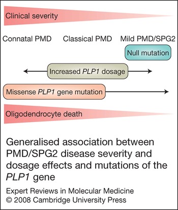
Although there is a wide range of mutations causing PMD/SPG2, some general phenotype–genotype correlations are emerging (Fig. 4) (Ref. Reference Nave and Boespflug-Tanguy97). Missense mutations that result in nonconservative amino acid substitutions (Ref. Reference Cailloux32) are mostly associated with severe connatal PMD. Patients have severe dysmyelination, with an almost complete absence of myelin and a significant reduction of oligodendrocytes. By contrast, a deletion of PLP1 or a loss-of-function mutation is associated with mild disease and widespread demyelination in the CNS and PNS (Refs Reference Sistermans78, Reference Garbern79, Reference Garbern80). These findings suggest that the dysmyelination caused by PLP1 mutations and duplications is not due to the loss of functional protein but instead from a possible gain-of-function mutation or the cytotoxic effect of mutant proteins (as discussed below).
Generalised association between PMD/SPG2 disease severity and dosage effects and mutations of the PLP1 gene. Patients with PLP1 duplications mostly have a classical PMD phenotype but symptoms at the connatal and mild SPG2 end of the spectrum are possible. PLP1 triplications and a PLP1 quintuplication cause severe connatal PMD. Patients with missense mutations have a wide range of phenotypes, but are often more severely affected than the PLP1 duplication patients. Patients with loss of PLP1/DM20 function caused by a PLP1 deletion or a null mutation have mild PMD/SPG2 and peripheral system involvement.

Most patients with PLP1 duplications have classical PMD and a moderate phenotype (Refs Reference Sistermans16, Reference Inoue59) (Fig. 3), whereas patients with triplications or a quintuplication of the PLP1 gene have severe connatal PMD, indicating that disease severity is proportional to PLP1 copy number (Ref. Reference Wolf62). However, there are some patients with duplications who also have connatal PMD, and conversely, others who are only mildly affected. Genes surrounding PLP1 are often included within the duplicated segment, but the role of these other loci in contributing to the disease phenotype has not yet been clarified. It was proposed that an increase in the size of the duplication correlates with an increase in clinical severity (Ref. Reference Inoue59); however, examination of a larger cohort of patients suggested that this is not likely to be the case (Ref. Reference Woodward60). Some of the patients with the largest duplications have the mildest symptoms, indicating that the most significant dosage-sensitive gene in the vicinity of Xq22 is PLP1 and that neighbouring genes are usually unaffected by dosage alterations. The range in clinical severities may be explained by the different duplication structures causing the juxtaposition of different regulatory sequences, which could influence the expression of PLP1. The effect of modifier genes, either within the duplicated region or elsewhere in the genome, may also affect PLP1 expression, and consequently, disease pathogenesis.
Other mutations that lead to a mild phenotype include those within the PLP1-specific region that affect transport of PLP1 but not DM20, point mutations resulting in premature stop codons, and frameshift alterations that give rise to truncating mutations and mutant mRNA, which is degraded by the nonsense-mediated decay (NMD) pathway (Ref. Reference Inoue98). However, heterogeneity in severity exists even within families (Ref. Reference Inoue59), suggesting the influence of modifier genes and/or genetic background. Such modifying loci have been described in mice (Ref. Reference Al-Saktawi99) where the PLP1 rumpshaker mutation (rsh, I186T) (Ref. Reference Schneider100) results in wide variation in phenotypic severity depending on the genetic background. The molecular basis for the phenotype variability in PMD patients is not completely understood, but probably reflects the distinct cellular effects of the different genetic mechanisms involved. Our understanding of how the different mutational mechanisms underlie pathogenesis is becoming clearer by comparing human patients with animal models.
Mechanisms of disease pathogenesis: distinct cellular defects underlie the different genetic abnormalities
PLP1 duplication
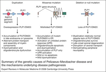
The underlying pathogenic mechanism of increased PLP1 gene dosage is thought to be a perturbation of protein transport and assembly of myelin components (Ref. Reference Simons51) (Fig. 5). PLP1 normally interacts with cholesterol (Ref. Reference Simons50) and other lipids to form myelin rafts, and it is subsequently transported to the plasma membrane. When PLP1 is duplicated, it is presumed to lead to overexpression of PLP1. In vitro studies have shown that overexpression of PLP1 results in accumulation of PLP1 in the late endosome/lysosome with cholesterol, and causes aberrant raft-membrane trafficking (Ref. Reference Simons51). The accumulation of PLP1 and cholesterol is suggested to interfere with myelination and to reduce viability of the oligodendrocyte.
Summary of the genetic causes of Pelizaeus–Merzbacher disease and the mechanisms underlying disease pathogenesis.

There are no spontaneous animal models for PLP1 gene duplication, but three lines of transgenic mice have been generated with autosomal copies of wild-type murine Plp1, and these also show that increased gene dosage has detrimental effects on oligodendrocyte function (Refs Reference Readhead24, Reference Kagawa25, Reference Inoue26). The mice show hypomyelination, and generally their age of onset and severity of disease is proportional to Plp1 gene dosage (Ref. Reference Anderson101). The mice with high Plp1 dosage (usually homozygous for the transgene) have severe early-onset dysmyelination, increased oligodendrocyte apoptosis and premature death. Pathogenesis is not completely understood, but autophagic vacuoles have been observed in the oligodendrocytes, suggesting that there is accelerated protein degradation (Refs Reference Anderson101, Reference Karim102).
By contrast, hemizygous mice with a lower transgene copy number develop normally and have no clinical signs until later in life. Late-onset demyelination plus axonal swelling and degeneration becomes apparent at around 15–18 months of age, suggesting that the oligodendrocytes are unable to maintain their myelin sheaths. The pathology is similar to that of the Plp1-knockout mouse, indicating that changes in gene dosage – either an increase or a decrease – may be causing the axonal changes.
Interestingly, a recent study of transgenic mice with high and low transgene copy number has provided further information on the mechanism underlying the dysmyelination associated with increased Plp1 gene dosage. The relationship between Plp1 gene dosage and the steady-state levels of PLP1 and DM20 protein is not straightforward and is affected by the myelination status within the cell (Ref. Reference Karim102). During myelination, Plp1 expression increases, and mice with a low transgene copy number were found to have a higher level of PLP1/DM20 protein in the oligodendrocyte soma (which probably correspond to the autophagic vacuoles), but the level in myelin was only minimally affected. By contrast, the cellular effects in mice with a high transgene copy number were significant, because a high level of PLP1/DM20 was retained in the soma and low levels of PLP1 were detected in myelin. Therefore, as the dosage of the Plp1 gene increases, the cell is less able to tolerate the increases in protein levels, indicating that the biosynthesis of PLP1 must be tightly controlled for normal myelin development.
Increased Plp1 gene dosage also affects other myelin genes, particularly MBP, and thus can alter the proportions of other components in the myelin sheath (Ref. Reference Karim102). MBP is the second most abundant protein in compact myelin and decreases in levels of its mRNA and protein were detected. The significance of this finding is currently unclear; however, as the dysmyelination associated with PMD is not caused by loss of functional PLP1 and DM20 proteins, it may be that the influence of PLP1 dosage on other genes could have an important role in disease pathogenesis.
Both PMD and CMT1A are caused by an increase in the dosage of a myelin gene. Increased peripheral myelin protein 22 gene (PMP22) dosage causes dysmyelination in the PNS, and expression of other myelin proteins is also reduced (Ref. Reference Sereda103). Together, these results suggest that the stoichiometry of myelin genes is important for maintaining normal myelin structure and function, and that the myelinating cells are sensitive to an imbalance in myelin protein components, possibly because of their high rate of myelin synthesis.
PLP1 deletion and null mutation
In contrast to PLP1 duplications and many missense mutations, a PLP1 gene deletion or null mutation allows myelination to proceed, and patients have a mild clinical phenotype, which is classified as mild PMD or complicated SPG2 (Refs Reference Raskind19, Reference Inoue20, Reference Sistermans78, Reference Garbern79, Reference Garbern80) (Fig. 5). Knockout mice with a functionally null Plp1 gene (Refs Reference Boison and Stoffel27, Reference Klugmann28, Reference Stecca29) do not develop classical signs of Plp1-related disease: their oligodendrocytes develop normally and synthesise compact myelin sheaths. The mice have only ultrastructural abnormalities, which include swellings in small-diameter axons (at 6–8 weeks) and late-onset axonal degeneration (Ref. Reference Griffiths104). Neither PLP1 nor DM20 is necessary for myelin assembly but they are required for myelin and axon maintenance, where they are probably involved in axon–glial interaction.
Length-dependent axonal degeneration has been found in humans and knockout mice with a functionally null PLP1 gene (Ref. Reference Garbern80). The degeneration is not associated with significant demyelination and is similar to that observed in the PNS of patients with CMT1A, who have an inherited demyelinating neuropathy. The disruption in PLP1-mediated axon–oligodendrocyte interaction is probably responsible for this axon degeneration. In addition, some patients with null mutations present with mild demyelinating peripheral neuropathy (Ref. Reference Garbern105). These patients demonstrate that although PLP1 is expressed at low levels in the PNS compared with the CNS, PLP1 or DM20 is required for peripheral myelin function.
PLP1 missense mutation
The mechanism underlying pathogenesis for most missense mutations is thought to be accumulation of mutant PLP1 in the rough endoplasmic reticulum (RER), presumably because of protein misfolding and failure in transport of PLP1 to the oligodendrocyte cell membrane (Refs Reference Roussel106, Reference Gow, Friedrich and Lazzarini107, Reference Gow and Lazzarini108, Reference Thomson109) (Fig. 5). The disruption of normal PLP1 processing and intracellular trafficking is considered to be a direct cause of premature oligodendrocyte death (Refs Reference Gow, Southwood and Lazzarini110, Reference Cerghet111, Reference Southwood112). Correlation between the abundance of PLP1 and DM20 at the cell surface with the different types of PMD has provided a cellular basis for the variation in disease severity (Refs Reference Gow, Friedrich and Lazzarini107, Reference Gow and Lazzarini108, Reference Thomson109). Mutations within the PLP1-specific region of the gene that lead to aberrant PLP1 and normal DM20, result in a milder clinical phenotype with less oligodendrocyte death compared with mutations that interrupt trafficking of both PLP1 and DM20, which give rise to severe PMD with increased oligodendrocyte death (Refs Reference Schneider100, Reference Gow and Lazzarini108, Reference Gow, Southwood and Lazzarini110). Exactly how the misfolded proteins trigger oligodendrocyte apoptosis and why they are harmful to the cell is currently uncertain. There could be a deleterious novel gain-of-function (Ref. Reference Gow, Southwood and Lazzarini110), which is supported by the inability of wild-type PLP1 to correct the naturally occurring jimpy mutation in mice with severe dysmyelination (Ref. Reference Schneider113). This, together with the results from the knockout mice experiments, suggests that the lack of PLP1 or DM20 function does not cause CNS dysmyelination.
Misfolded proteins in the ER can induce an unfolded protein response (UPR), which can modulate disease severity through a signalling cascade that includes transcriptional activation of chaperone genes and apoptosis (Ref. Reference Southwood112). Protein misfolding has been implicated in the pathogenesis of other neurodegenerative diseases (Ref. Reference Taylor114) and activation of the UPR has been described in Alzheimer disease and Huntington disease (Ref. Reference Hoozemans115, Reference Hoozemans116). Analysis of PLP1 and DM20 trafficking in oligodendroglial cells demonstrated that there are distinct trafficking defects for missense mutations associated with mild and severe disease and that cholesterol binding and lipid raft association was impaired for both (Ref. Reference Krämer-Albers117). Mutant PLP1 associated with severe disease was retained in the ER, whereas that associated with the mild rumpshaker disease phenotype reached the late endosomal/lysosomal compartment (Ref. Reference Krämer-Albers117). Interestingly, mutant PLP1 or DM20 was rapidly degraded compared with the wild-type protein, and there was an increased rate of turnover for mutant PLP1 associated with severe disease (Ref. Reference Krämer-Albers117). A similar finding of increased turnover of mutant PLP1 compared with the wild-type protein was reported by a second group of investigators (Ref. Reference McLaughlin118), but it is not thought that the reduction of PLP1 is the main cause of the dysmyelination. A recent study of a mouse model of PMD has shown that variability in disease severity may not be caused by differences in PLP1 processing, but possibly by a gain-of-function involving other components of the compacted myelin sheath (Ref. Reference McLaughlin119). A correlation between severe disease and activation of the transcription factor CHOP (C/EBP homologous protein 10) was detected, which maybe related to the increased level of apoptosis (Ref. Reference McLaughlin119). However, an earlier study contradicts this finding, suggesting that CHOP actually protects oligodendrocytes from apoptosis (Ref. Reference Southwood112). Therefore, further investigation is required to clarify the discrepancy.
The molecular mechanism that mediates the retention of mutant PLP1 in the ER is currently unclear. PLP1 missense mutations can cause a severe disease phenotype and result in premature oligomerisation of PLP1 in the ER of transfected cells (Ref. Reference Swanton120). Evidence from in vitro studies suggests that wild-type PLP1 is transported to the cell surface, where it slowly forms stable oligomers after a long time of maturation. By contrast, mutant PLP1 is assembled rapidly into oligomers within the ER, but has a similar conformation to mature, native PLP1. Rapid oligomer formation could possibly mask a signal for transport to the cell surface. Interestingly, mutations leading to severe disease are more likely to form rapid oligomers than mutations leading to a mild PMD phenotype (Ref. Reference Swanton120). The formation of stable oligmers in the ER instead of the cell membrane could affect oligodendrocyte function and may have an important role in disease pathogenesis rather than simply in the retention of misfolded protein (Ref. Reference Swanton120).
Although missense mutations occur throughout PLP1 and DM20, an over-representation has been found within the large second extracellular loop, suggesting that this region could have an important role in protein conformation and myelin stability. Recently, mutations in this extracellular loop of PLP1 and DM20 have been shown to perturb the formation of an intramolecular disulfide bridge in the lumen of the ER (Ref. Reference Dhaunchak and Nave121). The formation of a disulfide bridge is not essential for normal folding and trafficking of PLP1 and DM20. However, when the cysteine is unpaired, it becomes stearically exposed and forms abnormal intermolecular crosslinks, which result in retention in the ER, a failure to oligomerise and activation of the UPR (Ref. Reference Dhaunchak and Nave121).
A novel heptapeptide signal was recently found in the alternatively spliced PLP1-specific peptide that is involved in retention of mutant PLP1 in the ER (Ref Reference Southwood122). In vitro analysis of mutant PLP1 or DM20 trafficking demonstrated that the retention of mutant PLP1 associated with mild disease is partially dependent on the heptapeptide signal, but that the retention of mutant PLP1 associated with severe disease is independent. This suggestion that the PLP1-specific peptide regulates ER retention represents another possible mechanism for PMD pathogenesis.
Females affected with Pelizaeus–Merzbacher disease
Although PMD typically affects males, a small number of manifesting females have been described with heterozygous PLP1 point mutations (Refs Reference Nance123, Reference Hodes124, Reference Sivakumar125, Reference Hurst126). The affected females have mild to moderate symptoms of disease with a late onset, and are often found within families with mildly affected boys. A similar finding has been observed in animal models of PMD, where heterozygous female mice with a PLP1 mutation leading to mild disease in males show phenotypical signs of disease themselves (Ref. Reference Fanarraga127). The mechanism giving rise to affected females can be explained by random X-inactivation and oligodendrocyte differentiation and survival, depending on the severity of the PLP1 mutation (Ref. Reference Inoue35). Random X-inactivation in heterozygous female carriers results in half of the cells having an active X chromosome expressing mutant PLP1. Oligodendrocytes expressing a severe mutation that affects both PLP1 and DM20 will have a greater incidence of cell death and consequently, only those expressing the wild-type PLP1 allele will survive. By contrast, oligodendrocytes expressing a mild PLP1 mutation will survive and form abnormal myelin, which can lead to late-onset neurological problems (Refs Reference Nance123, Reference Hodes124, Reference Sivakumar125). Analysis of X-inactivation patterns in blood from female carriers supports this hypothesis. Female carriers of a PLP1 duplication are mainly asymptomatic and they show heavily skewed X-inactivation, with the X chromosome bearing the duplication being preferentially inactivated (Ref. Reference Woodward128). By contrast, point-mutation carriers show a random pattern of X-inactivation and cell fate is determined by the severity of the mutation. There have been only two reported cases of affected females with PLP1 duplications (Ref. Reference Inoue129). Both these unrelated girls had early-onset mild PMD or SPG2 followed by progressive clinical improvement. They did not show skewed X-inactivation, and their recovery was probably due to increased myelin production from the oligodendrocytes expressing the normal X chromosome with only one copy of PLP1. A recent report of female carriers with neurological signs, in a family with severely affected males, contradicts previous findings and cannot be easily explained (Ref. Reference Marble130). Random X-inactivation was detected in leukocytes, but this might not reflect the pattern of X-inactivation present in oligodendrocytes expressing the PLP1 gene.
Research in progress and outstanding research questions
Although there is currently no specific treatment available for PMD or SPG2 patients, the wide range of animal models analogous to the different molecular mechanisms of disease will facilitate the potential for novel approaches to prevention and therapy. Understanding the cellular and molecular defects underlying pathogenesis of PMD is essential, and animal models have had an important role in dissecting the mechanisms involved. Significant progress has been made but the essential functions of PLP1 and DM20, and the direct causes of pathogenesis and dysmyelination remain unknown.
Myelin function could possibly be restored by either gene therapy or somatic cell transplantation. However, there are great problems with gene therapy owing to the sensitivity of PLP1 gene dosage and the gain-of-function mutations. Gene delivery may be difficult, but it will be an even greater challenge to obtain PLP1 expression at the correct level for normal myelination and maintenance. Reduction of PLP1 expression may be more successful, possibly by antisense or RNAi therapy, because loss of PLP1 gives rise to a less-severe clinical phenotype than PLP1 duplications or missense mutations. Alternatively, somatic cell therapy may be an easier option, and transplantation of oligodendrocyte precursors into the CNS has shown potential for animal models (Refs Reference Duncan, Grever and Zhang131, 132). The sustained clinical improvement of two female PLP1 duplication carriers also supports this approach, demonstrating that certain oligodendrocyte lineages may proliferate and compensate for dysmyelination, even years after birth (Ref. Reference Inoue129). Other therapies aimed at maintaining the axon integrity may also be useful.
Acknowledgements and funding
I would like to acknowledge the Wellcome Trust for funding whilst I worked at the Institute of Child Health, London, UK. I would also like to thank Dr Gabern for Fig. 2 and the anonymous peer reviewers for their critical reading of the manuscript and their helpful comments.