Introduction
Transcanal endoscopic ear surgery is minimally invasive, and provides clear vision even in the narrow and deep surgical space in the middle ear.Reference Kozin, Gulati, Kaplan, Lehmann, Remenschneider and Landegger1 It has been gradually spreading worldwide because innovative devices and instruments have been developed.Reference Kakehata, Watanabe, Ito, Kubota and Furukawa2 There have been a number of recent advances in transcanal endoscopic ear surgery regarding cholesteatoma surgery.
According to Tarabichi,Reference Tarabichi3 the main region of a cholesteatoma is in the attic, close to the external auditory canal; the external auditory canal, as an access port for cholesteatoma surgery, is the main advantage of transcanal endoscopic ear surgery. Recently, several surgeons have resected cholesteatomas with transcanal endoscopic ear surgery, even in cases where the cholesteatoma extends to the mastoid cavity beyond the attic.Reference Imai, Nishiike, Oshima, Tanaka, Tsuruta and Tomiyama4 In order to remove a cholesteatoma in the mastoid under transcanal endoscopic ear surgery, it is necessary to perform transcanal endoscopic mastoidectomy.
There are, however, several problems with endoscopic transcanal mastoidectomy. For access in the narrow and deep space, generally, a shafted curved bur is attached to an otological drill system. The bur, however, does not provide saline perfusion, and bone dust and blood obscure the surgical field. In order to countermeasure against this disadvantage, we developed a novel endoscopic hydro-mastoidectomy technique, in which the operator performs the mastoidectomy ‘underwater’ using a lens cleaning system that provides saline perfusion in the surgical space (Figure 1). This provides a clear surgical view by washing out bone dust and blood from the surgical area.
Diagram demonstrating the endoscopic hydro-mastoidectomy technique. A curved bur with an otological drill, and a rigid endoscope with a lens cleaning sheath, are inserted into the external auditory canal. The endoscope and the drill are immersed in the saline provided by the lens cleaning system. The surgeon controls the infusion of saline solution by stepping on a power console footswitch for the lens cleaning system.

Endoscopic hydro-mastoidectomy equipment
The endoscopes (Hopkins; Storz, Tuttlingen, Germany) measure 2.7 mm in diameter and 18 cm in length, and have an angle of 0° and 30° (Table 1).Reference Imai, Nishiike, Oshima, Tanaka, Tsuruta and Tomiyama4 Illumination is generated by a light-emitting diode and transmitted to the endoscope via a fibre-optic light cable (Storz). High-resolution images are obtained with a full high-definition camera and viewed on a 26 inch (66 cm) monitor (Storz).
Instruments required for endoscopic hydro-mastoidectomy technique

HD = high-definition
For the endoscopic hydro-mastoidectomy technique, a curved round coarse diamond bur (with a diameter of 2.0 or 3.0 mm) is attached to a Visao otological drill (Medtronic, Minneapolis, Minnesota, USA) (Table 1). An Endo-Scrub lens cleaning sheath (Medtronic) is fitted to the endoscope. The bur and the tube of the sheath are connected to the integrated power console system (Medtronic). The surgeon controls the infusion of saline solution by stepping on a footswitch of the power console with the right foot, and controls the rotation of the drill by stepping on another footswitch with the left foot.
Generally, lens cleaning sheaths have been used in endoscopic sinus surgery, but we use these frequently in transcanal endoscopic ear surgery. A sheath delivers a high-power burst of irrigation to flush debris out of the endoscope lens tip when the surgeon taps the footswitch, so the surgeon maintains maximum visualisation at the surgical site during endoscopic sinus surgery, without removing the endoscope. For endoscopic hydro-mastoidectomy technique, when we step on the footswitch, the sheath delivers a continuous flow of saline and it fills the surgical space (Figure 1).
Endoscopic hydro-mastoidectomy technique
A 54-year-old male presented with right-sided hearing loss, ear fullness and aural thermalgia. A local otolaryngologist suspected cholesteatoma and referred him to our department. Fibre-optic findings demonstrated a cholesteatoma in the pars flaccida. An audiogram showed conductive hearing loss. Computed tomography images revealed soft tissue density in the attic and the mastoid.
During surgery, a meatal incision was made and the tympanomeatal flap was elevated. The chorda tympani was preserved. The external auditory canal bone was first resected with chisels and curettes to create a sufficient space. The tympanomeatal flap was widely shifted and touched the anterior wall of the external auditory canal to prevent it floating into the drill bur. Then, we started the endoscopic hydro-mastoidectomy technique.
The technique provided a good surgical view, by washing out bone dust and blood from the surgical area (see the short video, available on The Journal of Laryngology & Otology website; Appendix 1). The operator easily controlled the flow of saline perfusion by stepping on a foot pedal. The saline perfusion overflowed out of the external auditory canal to the cavity of concha, and an assistant collected it using a sucker.
The extent of the cholesteatoma within the mastoid cavity was determined, and then it was removed from the inferior to superior direction. In this case, the cholesteatoma invaded the medial part of the malleus head and the incus, which were removed. The ossicular chain was reconstructed with a piece of the auricular cartilage. The resected area of the external auditory canal was measured using a paper template. Then it was reconstructed with multiple pieces of the cartilage. The external auditory canal was packed with a gelatine sponge and then Merocel. After 12 months, the patient's hearing level was improved and the pars flaccida was firmly constructed.
Discussion
Transcanal endoscopic ear surgery represents a more minimally invasive approach than microscopic ear surgery, because neither a wide incision nor a broad mastoidectomy is required.Reference Tarabichi3, Reference Migirov, Yakirevitch and Wolf5 In a previous study,Reference Imai, Nishiike, Oshima, Tanaka, Tsuruta and Tomiyama4 we evaluated the resected areas of the external auditory canal during transcanal endoscopic ear surgery for cholesteatoma, and these were much smaller than those of microscopic surgery.
As TarabichiReference Tarabichi3 reported, the main region of a cholesteatoma is in the tympanic cavity, but if the mastoid cavity is involved, the external auditory canal has to be aggressively removed superiorly and posteriorly under endoscopy, in order to access the mastoid cavity.
Mastoidectomy has generally been performed under microscopy. The operator holds a drill with the irrigation device in the right hand and a suction tube in the left. Saline is intermittently infused into the surgical field, and overflow is removed by suctioning. A shafted curved bur is generally attached to an otological drill system during endoscopic mastoidectomy, in order to prevent injuring neighbouring tissues and to observe the drill tip in the deep and narrow space. The bur, however, does not have irrigation capabilities, and bone dust and blood obscure the surgical field. There is also a possibility of undesired heat damage on surrounding tissues.
We utilise an underwater endoscopic hydro-mastoidectomy technique to overcome these problems. We apply a lens cleaning sheath to a rigid scope, and saline perfusion is provided by the lens cleaning system. This system was originally used for endoscopic sinus surgery worldwide, but we now also use it for ear surgery. It washes out bone dust and blood from the surgical field, improving the surgical view during mastoidectomy. Saline perfusion reduces the risk of heat damage. Additionally, the operator can easily control the flow of the saline perfusion. The setup for endoscopic hydro-mastoidectomy technique is easy and the operator needs only to buy sheaths if they already own the power console, as many otological and rhinological surgeons do.
The benefit of underwater endoscopic ear surgery in cases of labyrinthine fistula closure and superior semicircular canal dehiscence plugging was reported by Yamauchi et al.Reference Yamauchi, Yamazaki, Ohta, Kadowaki, Nomura and Hidaka6 The purpose of underwater surgery was to preserve inner-ear function during cholesteatoma removal from the labyrinthine fistula. Saline was perfused using a syringe by an assistant.
In the case of the endoscopic hydro-mastoidectomy technique reported here, underwater surgery was performed mainly for the purpose of a better surgical view. Our underwater condition was created using both an otological drill and a lens cleaning system, the latter of which provides the saline perfusion.
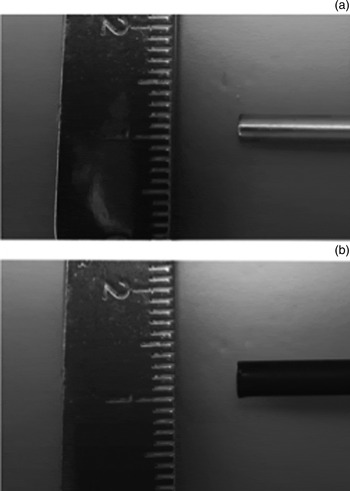
One of the disadvantages of the endoscopic hydro-mastoidectomy technique is poorer controllability of the endoscope, particularly in the narrow external auditory canal, because the sheath increases the width of the endoscope from 2.7 mm to 3.4 mm (Figure 2). For this reason, we first resect the external auditory canal bone with chisels and curettes to create sufficient space, and then begin the endoscopic hydro-mastoidectomy technique.
Width of endoscopes: the 0° endoscope measured 2.7 mm in diameter (a), but measured 3.4 mm in diameter when the lens cleaning sheath was applied (b).

Conclusion
We developed a novel endoscopic hydro-mastoidectomy technique for transcanal endoscopic ear surgery. The setup for the technique is easy. It provides a good surgical view, and the operator can easily control the flow of saline. Transcanal endoscopic ear surgery is minimally invasive and could be beneficial for patients with cholesteatoma.
Competing interests
None declared
Appendix 1. Supplementary video material
A short video demonstrating the endoscopic hydro-mastoidectomy technique is available online at The Journal of Laryngology & Otology website, at https://doi.org/10.1017/S002221511900046X.