INTRODUCTION
Marine gastrotrichs are microscopic, worm-like invertebrates that inhabit the interstices of littoral and subtidal sediments in all oceans and seas. While they are rarely the most abundant form of meiofauna (but see Coull, Reference Coull1985; Hochberg, Reference Hochberg1999), their morphological diversity can be overwhelming as a result of the various and often exquisite elaborations of their cuticle. The Thaumastodermatidae is the most speciose family of macrodasyidan gastrotrichs and one that shows the largest diversity in cuticular morphology and ultrastructure (see Ruppert, Reference Ruppert, Higgins and Thiel1988). The construction of the cuticle is highly elaborate, resulting from modifications of both endo- and exocuticle, and forming structures such as flat and bowl-shaped scales, spines, and multi-pronged hooks called ancres (Rieger & Rieger, Reference Rieger and Rieger1977). The shape of the cuticular structures may have strong phylogenetic signal, and some taxa within the Thaumastodermatidae can be grouped based on cuticular sculpture (Rieger & Rieger, Reference Rieger and Rieger1977; Hochberg & Litvaitis, Reference Hochberg and Litvaitis2001). The diversity of cuticular ornamentation (spines, scales, triancres, tetrancres and pentancres) is well-characterized from over 100 species distributed among five genera. Ruppert (Reference Ruppert1970) added a unique form of sculpture to our knowledge of cuticular diversity when he described Pseudostomella plumosa Ruppert, Reference Ruppert1970 from North Carolina, USA. The dorsal surface of P. plumosa is composed of highly modified triancres that form feather-like scales (feathered triancres). Since then, three species of Pseudostomella have been described with a similar cuticular covering; P. klauserae and P. megapalpator from Australia (Hochberg, Reference Hochberg2002b) and P. faroensis from the Faroe Bank (Clausen, Reference Clausen2004). No comparable cuticular structures have been described from other species in the family, and to date, nothing is known about their ultrastructure.
Research on marine interstitial gastrotrichs of Australia has yielded several new species in the past few years (Hochberg, Reference Hochberg2002b, Reference Hochbergc, Reference Hochberg2003; Nicholas & Todaro, Reference Nicholas and Todaro2005, Reference Nicholas and Todaro2006). Among them is a species of Tetranchyroderma with a cuticular sculpture similar to the feathered triancres of species of Pseudostomella. Here, I describe this new species of Tetranchyroderma and provide an ultrastructural account of the cuticle. These observations are compared to the ultrastructure of feathered triancres in P. megapalpator to arrive at a better understanding of the fine structural differences in their construction.
MATERIALS AND METHODS
Sediment samples from the littoral zone of Cylinder beach, North Stradbroke Island (27º 26′S 153º 32′E) were collected in July and August, 2001. Four sediment samples (100 cm3 each) were taken from the upper 10 cm of sand in the low and middle tide region. Fauna was extracted using the anaesthetization/decantation technique with 7.5% MgCl2 (Pfannkuche & Thiel, Reference Pfannkuche, Thiel, Higgins and Thiel1988). Gastrotrichs were analysed with DIC/Nomarski optics on an Olympus BH2 microscope equipped with ocular micrometer and a Sanyo HiRes Color CCD camera. Approximately 12 specimens of a new species of Tetranchyroderma were measured and photographed. The positions of organs are expressed in reference to percentage body units (total body length = 100 units (U)). Two specimens were fixed in 5% formalin, mounted whole in de Faures fluid, and deposited as types in the Queensland Museum.
Specimens of Tetranchyroderma and additional gastrotrichs were prepared for electron microscopy. Specimens of Pseudostomella megapalpator Hochberg, 2002 were collected from Frenchman's beach, Australia (27º 01′S 153º 35′E) as previously described (Hochberg, Reference Hochberg2002b). Tetranchyroderma cf. polypodium Luporini, Magagnini & Tongiorgi, 1971 was collected from Green Turtle beach, South Hutchinson Island, Fort Pierce, FL, USA (27º 25.177′N; 80º 16.368′W) in September 2004. All gastrotrichs were prepared for transmission electron microscopy (TEM) after relaxation in 7.5% MgCl2 for 20 minutes. Specimens were fixed in 3% glutaraldehyde in 0.1 M cacodylate buffer (pH 7.2) for 24 hours, followed by three buffer rinses (15 minutes each) and post fixation in 1.5% OsO4 in 0.1 M cacodylate buffer for 1 hour. After three more buffer rinses (15 minutes each), gastrotrichs were dehydrated in a graded ethanol series, transferred to propylene oxide and embedded in an Araldite/Epon mixture. Resin blocks for TEM were sectioned at 60–70 nm with a Diatome diamond knife on a Reichert or Sorvall ultramicrotome, collected on coated grids, and stained with uranyl acetate and lead citrate. Grids were examined with a JEOL 1010 TEM at 60 kV at the University of Queensland's Centre for Microscopy and Microanalysis and with a 100 CX II TEM at 60 kV at the Smithsonian Marine Station in Fort Pierce, Florida. One specimen of T. cf. polypodium was examined with scanning electron microscopy (SEM). After dehydration as in the above protocol, the specimen was dried in a critical point dryer, sputter coated with gold, and examined on a JEOL 6400 SEM at 10 kV at the Smithsonian Marine Station.
The descriptive terminology of Todaro (Reference Todaro2002) is adopted. Abbreviations used in the text are the following: ci, cilium; co, caudal organ; CrD, dorsal cirrata; en, endocuticle; eg, epidermal gland; ep, epidermis; ex, exocuticle; fo, frontal organ; mr, midrib of scale; mi, mitochondrion; mu, muscle; mv, microvilli; ph, pharynx; sb, scale base; sc, feathered scale; TbA, anterior adhesive tubes; TbD, dorsal adhesive tubes; TbL, lateral adhesive tubes; TbP, posterior adhesive tubes; tc, tactile cilia; te, testis; ti, tine.
RESULTS
TYPE MATERIAL
Holotype: an adult specimen 448 µm long, formalin-de Faure's wholemount, collected from the low tide region of Cylinder beach, North Stradbroke Island, Queensland, Australia (27° 01′S 148° 35′E) on July 10, 2001. Repository: Queensland Museum (G218766).
Paratype: an adult 333 µm long, wholemount, same data as holotype. Repository: Queensland Museum (G218772).
Other material: twelve specimens studied alive and measured. One slightly damaged specimen prepared for TEM.
ETYMOLOGY
The species is dedicated to my wife.
ECOLOGY AND DISTRIBUTION
Tetranchyroderma adeleae sp. nov. has only been recorded from the type locality. Cylinder beach is located on the north-east side of North Stradbroke Island, Australia. It is a long stretch of sand beach open to high-energy swells from the north-east. The beach consists of well-oxygenated and clean, medium-grained sand and containing some shell fragments. Sympatric interstitial fauna included: Amphipoda and Ostracoda (Crustacea), Diurodrilus sp. and Protodrilus sp. (Annelida), Nematoda, Neodasys sp., Paraturbanella sp., Tetranchyroderma sp., Turbanella sp., and Xenotrichula sp. (Gastrotricha), and several species of Kalyptorhynchia and Proseriata (Platyhelminthes) and Acoela.
DIAGNOSIS
Tetranchyroderma with body up to 448 µm long and 76 µm wide. Head with fringed oral hood bearing four epidermal glands. Mouth to 35 µm wide, its margin surrounded by small papillae. Pharynx to 104 µm long and 38 µm wide. Four pairs of epidermal glands from mid-pharynx to caudal end along lateral margin of body. Four TbA per side, 9–10 µm long; one pair of tubes at midline. Fourteen to sixteen TbL per side, 6–9 µm long. Three pairs of CrD (dorsal cirrata) to 32 µm long. Three TbP per side, 5–6 µm long. Cuticular sculpture consists of ordinary triancres and feathered triancres, extending from head to caudal end. Tactile cilia present sparsely at the mouth margin and along lateral margin of body. Transverse rows of locomotory cilia cover most of the ventral surface. Single testis on right side with vas deferens connecting to caudal organ. Frontal organ present.
DESCRIPTION
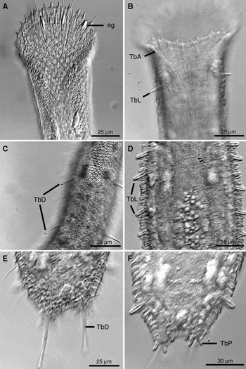
General appearance: the description is based on an adult specimen with a body length of 448 µm (Figures 1–3). The body is highly opaque and somewhat reflective under transmitted light due to the cuticle. The oral hood is up to 48 µm long and bears small papillae with cilia on its anterior margin (Figure 1A). Four elongate epidermal glands, 6–7 µm long, are present at the anterior portion of the hood (Figure 1A). Ventrally, the mouth margin bears few small papillae and is lined with short tactile cilia (2–3 µm; Figure 1B). In the trunk region, four pairs of epidermal glands are present dorsolaterally beginning at ~U24 and extending to U85. All glands are slightly elongate, asymmetrical in shape, and up to 11 µm long. Tactile cilia are present on the lateral margin of the body beginning at the base of the oral hood.
Light micrographs of Tetranchyroderma adeleae sp. nov. Holotype: (A) dorsal view of anterior end; (B) ventral view of antrerior end; (C) dorsal view of trunk; (D) ventral view of trunk; (E) dorsal view of caudal end; (F) ventral view of caudal end TbD mislabelled (=CrD).

Digestive tract: the digestive tract begins with a large terminal mouth to 35 µm wide. The pharynx, from the ventral mouth margin to the pharyngeointestinal junction, is 104 µm long and 18 µm wide. Pharyngeal pores are present at U32 and the pharyngeointestinal junction is present at U34. The intestine is up to 24 µm wide and glandular in appearance. A ventral anus is present at U88.
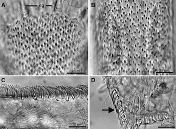
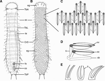
Cuticle: two types of cuticular structures are present (Figures 2 & 3). Ordinary triancres with a slight posterior curvature are present on the ventrolateral margins of the body (Figures 2C, D & 3E). Ordinary triancres consist of three tines that arise from a common base. Each tine is of equal length, with shorter tines ~3 µm long close to the lateral margins of the oral hood, and longer tines ~5 µm long on the lateral margins of the main trunk. All tines display a caudal bend but do not broaden into feather-shaped scales (Figures 2C & 3E). Ordinary triancres close to the caudal lobes have a more pronounced caudal bend than those at the anterior end of the body (Figure 2D). Feathered triancres are present on the dorsal surface from the middle of the oral hood to the caudal end (Figure 1A, B). Feathered triancres consist of three caudally-directed tines that arise from a common base (Figures 1A, B & 3C, D). On the trunk, each tine is made of a central shaft that rises vertically for ~3 µm and immediately broadens as it bends caudally to form a feathered scale ~5 µm long (Figure 3D). A midrib is apparent down the centre of each feathered scale (Figure 3D). The length of feathered scales is noticeably shorter at the anterior end of the body (2–3 µm). There are approximately sixteen longitudinal columns of feathered triancres on the dorsal trunk.
DIC micrograph of the cuticle of the paratype of Tetranchyroderma adeleae sp. nov. (A) Dorsal portion of oral hood; (B) dorsal view of trunk; (C) view of the ventrolateral body wall; (D) view of the ventrolateral body wall at the posterior end of the trunk. Arrow points to ordinary triancres with significant caudal bend.

Adhesive organs: four anterior adhesive tubes (TbA) per side are present ventrally at the lateral mouth margin (Figures 1B & 3A). All tubes are duogland in appearance and of equal length, ~8 µm. Two tube-like structures to 7 µm long are present at the midline of the mouth margin, the others being placed laterally. Sixteen lateral adhesive tubes (TbL) are slightly sublateral in position. The first pair of tubes, at U22, does not appear to be duogland (Figure 1B). All other lateral adhesive tubes are duogland and begin at U33 (Figures 1D & 3A). The last pair of adhesive tubes is highly cuticular and broader than the rest (Figure 1F). Three pairs of dorsal cirrata (CrD), ~32 µm long, occur along the trunk and contain small refractive secretions (Figures 1C, E & 3B). The first pair of tubes is present at U35, second pair at U46, and last pair inserts between and dorsal to the paired posterior adhesive tubes (TbP) on the caudal lobes. The caudal lobes are short and bear two adhesive tubes (TbP) ~4 µm long. A third pair of adhesive tubes, 4 µm long, is positioned between the caudal lobes (Figure 1F).
Schematic illustration of Tetranchyroderma adeleae sp. nov. (A) Ventral view; (B) dorsal view; (C) dorsal view showing the organization of the feathered triancres; (D) hypothetical lateral view of feathered triancre; (E) lateral views of the ordinary triancres present on the ventrolateral body wall.

Ciliature: ventral locomotory cilia begin below the mouth margin and form a continuous field of transverse rows down to the base of the trunk (Figures 1B & 3A). Tactile cilia are present on the oral hood and trunk. Up to 25 pairs of lateral tactile cilia, to 20 µm in length, protrude from the ventrolateral body margin (Figure 3B).
Reproductive system: all specimens are simultaneous hermaphrodites (Figure 3A). A single testis is present on right side beginning at the pharyngeointestinal junction and leading caudally into an elongate vas deferens. The vas deferens curves medially to connect to the base of the male caudal organ. The male caudal organ (co, Figure 3A), at ~U84, is an inverted pear-shape containing refractive spheres and wrapped in circular muscles. Anterior to the caudal organ at U73 is the frontal organ (fo, Figure 3A), 15 µm in diameter, and containing sperm. Paired ovaries are present at U88.
CUTICLE ULTRASTRUCTURE OF T. ADELEAE SP. NOV.
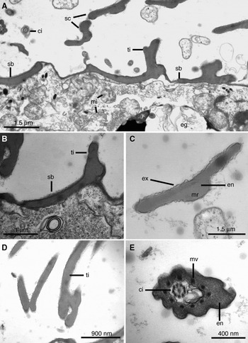
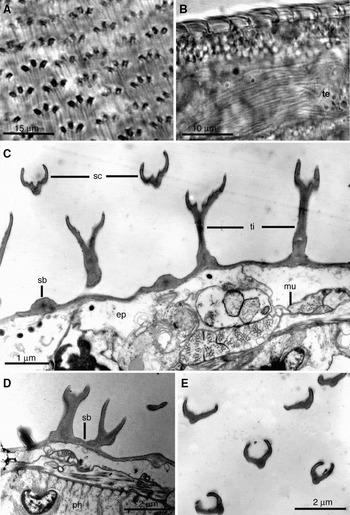
The following descriptions of the cuticle are based on a single, slightly damaged specimen. Sections through individual triancres are at an oblique angle relative to the longitudinal axis of the triancres (Figure 4). Most TEM sections were taken through the middle of the trunk region. The cuticle is divided into an outer lamellar layer, or exocuticle, and a basal layer, or endocuticle (Figure 5; see terminology by Rieger & Rieger, Reference Rieger and Rieger1977; Ruppert, Reference Ruppert, Harrison and Ruppert1991). The thickness of the entire cuticle excluding the ancres is 165–226 nm depending on the body region (min: 165 nm in the ventral trunk; max: 226 nm in the dorsal trunk). The maximum thickness of the exocuticle in the dorsal and dorsolateral trunk is ~42 nm, with thinner regions ~32 nm around sensory and locomotory cilia on the ventral and lateral body walls. Individual locomotory cilia are covered with a single layer of exocuticle ~9 nm thick. The organization of the exocuticular layers into bilayers (sensu Rieger & Rieger, Reference Rieger and Rieger1977) could not be determined, nor could the number of bilayers on the trunk.
Schematic illustration of the approximate TEM plane of section (shaded rectangle) through the feathered triancres.

TEM photomicrographs of the cuticle of Tetranchyroderma adeleae sp. nov. (A) Section revealing multiple feathered scales and their bases; (B) single scale base with a single tine in longitudinal section; (C) cross-section through a feathered scale; (D) longitudinal sections through ordinary triancres; (E) cross-section of a sensory cilium from the ventrolateral body wall.

The endocuticle has a slightly granular and homogeneous appearance; it is ~108–169 nm thick in the ventral midbody region and ~121–182 nm thick in the dorsal midbody region. All ancres (ordinary triancres and feathered triancres) are specializations of the endocuticle and appear as slightly lighter regions embedded within it (Figure 5A, B). The entire base of an ordinary triancre was not captured in section. Individual triancre tines have a maximum width of 384 nm, which consists mostly of endocuticle; only a single exocuticular layer is observed around individual tines (Figure 5D). Feathered triancres consist of three individual tines (shafts) with a distinct base that is embedded within the endocuticle (Figure 5A). The ancre base is up to 3.95 µm wide in cross-section and has a maximum height of 398 nm (max exocuticule of 37 nm; max endocuticle of 361 nm). All ancre bases narrow at their posterior apex to ~180–205 nm high. The tines that arise from the base and support the feathered scales are ~550–562 nm wide. The connection between the tine and an individual feathered scale was not captured in section. Feathered scales are similar in appearance to the tine and base, with a distinctly homogeneous appearance and composed of both endocuticle and exocuticule (Figure 5A, C). There are at least two exocuticular sheets around each feathered scale. The feathered scales have a slight concavity on their outer surface (Figure 5A, C) and a maximum width (edge to edge) of 2.4 µm, though this might be an overestimation due to the oblique plane of section. Each feathered scale has a distinct midrib on its ventral surface (Figure 5C). The maximum height of a feathered scale (total cuticle) from the ventral base of the midrib to the dorsal surface of the feathered scale is ~562 nm. The outer edges of the feathered scales are ~200–226 nm high.
Sections of several sensory cilia that project from the lateral body wall revealed a robust construction (Figure 5E). These structures are composed from inside to outside of the following: a central cilium with a 9 × 2 + 2 axoneme; a space with membrane-like structures; a homogeneous endocuticule up to 450 nm thick; and a single layer of exocuticle. Embedded within the endocuticle are ten symmetrically disposed microvilli.
CUTICLE ULTRASTRUCTURE OF PSEUDOSTOMELLA MEGAPALPATOR
The following description is based on a single specimen sectioned transversely. The sculptured cuticle consists of both excocuticle and endocuticle. Individual exocuticle bilayers are ~7–9 nm thick. The exocuticule on the dorsal and ventral sides of the body has a thickness of 30–45 nm. Up to six exocuticular bilayers surround each locomotory cilium, with a maximum thickness to 40 nm. On the dorsal surface of the trunk are feathered triancres similar in appearance to those of T. adeleae sp. nov. The feathered triancres are constructed of individual tines that are supported by an ancre base embedded in endocuticle (Figure 6B, C & D). The base has a similar granular and homogeneous appearance to the endocuticle of the trunk. Sections through the base reveal its maximum height to be 320 nm. Individual tines arise from the base and are ~160–180 nm wide and 1.6–1.9 µm high. The tines are similar in appearance to the base. Feathered scales arise from individual tines (Figure 6C). All scales are concave on their outer surface; this concavity gives each scale a distinct u-shaped appearance (Figure 6C, E). A single exocuticular sheet surrounds each feathered scale. At the centre of each scale is a small depressed region. This depression is observed in all sections of scales where the scale is connected to its respective tine (Figure 6C); however, sections that show free scales may or may not have this depression evident (Figure 6E). Individual scale widths (edge to edge) and scale contours are highly variable due to the plane of section and their orientation. Some scales are extremely concave on their outer surface while others appear relatively flat.
Cuticle of Pseuodostomella megapalpator. (A) Light micrograph of dorsal cuticle; (B) light micrograph of lateral body wall; (C) TEM cross-section through the dorsal body wall; (D) TEM cross-section through the lateral body wall; (E) cross-section through individual feathered scales.

TAXONOMIC REMARKS
Tetranchyroderma adeleae sp. nov. is the third species of Tetranchyroderma and the tenth species of marine Gastrotricha described from Australia (Hochberg, Reference Hochberg2002b, Reference Hochbergc, Reference Hochberg2003; Nicholas & Todaro, Reference Nicholas and Todaro2005, 2006). To date, there is a total of 62 described species of Tetranchyroderma reported from around the globe including all continents except Antarctica; 29 species possess five-pronged spines (pentancres), 29 species possess four-pronged spines (tetrancres), one species possesses three-pronged spines (triancres), and three species possess a mixture of ancre types (Nicholas & Todaro, 2006). Within Australia, only two species of Tetranchyroderma have been described previously: T. australiense Nicholas & Todaro, 2006 and T. pentaspersus Nicholas & Todaro, 2007. Tetranchyroderma adeleae sp. nov. is the first described species in the genus to possess feathered triancres. Ruppert (Reference Ruppert1970) reports that feathered triancres are known from other species in the genus, but no formal descriptions are published. The presence of both ordinary and feathered triancres is a consistent feature of this new species.
Thus far, the only other species of Tetranchyroderma with a triancrous covering is T. tribulosum Clausen, 1965 described from Norway. Apart from the difference in cuticular morphology, T. adeleae sp. nov. differs from T. tribulosum in possessing a larger body size, different distribution of adhesive tubes and epidermal glands, absence of piston pits, and presence of papillae and epidermal glands on the oral hood. In general appearance, T. adeleae sp. nov. most closely approaches the pentancrous T. polyprobolostomum Hummon, Todaro, Balsamo & Tongiorgi, 1996. Both species possess elongate dorsolateral cirrata (CrD) and an oral hood with papillae along the free margin. However, T. adeleae sp. nov. lacks the TbD that are prevalent along the midline of the body in T. polyprobolostomum.
The most distinctive feature of T. adeleae sp. nov. is the dorsal cuticle, which under transmitted light gives a sheen-like appearance due to the imbricated feather-like scales. The gross morphology of the feathered triancres bears strong resemblance to similar ancres described for Pseudostomella plumosa Ruppert, Reference Ruppert1970, P. klauserae Hochberg, 2002, P. megapalpator Hochberg, 2002 (Figure 6), and P. faroensis Clausen, Reference Clausen2004. However, differences are present in the relative shape of the feathered scales and the organization of their tines. For example, the scales of both P. plumosa and P. faroensis are slightly rounded at their apex while those of P. klauserae, P. megapalpator (Figure 6A), and T. adeleae sp. nov. have a more pointed appearance. Also, the latter four species appear to have feathered scales that arise independently from the base (on individual tines) as opposed to the condition in P. plumosa where all three feathered scales arise from a common tine.
DISCUSSION
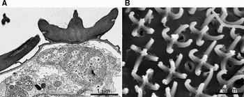
The morphology of the cuticle in the Thaumastodermatidae is perhaps the most structurally complex within the Gastrotricha, only rivalled by members of the Chaetonotidae (order Chaetonotida). Rieger & Rieger (Reference Rieger and Rieger1977) examined the ultrastructure of the cuticle in thaumastodermatids and hypothesized that the current diversity in forms (scales, spines, triancres, tetrancres and pentancres) was derived from an ancestral bowl-shaped scale with a central spine (Figure 7A). Evolutionary modification of the bowl into a circlet of spines around a central spine (pentancre, Figure 7B) is hypothesized to be the plesiomorphic condition for the subfamily Thaumastodermatinae Ruppert, 1978. Within the subfamily is a monophyletic clade containing Pseudostomella, Tetranchyroderma and Thaumastoderma (Hochberg & Litvaitis, Reference Hochberg and Litvaitis2001). Species of Thaumastoderma only possess tetrancres, while species of Pseudostomella and Tetranchyroderma possess pentancres, tetrancres, ordinary triancres or feathered triancres. Few species show mixed ancre types, though some show variation in the amount of ancre coverage (e.g. Tetranchyroderma hypopsylancrum Hummon, Tongiorgi & Todaro, 1993) and tine length (e.g. Tetranchyroderma tanymesatherum Hummon, Todaro, Balsamo & Tongiorgi, 1996).
Pentancres of Tetranchyroderma cf. polypodium. (A) TEM micrograph of a pentancre. Note the bowl-shaped appearance and central spine; (B) SEM micrograph revealing multiple pentancres.

The presence of more than one ancre type in species of Tetranchyroderma is exceptional, with only three other species known to possess mixed ancres: T. paradoxum Thane-Fenchel, 1970 possesses pentancres, tetrancres and ordinary triancres, and T. paralittoralis Rao, 1991 and T. tentaculatum Rao, 1993 possess pentancres and tetrancres. Tetranchyroderma adeleae sp. nov. is now the fourth known species to possess at least two different ancre types. The significance of mixed ancre types in a single species, or different ancre types within the Thaumastodermatinae, is difficult to ascertain without a better knowledge of species-level relationships within the subfamily. Important questions regarding the origins of the different ancre types include: (1) what is the ancestral ancre type of the subfamily?; (2) what are the selective pressures that favour different ancre types?; and (3) is the presence of mixed ancres in a single species a more primitive or derived trait relative to a single ancre type? Interestingly, no other species with feathered triancres also possess ordinary triancres; these include Pseudostomella plumosa (Ruppert, Reference Ruppert1970) from the USA (Ruppert, Reference Ruppert1970), P. klauserae and P. megapalpator from Australia (Hochberg, Reference Hochberg2002b), and P. faroensis from the Faroe Bank (Clausen, Reference Clausen2004).
To gain insight into the differences between ordinary and feathered triancres, I examined the fine structure of both types in T. adeleae sp. nov. and compared them to the feathered triancres in P. megapalpator. Results indicate that all triancres are broadly similar in fine structure to the variety of cuticular formations described for other species of Thaumastodermatidae (Rieger & Rieger, Reference Rieger and Rieger1977); all are solid modifications of the endocuticle with a finely granular appearance. Moreover, there is no distinctive substructure (e.g. striations and honeycombs) in the scales as present in species of Lepidodasys, which are hypothesized to be closely related to Thaumastodermatidae based on morphological grounds (Ruppert, Reference Ruppert, Harrison and Ruppert1991; Hochberg & Litvaitis, Reference Hochberg and Litvaitis2001). Instead, scale appearance is comparable to the tine (shaft) and base, though there are slight differences in staining quality between the feathered triancres and the rest of the endocuticle, i.e. the ancres are slightly more electron lucent than the general body endocuticle. In terms of cuticle thickness, both the exocuticle and endocuticle of T. adeleae sp. nov. fall into the range known for other species of Tetranchyroderma (Rieger & Rieger, Reference Rieger and Rieger1977). Moreover, the differences between species examined here with feathered triancres, T. adeleae sp. nov. and P. megapalpator, are relatively minimal, which is not unexpected considering the homogeneity of the sculptured cuticle within the family (see Rieger & Rieger, Reference Rieger and Rieger1977; Ruppert, Reference Ruppert, Harrison and Ruppert1991). However, the current investigation did reveal two differences worth noting: (1) the thickness of the tines between ordinary and feathered triancres; and (2) the contour of the feathered scales among species. First, ordinary tines appear much thinner and less robust than the tines that support feathered triancres, suggesting that the evolution of feathered scales involved more than just a caudal bend and flattening of ordinary tines. Second, the cross-sectional shape of the feathered scales examined in T. adeleae sp. nov. are relatively flat (Figure 5), while the feathered scales of P. megapalpator have a heavily convex surface (Figure 6). Significantly, these differences are not apparent at the light microscopic level (compare Figure 2A, B with Figure 6A), revealing that there is likely to be more variation in cuticular sculpture of these (and other) species than is currently reported. In addition, the structure and organization of the scale bases and tines are likely to be more variable than current assessments would indicate, warranting the future use of scanning electron microscopy to provide increased three-dimensional resolution.
In addition, transmission electron microscopy indicates that the stiff sensory cilia on the trunk of T. adeleae sp. nov. conform to the structure of sensory cilia described in other gastrotrichs by Rieger & Rieger (Reference Rieger and Rieger1977) and Hochberg (Reference Hochberg2002a). Individual sensory cilia are enwrapped in a sleeve of endocuticle at their base (and for several microns beyond the body wall), and within the endocuticle is a ten-part symmetrical arrangement of microvilli (Figure 5E). A similar condition is also noted for Lepidodermella squamata (Chaetonotida), which prompted Hochberg (Reference Hochberg2002a) to hypothesize that the structure of these cilia is an autapomorphy of Gastrotricha. Species of other taxa such as Loricifera (Kristensen, Reference Kristensen, Harrison and Ruppert1991), Kinorhyncha (Kristensen & Higgins. Reference Kristensen, Higgins, Harrison and Ruppert1991) and Platyleminthes (Rohde & Watson, Reference Rhode and Watson1995) have sensory cilia with an eight-part arrangement of microvilli.
ACKNOWLEDGEMENTS
I thank three anonymous referees for their critical comments that vastly improved the quality of this manuscript. I also thank Dr Lester R.G. Cannon of the Queensland Museum, Australia for the use of his research facilities. This research was supported by the National Science Foundation (DEB 0206517), the Smithsonian Marine Station at Fort Pierce, FL, and University of Massachusetts Lowell. This is Smithsonian Marine Station contribution No. 723.