Introduction
The 2-cell stage is a critical period of preimplantation development in mouse embryos. During the 2-cell stage, transition from maternal to embryonic control of development takes place. Following fertilization the early mouse embryo is essentially transcriptionally silent and its early development is directed by the complement of maternally-inherited mRNAs and proteins. The onset of embryonic gene activation usually does not occur until the 1-cell embryo undergoes cleavage to form a 2-cell embryo (reviewed in Schulz, Reference Schultz1993). This period may be the time when environmental factors have a great impact on embryonic health. In mice, with the exception of some inbred and F1 hybrid strains, a lot of in vitro cultured embryos are arrested at the 2-cell stage, a phenomenon which has been termed ‘2-cell block’ (Whitten & Biggers, Reference Whitten and Biggers1968).
It has been suggested that the 2-cell block and other forms of early developmental failure or early pregnancy loss in mouse correlate with aberrant methylation patterns or methylation reprogramming defects (Li et al., Reference Li, Bestor and Jaenisch1992; Neganova et al., Reference Neganova, Augustin, Sekirina and Jockusch1998; Patkin et al., Reference Patkin, Kustova and Perticone1998; Shi & Haaf, Reference Shi and Haaf2002). DNA methylation belongs in the group of epigenetic modifications, which mark genomic regions and act as heritable and stable instructions for the specification of chromatin organization and structure, which in turn dictate transcriptional states. It is characterized by covalent addition of a methyl group mainly in the cytosine residues at position 5 in CpG dinucleotides, becoming 5-methylcytosine (reviewed in Santos & Dean, Reference Santos and Dean2004). Although stable in somatic cells, DNA cytosine methylation changes distinctively during early embryo development. Mouse embryos undergo genome-wide methylation reprogramming by demethylation in early preimplantation development, followed by remethylation thereafter. An active cytosine demethylation wave arises in the zygote in the male pronucleus within hours of fertilization. Female pronucleus demethylation occurs passively in stepwise manner during subsequent cleavage divisions from the 2-cell to the morula stage (Santos et al., Reference Santos, Hendrich, Reik and Dean2002; Ribas et al., Reference Ribas, Taylor, McCorquodale, Maurício, Sousa and Wilmut2006). The genome-wide DNA methylation level declines to 30% of the typical somatic level during this period and the embryo is largely demethylated by the time it reaches the blastocyst stage. At the blastocyst stage, cytosine methylation occurs de novo and is observed predominantly in the inner cell mass (reviewed in Razin & Shemer, Reference Razin and Shemer1995; Santos et al., Reference Santos, Hendrich, Reik and Dean2002).
Apoptosis is a physiological process based on a genetic mechanism occurring spontaneously in the majority of cell populations. The main function of programmed cell death during preimplantation development is elimination of the minority of cells with abnormal or detrimental potential (reviewed in Pampfer & Donnay, Reference Pampfer and Donnay1999; Huppertz & Herrler, Reference Huppertz and Herrler2005). Apoptotic processes in blastocysts, including their genetic regulation, possible biochemical pathways and both beneficial and detrimental effects, have been reviewed several times in the past (Hardy, Reference Hardy1999; Levy et al., Reference Levy, Cordonier, Czyba and Guerin2001; Jurisicova & Acton, Reference Jurisicova and Acton2004; Fabian et al., Reference Fabian, Koppel and Maddox-Hyttel2005). However, little information on apoptosis in early embryos, including embryos at the 2-cell stage, has been assembled so far. In normally-developing 2-cell embryos, the presence of apoptotic features has not yet been documented (Fabian et al., Reference Fabian, Makarevich, Chrenek, Bukovská and Koppel2007b). Even 2-cell blocked embryos show no obvious signs of degradation which might characterize aged or dying cells within 3 days after they entered the block (Goddard & Pratt, Reference Goddard and Pratt1983; Bogolyubova et al., Reference Bogolyubova, Bogoliubova, Bogolyubov and Parfenov2006).
The aim of this study was to test the ability of normal 2-cell embryos and embryos arrested at the 2-cell stage to undergo apoptotic cell death and to correlate this to the level of cytosine demethylation in their genome. For the induction of developmental arrest and apoptosis, embryos were cultured in vitro and treated with the following chemicals: α-amanitin (a cytotoxic agent that inhibits RNA polymerase II-dependent transcription); actinomycin D [a cytotoxic agent which can cause damage to DNA, inhibit transcription from all three classes of RNA polymerase promoters and induce apoptosis (Kalousek et al., Reference Kalousek, Brodska, Otevrelova and Röselova2007)]; and tumour necrosis factor α [TNF-α; a pleiotropic cytokine that can induce apoptosis, mediated through its interaction with specific high-affinity cell surface receptors (Argilés et al., Reference Argilés, Carbó and López-Soriano1997)]. Possible correlation between the DNA cytosine methylation status and the functioning of apoptotic machinery was also tested in additional experiment with blastocysts containing constant proportion of apoptotic cells.
The obtained results produce new information on two important physiological events in developmentally competent and incompetent 2-cell embryos. Study elucidates their role in experimentally induced developmental failures and describes abilities of arrested embryos to perform these mechanisms.
Materials and Methods
Embryo recovery and culture
Female mice (ICR strain, Velaz, Prague, Czech Republic; 4 weeks old) underwent superovulation treatment with pregnant mare's serum gonadotropin (PMSG 5 IU ip; Foligon, Intervet international, Boxmeer, Holland), followed 47 h later by administration of human chorionic gonadotropin (hCG 5 IU ip; Pregnyl, Organon, Oss, Holland). Females were mated with males of the same strain overnight and mating was confirmed by identification of a vaginal plug. The mid-2-cell stage embryos were flushed from the oviduct of animals killed by cervical dislocation, using a flushing-holding medium [FHM (Lawits & Biggers, Reference Lawits, Biggers, Wassarman and DePhamphilis1993)] 40 h after hCG administration. The embryos were then pooled, washed in KSOM culture medium (Specialty Media Group), transferred to culture drops (1 embryo per 1 μl KSOM) and cultured at standard conditions (5% CO2, atmospheric O2 and 37 °C).
All animal experiments were reviewed and approved by the Ethical Committee for Animal Experimentation of the Institute of Animal Physiology, approved by the State Veterinary and Food Administration of the Slovak Republic and were performed in accordance with Slovak legislation based on Directive 86/609/EEC on the protection of animals used for experimental and other scientific purposes.
Experimental design
In the first experiment, isolated and pooled 2-cell embryos were randomly divided into five groups, washed and transferred into culture medium containing no additional supplement (control group) or one of following chemicals (experimental groups): α-amanitin (Sigma-Aldrich; dissolved in water) at 25 μg ml−1, actinomycin D (Sigma-Aldrich, dissolved in 100% ethanol) at 0.05 μg ml−1, TNF-α (tumor necrosis factor α; Sigma-Aldrich, dissolved in water) at 0.05 μg ml−1, or both actinomycin D and TNF-α (at the same concentrations). Embryos were then cultured for 24 h, 48 h and 72 h and subjected to cell death assays.
In the second experiment, isolated and pooled 2-cell embryos were randomly divided into four groups, washed and transferred into culture medium containing no additional supplement (control group) or one of following chemicals (experimental groups): α-amanitin (25 μg ml−1), actinomycin D (0.05 μg ml−1), or both actinomycin D and TNF-α (at the same concentrations). Embryos were then cultured for 24 h, 48 h and 72 h and subjected to genome methylation assay.
In the third experiment, isolated and pooled blastocysts (90 h post hCG) were randomly divided into two groups, washed and transferred into culture medium containing no additional supplement (control group) or actinomycin D at 0.05 μg ml−1 (experimental group). Embryos were then cultured for 24 h and subjected to genome methylation assay. Each experiment was performed three or more times.
The ability of α-amanitin to arrest embryonic development in mouse early embryos (Han et al., Reference Han, Chung, Gao and Latham2005; Zeng & Schultz, Reference Zeng and Schultz2005) and the ability of both actinomycin D (Pivko et al., Reference Pivko, Grafanau and Kubovicová2002; Ju et al., Reference Ju, Kwak, Lee, Kim, Kim, Kim, Choi, Jung, Lee, Do, Park and Choo2005; Fabian et al., Reference Fabian, Juhás, Il'ková and Koppel2007a) and TNF-α (Pampfer et al., Reference Pampfer, Wuu, Vanderheyden and De Hertogh1994; Wuu et al., Reference Wuu, Pampfer, Becquet, Vanderheyden, Lee and De Hertogh1999) to induce apoptosis in mouse blastocysts have been demonstrated previously.
Cell death assessment
For the detection of cell death, three staining methods were used. After each staining procedure, embryos were mounted on slides with Vectashield (Vector Laboratories), covered with coverslips and observed under fluorescence microscope at X400 magnification (BX 51 Olympus). Colour images were recorded with high-resolution CCD camera using Olympus DP-Soft image analysis software.
Fluorescence staining for annexin V
A staining kit (annexin-V-FLUOS; Roche Diagnostics) was used for labelling embryonic cells, according to the manufacturer's instructions. Embryos were washed in PBS supplemented with bovine serum albumin (BSA; Sigma-Aldrich), incubated in a mixture of binding buffer, fluorescein isothiocyanate (FITC)-conjugated annexin V and propidium iodide for 10 min at room temperature and rewashed in PBS (with BSA). Afterwards, the nuclei of embryonic cells were counterstained with Hoechst 33342 (20 μg ml−1 in PBS; Sigma-Aldrich) for 5 min at room temperature. Staining enabled morphological assessment of nuclei, assessment of cell viability and detection of phosphatidylserine redistribution in the plasma membrane.
Fluorescence staining for TUNEL/propidium iodide
Embryos were washed in KSOM, incubated for 5 min at 37°C in a culture medium containing 10 μg ml−1 propidium iodide (Sigma-Aldrich) and rewashed at room temperature in PBS (with BSA). Embryos were then fixed for 1 h in 4% paraformaldehyde (Merck). Fixed embryos were washed in PBS (with BSA), permeabilized for 1 h in PBS with 0.5% Triton X-100 (Sigma-Aldrich) and rewashed in PBS (with BSA). Then they were incubated in 10 μl of terminal deoxynucleotidyl transferase and 90 μl of fluorescein-conjugated dUTP (TUNEL, In Situ Cell Death Detection Kit; Roche) for 60 min at 37°C in the dark. After the TUNEL reaction, the embryos were washed in PBS (with BSA) and counterstained with Hoechst 33342 (20 μg ml−1 in PBS) for 5 min at room temperature (Fabian et al., Reference Fabian, Juhás, Il'ková and Koppel2007a). Control staining was performed by pre-incubation of additional embryos in DNase I (deoxyribonuclease I 100 IU ml−1; Invitrogen) for 30 min at 37°C. Staining enabled morphological assessment of nuclei, assessment of cell viability and detection of specific DNA degradation in the nucleoplasm.
Fluorescence staining for TUNEL/active caspase-3
Embryos were washed in PBS (with BSA), fixed in 4% paraformaldehyde, rewashed in 0.3% Triton X-100 and permeabilized in 1% Triton X-100 at room temperature for 2 h. Afterwards, they were washed in PBS (with BSA) and subjected to the TUNEL reaction with fluorescein-conjugated dUTP. TUNEL-reacted embryos were then subjected to immuno-histochemical staining with a wash in PBS (with BSA), blocking in a PBS buffer with 5% normal goat serum (Invitrogen) and 0.1% Triton X-100, followed by overnight incubation with a primary anti-active-caspase-3 antibody (polyclonal rabbit Cleaved Caspase-3 (Asp175) antibody; Cell Signaling Technology; 1:100) (Fabian et al., Reference Fabian, Juhás, Il'ková and Koppel2007a). Specimens were then rewashed and incubated with a Texas Red-conjugated secondary antibody (goat anti-rabbit IgG; Jackson ImmunoResearch). Finally, embryos were washed in 0.3% Triton X-100 and PBS (with BSA) and counterstained with Hoechst 33342 (20 μg ml−1).
Immunocytochemical control staining was performed in two ways: (i) pre-absorption of the primary antibody with specific antigen [Cleaved Caspase-3 (Asp175) Blocking Peptide; Cell Signaling Technology]; and (ii) substitution of primary antibody with non-immune rabbit IgG (Rabbit Immunoglobin Fraction–Negative Control; DakoCytomation). Staining enabled morphological assessment of nuclei, detection of specific DNA degradation in the nucleoplasm and detection of active caspase-3 in the cytoplasm.
5-Methylcytosine immunodetection
Nuclear DNA methylation was visualized using indirect immunofluorescence with an antibody to 5-methyl cytosine (Santos et al., Reference Santos, Hendrich, Reik and Dean2002; Ribas et al., Reference Ribas, Taylor, McCorquodale, Maurício, Sousa and Wilmut2006). Embryos were washed in PBS (with BSA), fixed for 1 h in 4% paraformaldehyde, rewashed in 0.05% Tween 20 (Sigma-Aldrich) and permeabilized in 0.3% Triton-X in PBS for 30 min at room temperature. After extensive washes in 0.05% Tween 20, the embryos were subjected to 2 M hydrochloric acid (Sigma-Aldrich) for 30 min at 37°C to denature the DNA, subsequently neutralized for 10 min with 100 mM Tris/HCl buffer (pH 8.5) and then washed again in 0.05% Tween 20. The embryos were blocked overnight at 4°C in the blocking solution [2% bovine serum albumin (Roche) in PBS] and methylated DNA was visualized with a mouse monoclonal antibody against 5-methylcytosine (Calbiochem). Incubation with this antibody was performed at 37°C for 1 h (1:400 in the blocking solution). The embryos were then washed four times in blocking solution for 15 min each and incubated in goat anti-mouse IgG conjugated to Texas Red (Jackson ImmunoResearch) at a concentration of 1:200 for 1 h at room temperature. Afterwards, embryos at early stages were counterstained with YOYO-1 iodide (Invitrogen) at 100 nM for 25 min and blastocysts were counterstained with DAPI (Sigma-Aldrich) at 5 μg ml−1 for 5 min. Finally, the embryos were mounted on slides and observed under fluorescence microscope. For each image recording, the same exposure parameters were used.
An antibody staining control was performed for each immunofluorescence experiment. Omission of primary or secondary antibody in the staining procedure resulted in complete absence of immunofluorescence staining. The heavily methylated, brightly fluorescing second polar body did not undergo active or passive demethylation during preimplantation development, so it served as a positive staining control (Shi & Haaf, Reference Shi and Haaf2002).
Morphological analysis
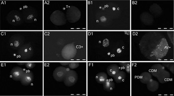
Depending on their nuclear morphology (M; evaluated by Hoechst 33342, DAPI, or YOYO-1 iodide staining), PI positivity/negativity (PI) and the presence of phosphatidylserine on the cell surface (AV; annexin V staining), stained cells were classified as: normal (oval, without morphological changes in nuclei, without PI inclusion into the nucleoplasm and without annexin V); apoptotic [showing typical fragmented or condensed nuclear morphology (M+; Fig. 1b1, d1), usually labelled with annexin V (AV+; Fig. 1d2), PI negative in early stages and PI positive in later stages of the apoptotic process]; and necrotic (showing PI positive nuclei without specific morphological changes).
Illustrative fluorescence micrographs of normal and arrested mouse in vitro cultured embryos at the 2-cell stage (original magnification ×400, bar = 50 μm). Nuclear morphology is visualized by chromatin staining with Hoechst 33342 (a1, b1, c1, d1) or YOYO-1 iodide (e1, f1). (a) control embryo (24 h culture) containing (a1) two normal nuclei (n) and polar body (pb) with (a2) specifically degraded DNA, visualized by TUNEL labelling (T+); (b) embryo treated with actinomycin D (48 h culture) containing (b1) one condensed nucleus (c) and showing no other apoptotic features (b2 for TUNEL\C3 assay); (c) embryo treated with α-amanitin (72 h culture) containing (c1) one condensed nucleus and showing (c2) the presence of active caspase-3 in the cytoplasm, visualized by immunohistochemical staining (C3+); (d) embryo treated with actinomycin D (72 h culture) containing (c1) one condensed nucleus and showing (d2) phosphatidylserine redistribution on plasma membrane, visualized by annexin V staining (AV+); (e) control embryo at the 2-cell stage (fresh isolate) showing (e2) methylated DNA in both nuclei, visualized by 5-methylcytosine immunostaining; (f) embryo arrested at the 2-cell stage showing (f2) partially demethylated DNA cytosine (PDM) and embryo arrested at the 2-cell stage containing (f1) two condensed nuclei and showing (f2) complete demethylation (CDM) (72 h culture, actinomycin D treated).

Depending on their nuclear morphology (M), PI positivity/negativity (PI) and the presence of specific DNA degradation in the nucleoplasm (T; TUNEL labelling; Fig. 1a2), stained cells were classified as: normal (without morphological changes in nuclei, PI−, T−; Fig. 1a1); apoptotic [showing typical fragmented or condensed nuclear morphology (M+; Fig. 1c1), usually labelled with TUNEL (T+), PI− in early stages / PI+ in late stages]; and necrotic (PI+, without specific morphological changes).
All cells containing the active form of the main apoptotic enzyme caspaze-3 (C3; Fig 1c2) in their cytoplasm were classified as apoptotic. Pyknotic clusters of chromatin extruded to the embryo surface were classified as polar bodies (Fig. 1a–f).
Depending on the presence and the amount of immunochemically visualized 5-methylcytosine in the nucleoplasm, embryonic nuclei were classified as: methylated (showing clear, contrast and uniform red staining), partially demethylated (PDM; showing very low, but still recognizable red staining) and completely demethylated (CDM; showing no red staining; i.e. the fluorescence intensity of the nucleus and cytoplasm was the same) (Fig. 1e, f). Staining signals in the nucleoplasms of freshly isolated 2-cell embryos were taken as a standard.
Statistical analysis
Statistical analysis was performed using Statistica software (StatSoft). The percentage of embryos with apoptotic cells represented the number of embryos containing at least one apoptotic cell relative to the total number of evaluated embryos. The percentage of embryos with demethylated nuclei represented the number of embryos containing at least one (partially or completely) demethylated cell relative to the total number of evaluated embryos.
Standard chi-squared tests (with one and two degrees of freedom) were used to detect differences in the incidence of apoptotic embryos and cells and of demethylated embryos and cells between experimental and control groups (or between two experimental groups). Differences in the distribution of embryonic stages were also evaluated by chi-squared test (with degrees of freedom dependent on the number of developmental stages reached by embryos in the compared groups). Values p < 0.05 were considered as significant. Student's t-test was used to detect differences in mean numbers of cells per blastocyst between experimental and control groups. These results were expressed as mean values ± SD.
Results
Embryonic growth
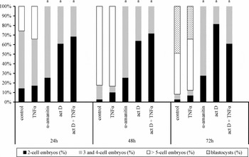
The majority of control embryos showed very good ability to grow and differentiate in in vitro conditions. After 24 h culture, only 14% of controls remained at the 2-cell stage, whereas the rest of them underwent one (60%) or more (26%) cleavage cycles. After 48 h culture, 3% of control embryos were definitively arrested at the 2-cell stage, 15% showed retarded development and 82% successfully reached higher stages of development (8-cells, 16-cells or morulae). After 72 h culture, 8% of embryos showed arrested development, 43% reached cleavage stages (16-cell or more) and the rest of them (49%) reached the blastocyst stage (average number of nuclei: 48.25 ± 13.49).
As shown in Fig. 2, the distribution of embryos treated with TNF-α was very similar to that of control embryos and it did not show significant differences at any time of collection (p > 0.05). The average number of nuclei in blastocysts developed from TNF-α influenced embryos was also comparable to controls (50.20 ± 13.72; p > 0.05).
Embryonic growth in untreated and treated mouse 2-cell embryos after 24 h, 48 h and 72 h culture. The graphs show the proportion of embryos which reached the 2-cell stage, the 3- and the 4-cell stages, the 5-cell or higher stage & the blastocyst stage of preimplantation development in control and experimental groups evaluated by fluorescence counterstaining of nuclei. ap < 0.001; act D, actinomycin D.

All other chemicals used showed highly detrimental effects on embryo development (p < 0.001). Approximately one-quarter of embryos cultured in media with the addition of α-amanitin were already arrested at the 2-cell stage after 24 h culture. The rest of them (around 75%) underwent one cell cycle and became arrested at the 4-cell stage. Later (at 48 and 72 h), no further development was observed in these embryos. During the first 24 h of culture, the presence of actinomycin D or both actinomycin D and TNF-α caused developmental arrest in almost two-thirds of the 2-cell embryos. Only a relatively small part of such treated embryos was able to cleave and reach the 3- or 4-cell stage (around 30%). The addition of TNF-α into media containing actinomycin D did not affect its action (p > 0.05).
Apoptosis occurrence and incidence
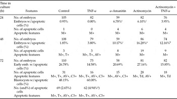
After 24 h culture, only one cell showing apoptotic morphology (nuclear condensation) was observed in control group embryos. The treatment of embryos with TNF-α did not affect the incidence of apoptosis at this time. The addition of α-amanitin and of actinomycin D slightly increased the proportion of embryos containing apoptotic cells (p < 0.05), but their numbers also remained low (Table 1). Apart from nuclear condensation, no other features of apoptosis were observed in any group of embryos cultured for 24 h.
The occurrence and incidence of apoptotic cells and apoptotic features in untreated and treated mouse 2-cell embryos after 24 h, 48 h and 72 h culture.

ap < 0.001; bp < 0.01; cp < 0.05 (compared with control); M+, morphologically changed (fragmented or condensed) nuclei; T+, positive TUNEL labelling; Td, dubious (weak) TUNEL labelling; AV+, positive annexin V labelling; C3+, presence of active caspase-3 in cytoplasm.
After 48 h culture, the proportion of cleaving control embryos containing apoptotic cells was still very small (2%). In these few cells, nuclear condensation and the presence of specific DNA degradation were observed. Cleaving embryos treated with TNF-α showed a similar incidence of apoptosis again (p > 0.05). However, in addition to the already-mentioned apoptotic features, the presence of phosphatidylserine redistribution in the plasma membrane was recorded in this group. Arrested embryos in all other experimental groups showed increased percentages of embryos with apoptotic cells (p < 0.05, p < 0.001, p < 0.01 respectively for α-amanitin, actinomycin D and actinomycin D + TNF-α group). Even so, these cells still showed only one apoptotic feature–nuclear condensation.
After 72 h culture, approximately 1/4 of arrested and slowly-growing control embryos and approximately 1/2 of control blastocysts showed the presence of at least one apoptotic cell. The average percentage of dead cells per blastocyst reached 2.65%. In these cells, all evaluated features of apoptosis were observed (i.e. nuclear fragmentation and condensation, specific DNA degradation, phosphatidylserine redistribution and the presence of active caspase-3). The addition of TNF-α did not affect apoptosis incidence in the arrested or slowly-growing embryos. On the other hand, it significantly increased the percentage of dead cells in blastocysts (4.94%; p < 0.001). In α-amanitin, actinomycin D and actinomycin D + TNF-α groups, the proportions of embryos containing apoptotic cells were comparable with the proportions recorded in arrested and slowly-growing embryos from the control group (p > 0.05). At this time, arrested embryos also showed some other apoptotic features besides nuclear condensation–phosphatidylserine redistribution, the presence of active caspase-3 and dubious (weak) TUNEL-labelling, suggesting the presence of (at least) partial DNA degradation.
Genome methylation status
All freshly-isolated 2-cell embryos (n = 52) showed homogeneous methylation staining throughout both blastomere nuclei.
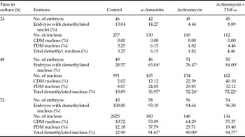
First signs of the demethylation process were recorded after 24 h culture. Still, the proportion of embryos with reduced DNA methylation level did not exceed 15% in any of the control or experimental groups and no nuclei with completely demethylated genome were observed (Table 2).
The proportion of demethylated nuclei in untreated and treated mouse 2-cell embryos after 24 h, 48 h and 72 h culture.

ap < 0.001 (compared with control); CDM, nuclei with completely demethylated DNA cytosine; PDM, nuclei with partially demethylated DNA cytosine.
After 48 h culture, the percentage of cleaving control embryos with demethylated genome had increased. Interestingly, the majority of arrested embryos treated with α-amanitin (63%), actinomycin D (77%) or both actinomycin D and TNF-α (84%) also showed partial or complete demethylation in one or more nuclei (p < 0.001, when compared with controls).
After 72 h culture, demethylated nuclei were present in all slowly-growing control embryos and all control blastocysts and their average incidence reached 22.90%. In arrested embryos, the genome demethylation process was almost completed and no de novo methylation was observed. The proportion of demethylated nuclei was higher than 90% and the number of nuclei with completely absent methylation signal represented more than half of all evaluated nuclei–α-amanitin (53.89%), actinomycin D (64.29%), actinomycin D + TNF-α (75.37%; p < 0.001 when compared with control).
Methylation status of apoptotic cells
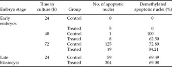
As shown in Table 3, the genomes of all those few nuclei with apoptotic morphology which were present in early embryos after 24 h culture, were methylated. After 48 h culture, the situation changed and the majority of observed apoptotic nuclei had undergone demethylation. More than two-thirds of apoptotic cells present in both untreated and treated embryos cultured for 72 h also showed weak or absent methylation signal.
Methylation status of apoptotic nuclei in untreated (control) and treated (α-amanitin, actinomycin D, actinomycin D + TNF-α) mouse 2-cell embryos after 24 h, 48 h and 72 h culture and in untreated and treated (actinomycin D) mouse blastocysts after 24 h culture.

Late blastocysts collected after 24 h culture with or without actinomycin D contained 36.94% and 2.75% respectively of cells showing apoptotic changes in nuclei, for treated and control groups alike (p < 0.001). Again, a relatively high proportion of these cells showed partial or complete reduction in DNA cytosine methylation (69.49% in controls, 69.08% in actinomycin D-treated embryos).
Discussion
The presence of various molecular components of the apoptotic cascade has been widely documented in embryos at all stages of preimplantation development (Jurisicova et al., Reference Jurisicova, Latham, Casper and Varmuza1998; Warner et al., Reference Warner, Exley, McElhinny and Tang1998; Exley et al., Reference Exley, Tang, McElhinny and Warner1999; reviewed in Levy et al., Reference Levy, Cordonier, Czyba and Guerin2001). Nevertheless, our analysis of the occurrence and incidence of apoptosis has indicated evident stage- and species-dependent specificity (Fabian et al., Reference Fabian, Makarevich, Chrenek, Bukovská and Koppel2007b). Our previous studies described the presence of cells undergoing both spontaneous and induced apoptotic cell death in mouse in vitro-cultured embryos at all stages following embryonic genome activation (4-cell and later) (Baran et al., Reference Baran, Fabian, Rehak and Koppel2003; Fabian et al., Reference Fabian, Makarevich, Chrenek, Bukovská and Koppel2007b). However, in normal 2-cell embryos and 2-cell embryos evaluated up to 24 h after treatment with inducers of apoptosis (actinomycin D or staurosporine), no signs of apoptosis were observed in mouse (Fabian et al., Reference Fabian, Makarevich, Chrenek, Bukovská and Koppel2007b), rabbit (Bellier et al., Reference Bellier, Chastant, Adenot, Vincent, Renard and Bensaude1997; Fabian et al., Reference Fabian, Makarevich, Chrenek, Bukovská and Koppel2007b) or cow (Gjørret et al., Reference Gjørret, Fabian, Avery and Maddox-Hyttel2007) embryos. Unusual resistance of embryos at very early stages of development to the lethal effects of various inducers has also been observed in other studies. Mouse zygotes treated with diamine were arrested at the 1-cell stage and did not display apoptotic-like features (blebbing of plasma membrane, cell shrinkage and condensation of pronuclei) until 48 h after treatment (Liu et al., Reference Liu, Trimarchi and Keefe1999). In mouse embryos at the 1-cell and the 2-cell stage treated with staurosporine, one apoptotic-like feature (nuclear pyknosis) was observed only after very long culture (37 h) with the inducer (Weil et al., Reference Weil, Jacobson, Coles, Davies, Gardner, Raff and Raff1996). Nevertheless, definitive proof of the ability of 2-cell embryos to undergo and finish the apoptotic cascade has not been presented so far.
In our study, all freshly-isolated 2-cell embryos showed normal morphology without any sign of apoptosis. In a small portion of untreated embryos which remained at the 2-cell stage after 24 h or longer culture (i.e. in 2-cell blocked embryos), only one apoptotic-like cell (with condensed nucleus and without PI staining in the nucleoplasm) was observed. Generally, the developmental capacity of control in vitro embryos was high. At 120 h post hCG (day 5), the majority of them (87%) reached the blastocyst stage and showed 80.81±17.13 nuclei per embryo on average (preliminary experiment).
The presence of specific apoptotic inducer TNF-α did not trigger apoptotic processes in any of the 2-cell embryos and it did not affect their growth either. However, TNF-α induced earlier occurrence of phosphatidylserine redistribution in the plasma membrane of several embryos at later stages of development and significantly increased the incidence of apoptotic cells in blastocysts. Our results show that although the TNF-receptor member 1a mRNA is present in embryos at the 2-cell stage (Kawamura et al., Reference Kawamura, Kawamura, Kumagai, Fukuda and Tanaka2007), TNF-α is not able to trigger apoptotic cascade in their cells.
Both actinomycin D and α-amanitin arrested embryonic development at the 2-cell or the 4-cell stage. The cytotoxic effects of both chemicals on embryos arrested at the 2-cell stage showed comparable design. A small proportion of 2-cell embryos displayed one or two apoptotic-like cells with condensed nuclear morphology after 24 h and 48 h culture. After 72 h culture, their number increased and besides morphological changes some other apoptotic features were also observed in them–plasmatic phosphatidylserine flip (visualized by annexin V staining) in both groups, cytoplasmic presence of active caspase-3 (visualized by IHC staining) in α-amanitin group and weak TUNEL labelling (suggesting at least partial DNA degradation) in actinomycin D group. Viability assays (vital PI staining) proved that none of these cells were necrotic.
These results indicate that embryos at the 2-cell stage are occasionally, at specific conditions, able to undergo apoptotic processes and to display several different features of apoptosis. However, this ability is generally very low. Furthermore, the whole process apparently takes much longer than the apoptotic cascade in differentiated blastocyst cells or somatic cells. The phosphatidylserine flip, which is considered as an early event in the apoptotic process (Huppertz et al., Reference Huppertz, Frank and Kaufmann1999), was first observed after 72 h from the beginning of the treatment. It was followed shortly afterwards by the activation of caspase-3 and other enzymes causing specific degradation of DNA. Nevertheless, the occurrence of these features was very rare. Interestingly, the appearance of nuclear condensation preceded all another manifestations of apoptosis. Furthermore, in arrested 2-cell embryos only very few dying cells showed nuclear fragmentation, which is frequently observed in blastocysts (Fabian et al., Reference Fabian, Juhás, Il'ková and Koppel2007a) and no dying cells showed decay to apoptotic bodies, which is considered the terminal stage of the apoptotic process (Fabian et al., Reference Fabian, Koppel and Maddox-Hyttel2005).
Prolongation of apoptotic processes might be explained by the general inhibition of transcription. However, our recent results have proved that actinomycin D acts as a reliable inducer of apoptosis and despite its negative effect on transcription it can significantly increase the incidence of apoptotic cells in blastocysts even after 6 h treatment (Fabian et al., Reference Fabian, Juhás, Il'ková and Koppel2007a). Thus, in actinomycin D-treated cells, at least one biochemical pathway probably remains unaffected and the situation with α-amanitin might be analogous.
The manifestation of specific apoptotic features in arrested embryos came at the same time as the onset of the wave of spontaneous apoptosis in normally developing embryos that have reached the blastocyst stage. This observation suggests that the delay might be a physiological event conditioned by the maturation of specific cellular components necessary for the apoptotic machinery or by the release of specific repressing mechanisms preventing developing cells of early embryos from programmed cell death, the existence of which was hypothesized previously (Gjørret et al., Reference Gjørret, Fabian, Avery and Maddox-Hyttel2007).
The aim of this study was also to elucidate whether passive genome demethylation might be involved in these delaying processes. Previous studies have shown that cytosine demethylation is essential for normal mouse development and it plays an important role in the regulation of gene expression (Li et al., Reference Li, Bestor and Jaenisch1992; Santos & Dean, Reference Santos and Dean2004). Furthermore, in normally-developing mouse embryos, the timing of the onset and linear progress of passive genome demethylation [from 2-cell to morula stages (Rougier et al., Reference Rougier, Bourc'his, Gomes, Niveleau, Plachot, Pàldi and Viegas-Péquignot1998)] overlaps with the timing of the onset and progress of spontaneous apoptosis (Fabian et al., Reference Fabian, Makarevich, Chrenek, Bukovská and Koppel2007b).
However, no direct correlation between genome methylation and apoptosis was found in our study. The observed apoptotic cells showed either demethylated or methylated DNA cytosine in their nuclei. In early embryos, the methylation status of apoptotic cells usually reflected the general methylation status present in normal cells. Only in late blastocysts, approximately two-thirds of nuclei showing apoptotic morphology were highly demethylated, while the majority of normal cells presented de novo methylation. Based on this, we can propose that demethylated cells may probably undergo apoptotic processes more easily than methylated ones and that they might also be more sensitive to apoptotic induction. However, the possible effect of DNA methylation on the expression of pro- and anti-apoptotic genes in embryonic cells is highly hypothetical and remains to be elucidated in detail.
Nevertheless, it is still possible that cytosine demethylation might belong in the group of mechanisms which participate in the advancement of both normal and arrested embryonic cells to a state in which they are able to run apoptotic processes.
Our methylation assay showed that normally-developing control embryos underwent passive demethylation, followed by remethylation. When compared to previous findings (Santos et al., Reference Santos, Hendrich, Reik and Dean2002, Ribas et al., 2006), the onset of this process showed a slight delay, but this was probably caused by the embryo transfer from in vivo to in vitro conditions.
Interestingly, our results demonstrate that arrested embryos display reduction in DNA cytosine methylation too. Demethylation of non-developing 2-cell (or 4-cell) embryos showed a significantly wider range and finished in almost complete demethylation of the embryonic genome. In addition, in this case the whole process was apparently independent of DNA replication or cellular cleavage. As described previously, in normally developing embryos, decline in methylation occurs as a result of the absence of primary DNA methyl transferase (Dnmt1) during DNA replication in the nucleus. Thus, the newly replicated strand fails to become methylated and the level of methyl cytosine per nucleus declines (Bestor, Reference Bestor2000; Rougier et al., Reference Rougier, Bourc'his, Gomes, Niveleau, Plachot, Pàldi and Viegas-Péquignot1998). The decline in methylation in arrested (but still viable) embryos might be explained by the existence of other demethylation mechanism, independent on cellular cleavage. An example of a replication-independent process of active genome-wide demethylation was already documented in differentiating mouse myoblasts (Jost & Jost, Reference Jost and Jost1994). However, the effect of some degenerative changes in chromatin or DNA structure which diminish the amount of 5-methylcytosine antibody targets could not be excluded too.
In conclusion, arrested embryos treated with α-amanitin or actinomycin D show no signs of necrosis even after 72 h culture. Their cells are still alive and a portion of them is able to undergo apoptotic cell death, an active biochemical process. The timing and the level of this ability is probably influenced by various physiological mechanisms preventing both normally and abnormally developing cells of early embryos from untimely triggering of programmed cell death.
Acknowledgements
We thank Dana Čigašová for her technical assistance and Andrew Billingham for his English proof-reading. This work was supported by the Slovak Research and Development Agency (under Contracts No. APVT-51-006204 and No. APVV-0620-07) and by the Slovak Academy of Sciences (VEGA 2/0046/08).