Introduction
Dacryocystorhinostomy (DCR) is the standard treatment for nasolacrimal duct obstruction. Endoscopic DCR is increasingly acknowledged to be as successful as external DCR for the management of acquired nasolacrimal duct obstruction.Reference Hartikainen, Antila and Varpula1
We present a modified approach to powered endoscopic DCR that minimises bone exposure, with no post-operative stenosis of the sac, less scarring and early mucosalisation.
Materials and methods
In this retrospective, non-randomised study, 105 patients underwent modified primary endoscopic DCR at our centre, from May 2012 to March 2013, using powered instrumentation but with no stenting. The institutional ethical committee approved the study protocol.
Data collected included patient age, gender, laterality, pre-operative examination results, intra-operative findings and follow-up results.
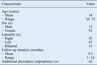
All patients underwent pre-operative rigid nasal endoscopy, and were assessed using nasolacrimal system syringing and dacryocystography. The patients’ characteristics are described in Table I.
Patient characteristics

The modified powered endoscopic DCR technique used is described here. After consent and pre-operative evaluation of the patient, surgery was performed under general anaesthesia. After the induction of anaesthesia, the patient was placed in a supine position, with 20–30-degree head-end elevation and their head slightly rotated towards the right side.
A 0-degree 4 mm endoscope was used for the procedure. Any cases of anterior septum deviation or other anatomical variations were addressed at the initial step in the procedure.
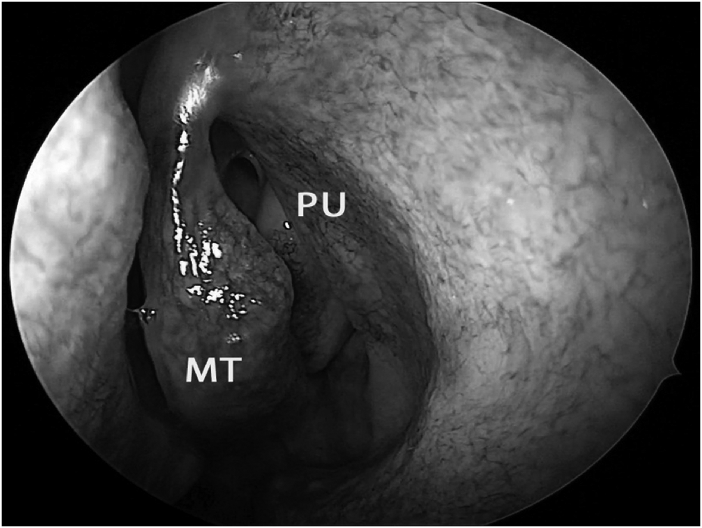
Nasal packing was achieved using 1:1000 adrenaline-soaked pledgets to decongest the nose (Figure 1). Local anaesthetic infiltration was avoided, as oozing can fog the endoscope, obscuring vision. The packing was placed medial to the axilla of the middle turbinate and anterior to the uncinate process.
Endoscopic image of left nasal cavity after decongestion with 1:1000 adrenaline. PU = uncinate process; MT = middle turbinate.
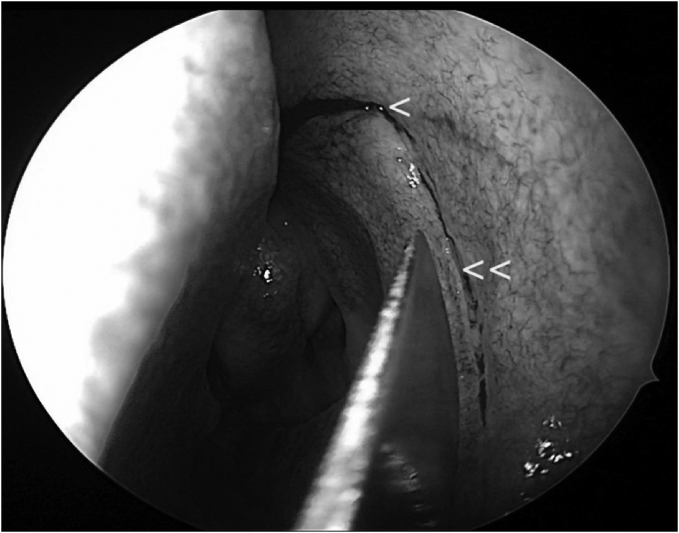
An inferiorly based flap was raised in the lateral nasal wall to expose the lacrimal bone and the frontal process of the maxilla using a knife. A superior incision was placed just above the axilla of the middle turbinate. An anterior incision was placed 8 mm anterior to the axilla of the middle turbinate, which was in front of the uncinate, posteriorly (Figure 2).
Endoscopic image showing superior and anterior incisions (arrows) for inferiorly based flap.
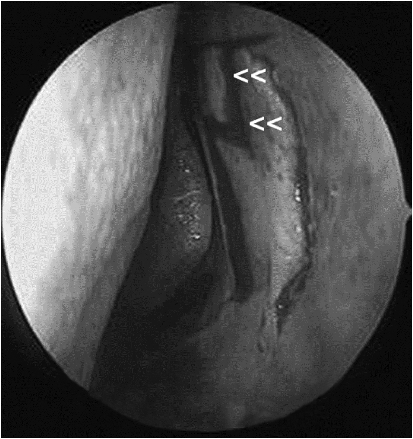
A 0.5 × 0.5 cm section of the mucosa in the flap in front of axilla was incised and removed using a Rosen elevator or a blade (Figure 3). This was the mucosa overlying the lacrimal sac. The Rosen elevator was used to gently elevate the flap from the bone in an anterior to posterior direction. From the posterior edge, the flap was gently pulled in the inferior direction to free it from nasal mucosa using Blakesley forceps. The use of suction was avoided.
Mucosa in the flap overlying the lacrimal sac was incised (0.5 × 0.5 cm incision inside the flap (arrows)) and removed.
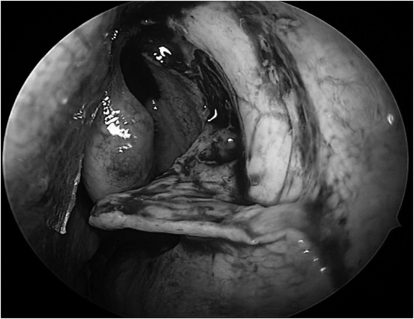
Pledgets were used to secure haemostasis during flap elevation (Figure 4). The inferiorly based flap was rotated on itself and placed on the inferior turbinate. Any adhesions to the bone or nasal mucosa were cut using scissors.
Endoscopic image of left nasal cavity showing elevation of the flap.
The frontal process of the maxilla was nibbled in a posterior to anterior direction, just above the flap, to the anterior edge, using a 2 mm Kerrison rongeur. The tip of the Kerrison rongeur was then engaged at the posterior-most edge of the frontal process of the maxilla and bone was nibbled in the same direction. Traction was maintained medially to prevent pressure injury to the lacrimal sac during this process.
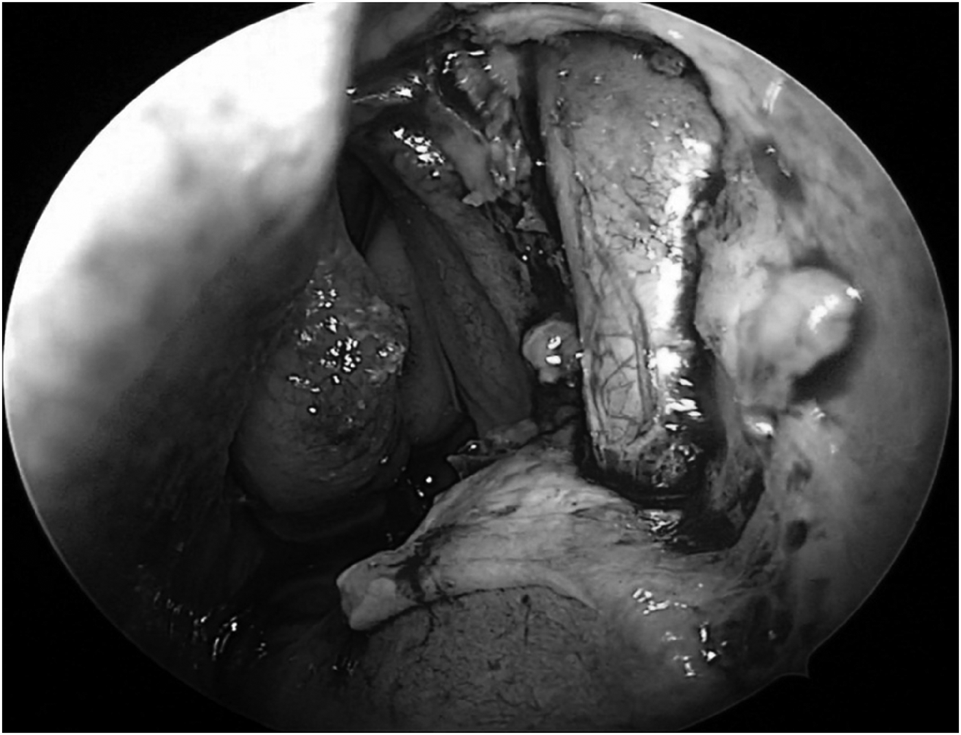
Drilling was conducted using a DCR burr to remove the superior thick bone and expose the fundus of the sac, without applying pressure, thereby avoiding lacrimal sac injury (Figure 5). The thin lacrimal bone was gently elevated from the lacrimal sac. Complete bone removal was ensured.
Endoscopic image showing bone removal with rongeur and drill using dacryocystorhinostomy burr.
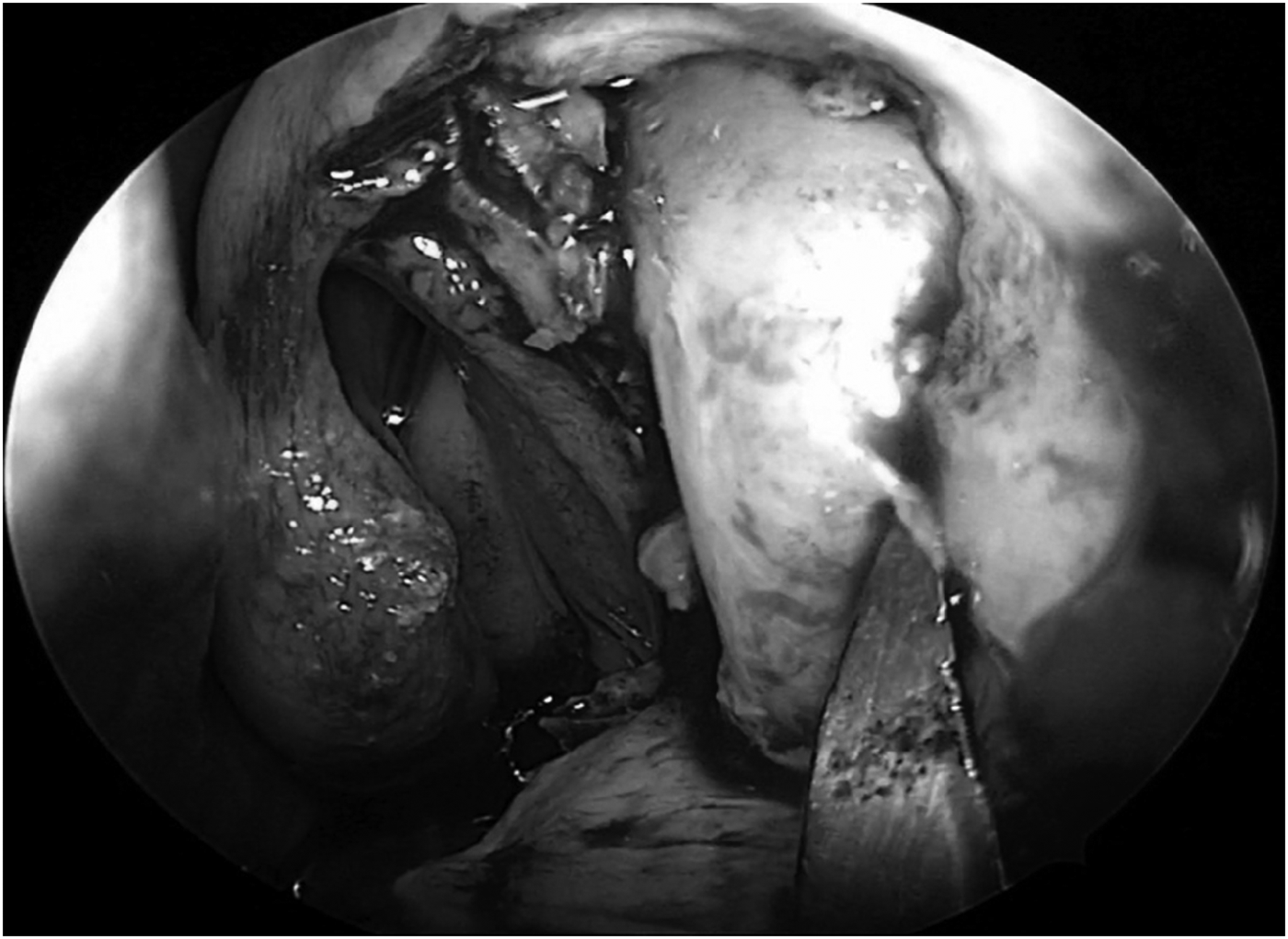
After removal of bone, the endosteal layer was observed on the medial wall of the sac (Figure 6).
Endosteal layer being removed.
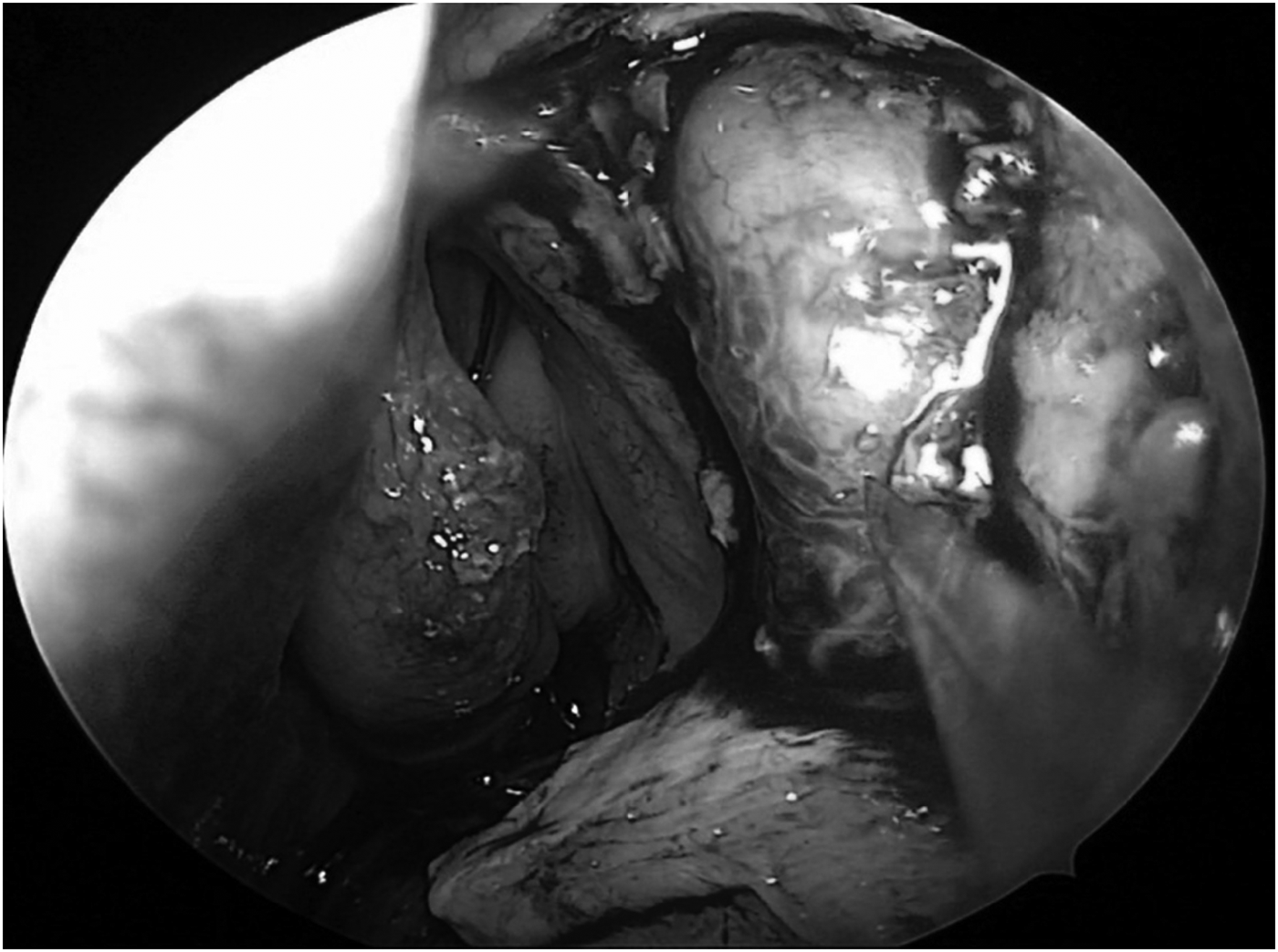
In the modified technique, removal of the endosteal layer was performed separately from that of the endothelial layer of the sac. The endosteal layer was removed with a sickle knife, gently and in a downward direction (Figure 7).
Endoscopic image showing endothelial layer of lacrimal sac completely exposed.
The medial wall was then incised using a number 11 blade, allowing the accumulated pus to drain. Anterior and posterior flaps of the medial wall of the sac were created, thus marsupialising the lacrimal sac. Canalicular openings were visualised. Syringing was performed to remove mucus plugs and ensure patency.
The inferiorly based flap was repositioned and approximated with the nasal mucosa so that no bone was left exposed. The anteriorly based flap was repositioned on the inferiorly based flap and the posterior flap was placed on the uncinate. Tissue glue was used to keep the flap in position. Nasal packing was avoided and antibiotic ointment was instilled using a syringe.
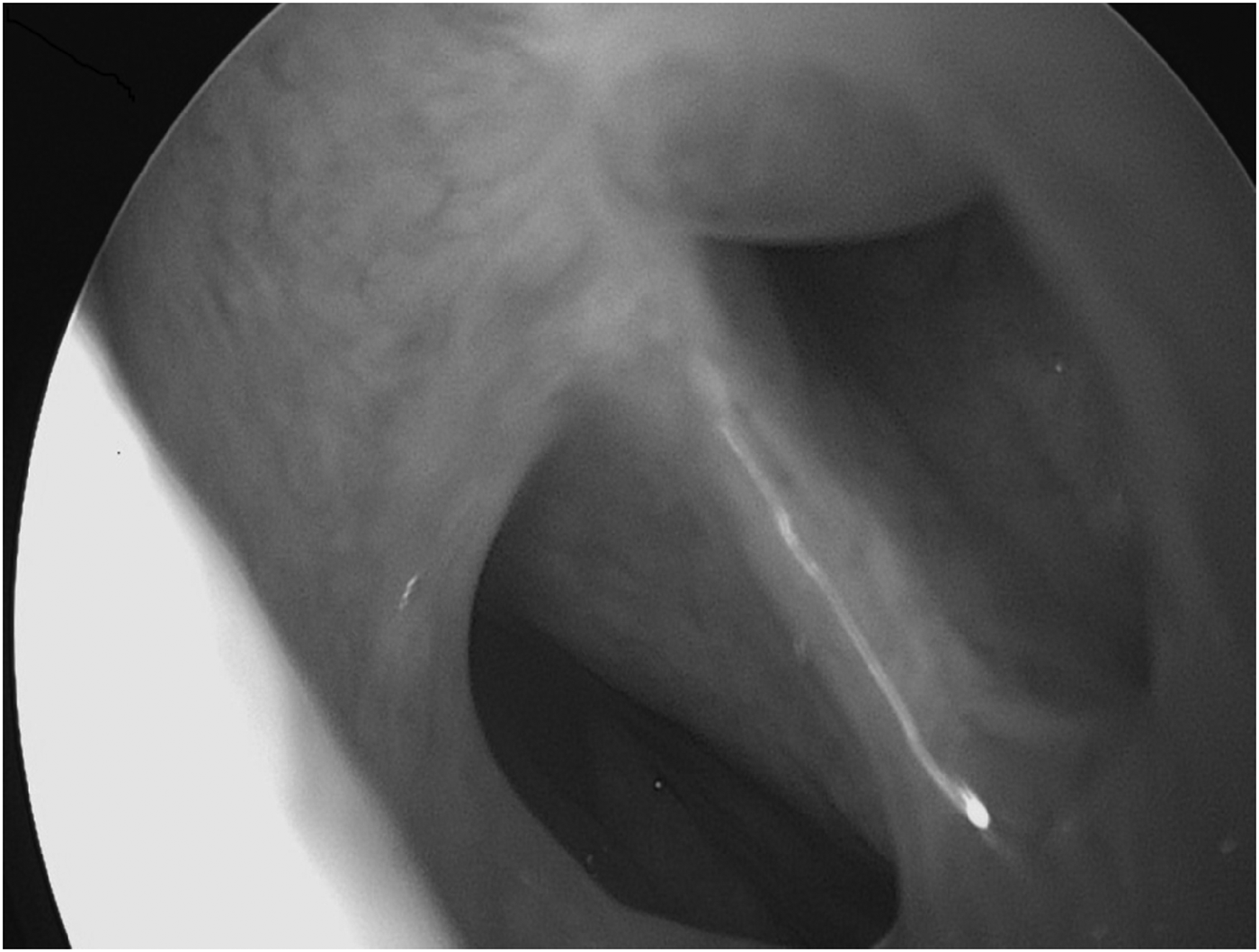
Post-operatively, the nose was irrigated using a sodium chloride solution. Antibiotic eye drops were instilled following irrigation. Intra-nasal debris was removed during the first post-operative visit. Syringing was conducted weekly for four weeks. Patients were followed up weekly for 1 month, then at 3 months, 6 months and 12 months (Figure 8).
Post-operative endoscopic visualisation of ostium.
Results
A total of 105 patients were enrolled during the study period. All patients had adequate anatomical patency on follow up, with an absence of presenting symptoms. This was confirmed on nasal endoscopy at follow up.
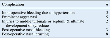
Complications were rare and are listed in Table II. None of the patients had orbital complications or underwent conversion to an open method. There were no cases of post-operative infection. Two patients had granulations in the initial learning phase where bone was encountered. One hundred of the 105 patients were completely asymptomatic after a follow-up period of 12 months. Of the five patients with persistent symptoms, one patient improved, while four patients had persistent symptoms related to granulations and synechiae intra-operatively. Hence, the overall success rate was 95.2 per cent for this study.
Complication data
Discussion
The advantages of the endoscopic DCR approach for nasolacrimal duct obstruction are well described in the literature.Reference Wormald and Nilssen2–Reference Tsirbas and Wormald4
Our use of the inferiorly based mucosal flap was different from the technique described by Wormold.Reference Wormald3 Placement of the mucosal flap inferiorly was crucial to the success of our technique, as it ensured full exposure of the sac. The size of this flap inhibited any chance of it getting sucked during drilling. It also promoted early mucosalisation, without hindering the procedure. During this procedure, no bone was left exposed and granulations were minimal; hence, excellent healing by primary intention was observed.
• This paper describes a modified powered endoscopic dacryocystorhinostomy technique
• An inferiorly based nasal mucosal flap is created
• The endosteal layer of the sac is separated and removed from the endothelial layer
• The anterior flap of the sac is stabilised with a nasal flap and posterior flap on the uncinate
• The was no bone exposure, granulations or stenosis of the ostium
Once the lacrimal sac was completely exposed, the endosteal layer was separated from the endothelial layer, and the endosteal layer was removed separately. This step reduced post-operative stenosis of the ostium.
It was also easier to elevate flaps of the medial wall of the sac after exposing the endothelial layer. A ball probe was used to remove adhesions in the endothelial layer. None of these modifications have been mentioned previously in the literature.
The stabilisation of flaps was conducted so that no bone was left exposed, unlike some reports that suggest exposing agger nasi cell mucosa to stabilise the posterior flap.Reference Wormald3
Conclusion
Modified powered endoscopic DCR is a successful technique for the management of acquired nasolacrimal duct obstruction. With this technique, no bone was left exposed; early mucosalisation of flaps was observed without post-operative stenosis of the ostium, even without the use of stents. The success rate achieved with our modified endoscopic DCR technique was hence better than earlier powered endoscopic DCR techniques that have been described. This finding needs to be validated with a prospective, randomised, appropriately powered study.