In palaeobotany, studies are dependent on the plant part considered, the preservation and the technique. Therefore, some palaeobotanists specialise in wood, and others in leafy megaremains or reproductive organs. This may result in some gaps in fossil plant taxonomy and systematics, which makes it difficult to erect natural taxa that gather all vegetative and reproductive features. Significant information about the whole histology of Cretaceous plants was provided by the study of American and Asian silicified fossils (e.g., Atkinson et al. Reference Atkinson, Stockey and Rothwell2016; Smith et al. Reference Smith, Stockey, Rothwell and Little2016; Stockey et al. Reference Stockey, Nishida and Atkinson2016 and numerous references therein). However, until recently, such materials were unknown in western Europe.
We report here a new locality called Ane Vert, with silicified permineralisations of plant megafossils from the upper Turonian of Colombiers, Vienne, western France. The specimens are discussed and compared with some contemporaneous plant impressions and compressions from the Turonian–Coniacian of France. We also discuss how these plant-fossil-bearing silicified nodules were formed based on both petrography and palaeobotany evidence.
1. Geological setting
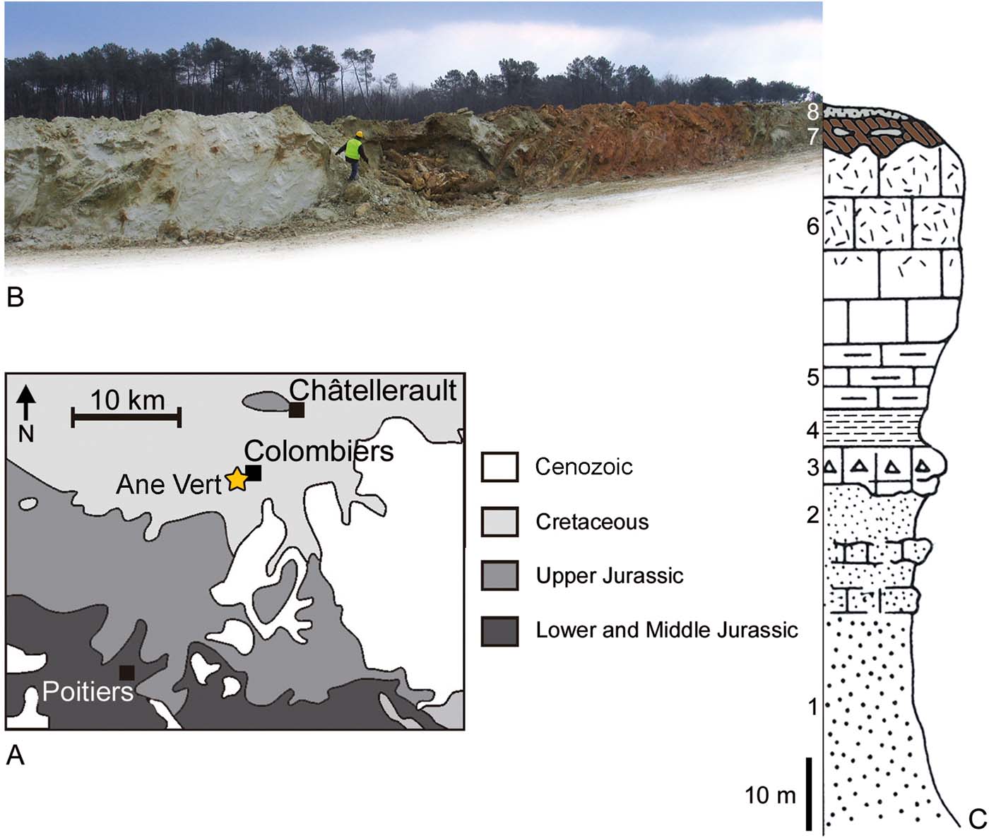
The Cenomanian–Turonian succession of Jaunay-Clan/Marigny Brizay has been described in detail by Valentin et al. (Reference Valentin, Gomez, Daviero-Gomez, Charbonnier, Ferchaud, Kirejtshuk, Licht, Néraudeau, Vullo and Garcia2014). The silicified nodules bearing the plant megafossils studied herein were collected from sandy claystones found at a depth of 2m, the latter also containing pieces of silicified limestones in the locality Ane Vert in the town of Colombiers, Vienne, Nouvelle-Aquitaine, western France (Figs 1, 2A). They were observed in situ in ferruginous sandstone joints intercalated in the uppermost claystone beds of the series (Fig. 2B, C, beds 7 and 8), which constitute tabular crusts of only up to 15cm high and 2m long. They are yellow ochre to light brown, and are rich in quartz grains. They also contain coastal marine fossils including sponge spicules, foraminifera and bryozoans. They may have been formed from the limestones and glauconitic sandstones that compose the yellow tufa, which is upper Turonian in age.
Similar silicified nodules with palaeontological inclusions have been reported from nearby colluvial deposits along the Valley of Envigne River. They were used in the archaeological industry during the Lower Palaeolithic (Hérisson et al. Reference Hérisson, Airvaux, Lenoble, Richter, Claud and Primault2012).
Geographical map of France with locations of selected Turonian–Coniacian plant fossil localities. (1) Upper Turonian of Le Beausset, Var (de Saporta & Marion Reference de Saporta and Marion1873). (2) Lower Coniacian of Bagnols-sur-Cèze, Gard (Zeiller Reference Zeiller1882). (3) Lower Coniacian of Torsac, Charente (Néraudeau Reference Néraudeau2014). (4) Upper Turonian of Claix, Charente (Moreau et al. Reference Moreau, Néraudeau, Plattel and Ravon2016). (5) Upper Turonian of Sainte-Mondane, Dordogne (Néraudeau et al. Reference Néraudeau, Saint-Martin, Batten, Colin, Daviero-Gomez, Girard, Gomez, Nohra, Polette, Plattel, Saint-Martin and Vullo2016). (6) Upper Turonian of Colombiers – Ane Vert, Vienne (the present study).

Geological setting. (A) Geological map with location of the silicified nodules from the upper Turonian of the locality Ane Vert in the town of Colombiers, western France. (B) Exposure of the trench where the plant fossils were collected. (C) Stratigraphic column: (1) claystones and white sandstones of Vierzon (lower Cenomanian); (2, 3) green sandstones and sandy-claystones of La Pallu (middle Cenomanian); (4) plastic claystones and white marlstones with oysters (upper Cenomanian); (5) clayey limestones and chalkstones with Inoceramus labiatus (lower Turonian); (6) white tufa (micaceous glauconitic chalk, middle Turonian); (7, 8) altered yellow tufa and clayey sandstones with silicified nodules (upper Turonian).

2. Material and methods
2.1. Petrography
Two silicified nodules were used as samples for petrography analysis. They were sliced with a diamond micro-saw. Thin sections were pasted with epoxy onto a glass slide, and then they were polished until necessary for examination under a transmitted light optical petrographic microscope.
2.2. Palaeobotany
In 2010, one of the authors, Patrice Cantinolle, collected two broken open silicified nodules showing plant megafossils in the field in the locality Ane Vert in the town of Colombiers. One bears a conifer twig and the other a very large fern-like compound leaf. The two hand-rock specimens will be housed in the collections of the Université de Poitiers under the numbers UP (for Université de Poitiers) + CAV (for Colombiers – Ane Vert) + year (2016) + specimen number: UP.CAV.16.001 (Geinitzia reichenbachii) and UP.CAV.16.002 (‘Lomatopteris' superstes). Photographs of the plant specimens were taken with a macro lens 100mm mounted on a Canon EOS60D camera. Detail photographs were taken with the same camera mounted on a stereomicroscope Olympus SZX10. Line drawings were made with a camera lucida mounted on the same stereomicroscope.
3. Results
3.1. Petrography
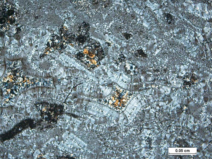
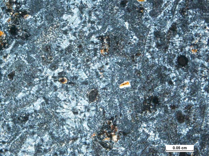
The first sample contains a low proportion of detrital particles, which are angular quartz, not bound together (Figs 3, 4), and therefore has a mudstone texture (Dunham Reference Dunham1962). According to the descriptions by Primault (Reference Primault2003) and Fernandes (Reference Fernandes2012), it can be identified as silcrete. It shows a few foraminifer clasts. X-ray diffraction confirms that the translucent matrix corresponds to microcrystalline drusy quartz (Figs 3, 4). The cortex is 8–10mm thick, and X-ray diffraction shows that it contains some geothite, which fill the porosity, along with quartz, and are responsible for the yellow ochre to brown colour. The silicification may have occurred by the flow of silica-rich water and the crystallisation of quartz.
Silcrete formed by precipitation of SiO2 from the solution. Cavities are partly filled by iron oxides.

Silcrete with microcrystalline drusy quartz.

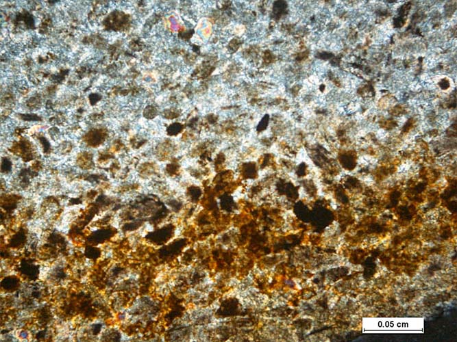
The second sample consists of a fine-grained flint with a translucent silicate matrix (Fig. 5). It contains a very low proportion of coarse detrital particles, and has a mudstone texture (Dunham Reference Dunham1962). The cortex is 4–5mm thick, and also has a brownish colour (Fig. 5). It may have been formed by the replacement of lime or mud by silica.
Flint with iron oxide patination in the cortex.

3.2. Palaeobotany
3.2.1. Conifers
Order Coniferales
Family Cupressaceae Gray, Reference Gray1822
Genus Geinitzia Endlicher, Reference Endlicher1847
Type species Geinitzia formosa Heer, Reference Heer1871
Geinitzia reichenbachii (Geinitz, Reference Geinitz1842)
Hollick and Jeffrey, Reference Hollick and Jeffrey1909
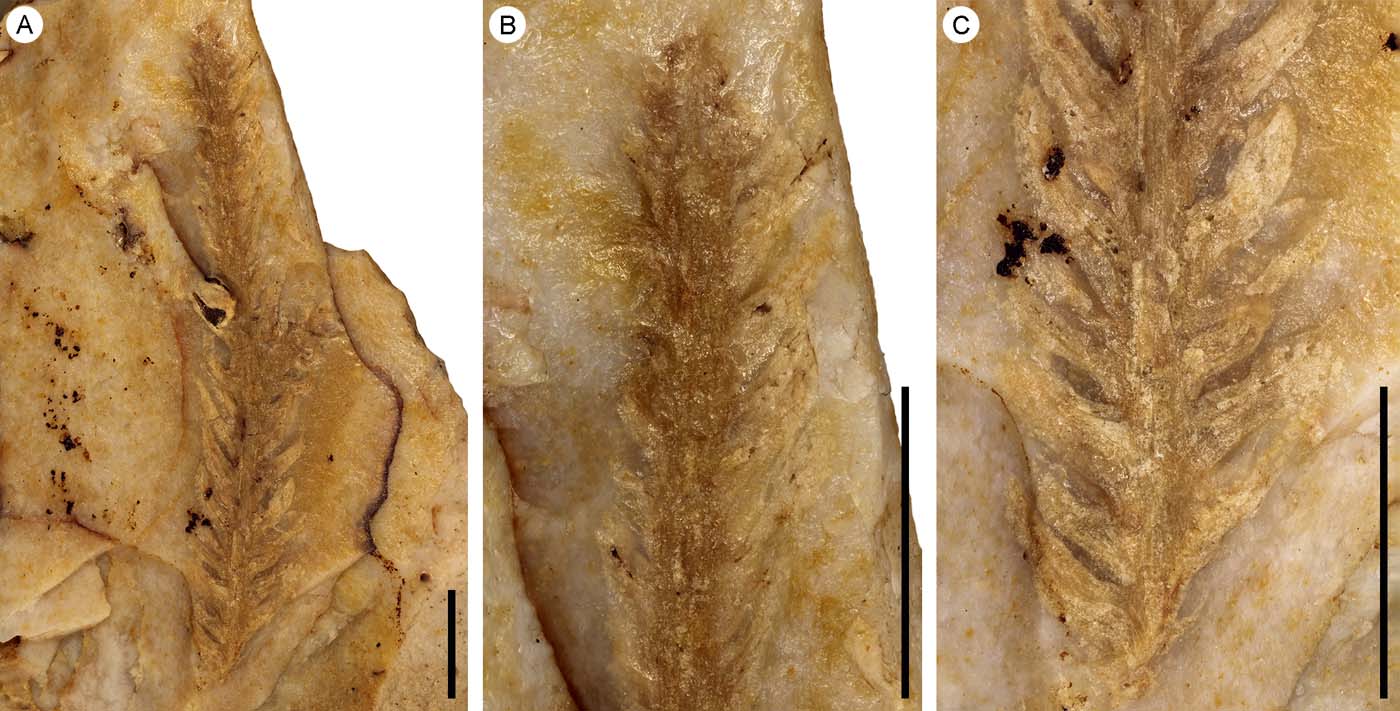
The specimen consists of once branched leafy stem (Fig. 6A–C). It branches at an angle of ca. 45° (Fig. 6A). The longest stem is 61mm long and up to 1.6mm wide. Leaves are spirally arranged and awl-shaped (Fig. 6B, C). They are up to 8.5mm long and 3.5mm wide, the widest part being at the base. The proximal part of the leaf base is decurrent onto the stem.
Geinitzia reichenbachii (Geinitz) Hollick and E. Jeffrey. (A) Once-branched leafy stem showing uniform, spirally arranged, falcate leaves. (B) Detail of the distal part. (C) Detail of the proximal part. Specimen no. UP.CAV.16.001. Scale bars=10mm.

3.2.2. Angiosperms
Clade Eudicots
Order Incertae sedis
Family Incertae sedis
‘Lomatopteris' superstes de Saporta, Reference de Saporta1878
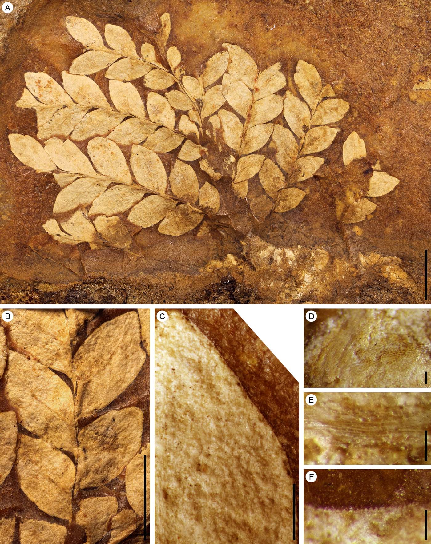
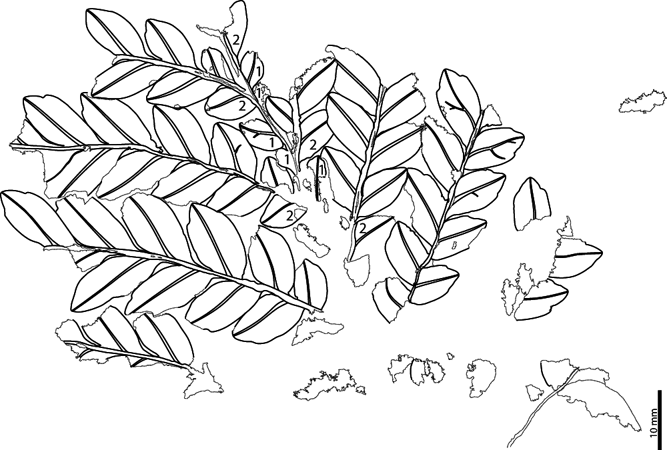
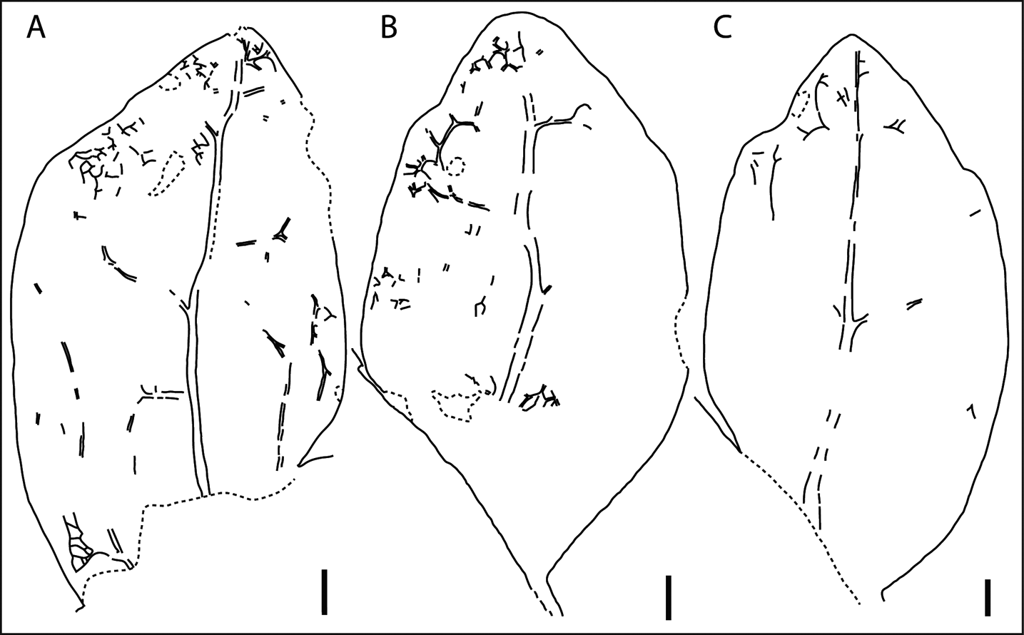
The specimen consists of the apical part of an at least bipinnately compound leaf (Figs 7A, 8). It is 89mm long and 101mm wide. Nine ultimate (secondary?) pinnae remain visible, and are arranged alternate distichous along the two sides of the penultimate (primary?) rachis. The rachis bears one or two intercalary leaflets between two successive ultimate (secondary?) rachillae. One leaflet is usually borne half on the rachis and half on the rachilla. Rachillae arise at angle of 30–50°, this angle decreasing toward the apex. They are slightly flexuous (Figs 7A, B, 8). Leaflets along the rachillae are arranged alternately. They arise at angle of 35–65° from the rachillae. They are 5.4–14.9mm long and 3.6–7.8mm wide. The largest leaflets along the rachillae are observed in the middle part. They are sessile and broadly attached by the lamina base. The margin is macroscopically entire except for two apical large teeth, more or less developed depending on the rachilla-bearing leaflet size, and being one on each side of the apex. At the leaflet base, the basal margin is decurrent, whereas the apical margin is constricted. The apex is acute. The lamina shows a single primary vein that runs into the apex and branches into at least two pinnate secondary veins in the middle of the leaflet lamina (Figs 9, 10). Other secondary and higher-order veins are very faint, but display a reticulate net venation pattern. The exmedial pattern of the visible secondary veins resembles a brochidodromous venation pattern.
‘Lomatopteris' superstes Saporta. (A) Bipinnate compound leaf, showing nine ultimate pinnae. (B) Portion of pinna showing slightly flexuous rachis bearing leaflets attached by a broad base, two more or less developed lobes on each side of the obtuse apex and a strong primary midvein. (C) Detail of the leaflet lamina showing entire margin and numerous stomata with mostly longitudinal (sometimes oblique, rarely transverse) oriented apertures. (D) Detail of the leaflet lamina showing polygonal cells of the mesophyll. (E) Detail of a vein showing elongate and rectangular cells. (F) Papillae over epidermal cells well visible along the margin, which appears micro-seriate. Specimen no. UP.CAV.16.002. Scale bars=10mm (A, B); 1mm (C); 200μm (D–F).

‘Lomatopteris' superstes Saporta. Line drawing of specimen no. UP.CAV.16.002. Light grey, intercalary leaflets borne on the penultimate rachis. Dark grey, leaflet borne half on the penultimate rachis and half on the ultimate rachis. Scale bar=10mm.

‘Lomatopteris' superstes Saporta. (A–C) Line drawings of selected leaflets showing the best preserved venation patterns with one primary midvein, a wide range of vein orders and a faint reticulate vein net. Specimen no. UP.CAV.16.002. Scale bars=1mm.

‘Lomatopteris' superstes Saporta. Ideal reconstruction of a leaflet showing one primary midvein and two veins raising from the rachis, two secondary veins raising from the primary vein at the upper part of the lower half lamina and a faint reticulate net venation pattern. Scale bar=10mm.

Stomata are numerous (Fig. 7C). Apertures are mostly longitudinally oriented, sometimes obliquely and rarely transversely. Epidermal cells bear papillae, which make the margin microscopically seriate (Fig. 7F). Papillae are 15–25μm wide and high. Cells at the level of the veins are rectangular and elongated following the direction of the veins (Fig. 7E). They are up to 50μm long and 20μm wide. Polygonal cells of the mesophyll are well visible in some areas (Fig. 7D). They are between 25 and 90μm long and wide.
4. Discussion
4.1. Palaeobotany and palaeogeography
Our specimen of Geinitzia reichenbachii shows the main features of the gross morphology in the neotype proposed by Kunzmann (Reference Kunzmann2010) based on materials from the upper Turonian of Germany and Czech Republic, including the type locality: uniform falcate leaves that spread abruptly from the axis, four-sided in cross section, decurrent leaf bases and a single central resin canal. Geinitzia reichenbachii was a widespread Cretaceous species and a common component of upper Cretaceous plant assemblages in Europe (e.g., Coiffard et al. Reference Coiffard, Gomez, Kvaček and Thévenard2006; Coiffard & Gomez Reference Coiffard and Gomez2010). Not surprisingly, it has been reported from many Cenomanian–Coniacian localities of western France (Néraudeau et al. Reference Néraudeau, Vullo, Gomez, Perrichot and Videt2005; Moreau et al. Reference Moreau, Néraudeau, Gomez, Tafforeau and Dépré2014a, Reference Moreau, Néraudeau, Gomez, Tafforeau and Dépréb, Reference Moreau, Néraudeau, Plattel and Ravon2016).
It is noteworthy that ‘Lomatopteris' superstes is reported from western France for the first time. At the end of the 19th Century, it had been reported first from the upper Turonian of Le Beausset, near the city of Toulon, Var (de Saporta Reference de Saporta1878), and second from the lower Coniacian of Bagnols-sur-Cèze, Gard (de Saporta Reference de Saporta1890), south-eastern France (Fig. 1). Lomatopteris Schimp., however, was a genus name used for Jurassic ferns, and is now a nomen nudum. Moreover, several features observed thanks to the good preservation of our specimen from the upper Turonian of Colombiers – Ane Vert – indicate, against its fern-like appearance, that it likely belongs to angiosperms and eudicots. These include the reticulate venation pattern, the marginal teeth and the bipinnately compound leaf, the latter being only known outside the eudicots in a few species in the family Hernandiaceae (e.g., Illigera Blume) within the Magnoliids (APG IV 2016).
Preliminary inventories of silicified plant megafossils have been previously reported from the upper Turonian or lower Coniacian of Claix and Torsac, Charente, western France (Néraudeau Reference Néraudeau2014; Moreau et al. Reference Moreau, Néraudeau, Plattel and Ravon2016). However, the descriptions and pictures provided in these studies do not allow for accurate identifications. Comparisons between these three plant assemblages are, thus, irrelevant until an in-depth revision of these materials is carried out. A further impediment to any comparison lies in the age uncertainty for the plant assemblage described by Néraudeau (Reference Néraudeau2014) (Moreau et al. Reference Moreau, Néraudeau, Plattel and Ravon2016), which was suggested as lower Coniacian based on the plant megafossil assemblage itself. This would also entail two significant sources of error: (1) the stratigraphic range of genera and species of Cretaceous plant megafossils, such as the conifers Frenelopsis, Glenrosa, Geinitzia and unidentified angiosperms, is, until now, far from well-constrained and conclusions are often misleading; (2) possible highly adapted plant taxa or relictual association in highly constrained environmental conditions such as those that prevailed during the upper Cretaceous in western Europe cannot be discarded, and so plant megafossil assemblages may not be representing a strict given time interval.
4.2. Silicification process of silicified nodules and plant fossils, and insights into the palaeoenvironment
The petrographic study has shown that silica is present everywhere in the nodules. The origin of this silica is most probably the bedrock itself, which was dissolved and re-precipitated in various places in the depositional bed in function of water flow. Our observations of an early crystallisation of quartz support that this silicification occurred contemporary to the earliest stages of fossil diagenesis, or even during the burying, of the plant megafossils.
Two hypotheses of formation can be proposed, which are not mutually exclusive. First, the coastal areas where freshwater meets seawater constitute places with conditions favourable for particular siliceous diagenesis. The silica solubility in freshwater is higher than in seawater, which implies that silica solubility decreases, and so silica tends to precipitate onto or exchange with the organic matter, when freshwater mixes with seawater. Second, the silicification can be a pedologic process related to the circulation of low-flow, meteoric or phreatic waters rich in dissolved silica. Indurated duricrusts or silcretes form when surface soil is cemented by silica. They usually form on the edges of plateaux or the low parts of hillsides. They occur in clayey sands by dissolution of silica at the top of the profile and concentration at the bottom. The material rich in silica is its own provider of dissolved silica. Important silicification occurs in a semi-arid climate because of the alternation of a wet season for the dissolution and circulation of silica-rich water with a drought season for the precipitation of silica in the porosity. This process can be amplified in a tropical climate, in which a higher quantity of silica can be transferred; a tropical climate for the palaeogeography of France during the Turonian is consistent with the trends in climate zone distribution described for the upper Cretaceous (Hay & Floegel Reference Hay and Floegel2012; Chaboureau et al. Reference Chaboureau, Sepulchre, Donnadieu and Franc2014). It is also noteworthy that a thermal maximum for the Cretaceous was reached during the Cenomanian–Santonian, which was at least significant in the Tethyan Realm (Martin et al. Reference Martin, Amiot, Lécuyer and Benton2014), and it coincides with the time interval during which these French silicified nodules were formed.
The silicification of plant fossils requires that silicate-rich fluid entered and filled the intercellular spaces and cells of some dead parts of the plant such as leaves and stems. After dehydration, an opaline crystal structure forms an internal cast or even completely replaces the cell walls. The two plant megafossils from the upper Turonian of Colombiers studied herein are fully permineralised, and show the excellent preservation of a wide range of cell types. Decay of the softest tissues, particularly of parenchyma cells consisting of thin-walled, pectocellulosic primary walls, is a very quick process. Thus, their preservation in these two specimens, in accordance with petrography evidence, firmly indicates that silicification occurred during the earliest stages of fossil diagenesis. This view also corresponds with the petrography.
In contrast, Néraudeau (Reference Néraudeau2014) and Moreau et al. (Reference Moreau, Néraudeau, Gomez, Tafforeau and Dépré2014a, Reference Moreau, Néraudeau, Gomez, Tafforeau and Dépréb, Reference Moreau, Néraudeau, Plattel and Ravon2016) suggested that the plant-fossil-bearing flints in western France were mostly, if not all, secondarily formed by leaching of upper Cretaceous rocks during warm climatic maxima of the Palaeogene, and especially of the Eocene, so that the silicification episode or episodes occurred up to 44 million years later. Néraudeau (Reference Néraudeau2014) and Moreau et al. (Reference Moreau, Néraudeau, Plattel and Ravon2016) noted that some flints from both the upper Turonian or lower Coniacian of Claix and Torsac, Charente, clearly show alternations of fine, sandy veins or lenses and fine, clayey-marly beds in a lignite facies, but these authors did not consider these sources of silica as a possible contemporaneous silicification in suitable environmental and climatic conditions. Moreover, Moreau et al. (Reference Moreau, Néraudeau, Gomez, Tafforeau and Dépré2014a, Reference Moreau, Néraudeau, Gomez, Tafforeau and Dépréb, Reference Moreau, Néraudeau, Tafforeau and Dépré2015) showed that the softest plant tissues of conifer inclusions were preserved inside the flints using X-ray synchrotron microtomography, but they did not consider these palaeobotanical and taphonomic evidence suggesting a rapid fossilisation.
5. Conclusions
Several silicification episodes most probably occurred during the Cenomanian–Coniacian interval, and allowed for the excellent preservation of plant fossils, including their most delicate histology. These silicifications were endogenic, occurred during the earliest stages of the fossil diagenesis and were promoted by palaeoenvironmental and climatic conditions. Geinitzia reichenbachii and ‘Lomatopteris' superstes are newly reported from the upper Turonian of Colombiers – Ane Vert, Vienne, western France. The latter taxon has only been previously reported in the late 19th Century from the Turonian–Coniacian of Le Beausset and Bagnols-sur-Cèze, Var and Gard respectively, south-eastern France. We have provided evidence for assigning this taxon into the angiosperm eudicots. Future research should be dedicated to revising the taxonomy and systematics of recently reported and described silicified plant fossils from the Cenomanian–Coniacian of western France, as well as to confirming that these silicifications were endogenic.
6. Acknowledgements
Funding for B. G., V. D.-G. and A. B. was provided by Unité Mixte de Recherche 5276 of Centre National de la Recherche Scientifique, projects CGL2013-42643-P, CGL2015-68363-P and CGL2015-60805-P of the Ministerio de Economía y Competitividad of the Spanish government and Project 2014SGR-251 funded by the Catalan government. This work is supported, in part, by the Liséa Fondation Biodiversité. G. G. and X. V. acknowledge the municipality of Colombiers (J. Gauthier and A. Vausselle), J. M. Boiteau, P. Ferchaud, C. Fontaine, B. Guével, C. Laforest and S. Riffaut for technical support and J. Primault (DRAC, Nouvelle Aquitaine), A. Decarreau and J. Airvaux for scientific discussions.