Introduction
One-third of the world’s deer species are classified as rare or endangered (IUCN, 2015). Given this scenario and the difficulties of conserving the natural habitats of these species, assisted reproduction techniques associated with genome banks have proven to be important tools for in situ and ex situ conservation programmes (Holt and Pickard, Reference Holt and Pickard1999). The collection of materials of endangered species is important, but collection should also be carried out on species that are not yet at risk of extinction, to maintain genetic diversity, which is frequently lost in rare species (Duarte and Garcia, Reference Duarte and Garcia1995).
Among the available techniques of assisted reproduction, in vitro production of embryos is the most efficient technique for the propagation of small populations (Comizzoli et al., Reference Comizzoli, Mermillod and Mauget2000). It allows broader knowledge of the physiological, biochemical and biotechnological processes, while supporting the development of other biotechnologies, such as cloning by nuclear transfer and transgenesis (Gonçalves et al., Reference Gonçalves, Visintin, Oliveira, Montagner and Costa2002). This technique allows the cryopreservation of oocytes and embryos (Bainbridge et al., Reference Bainbridge, Catt, Evans and Jabbour1999) and requires fewer viable sperm for fertilization compared with artificial insemination or natural mating (Loskutoff et al., Reference Loskutoff, Bartels, Meintjes, Godke and Schiewe1995). These characteristics mean that in vitro production of embryos could be particularly interesting for deer species of the genus Mazama, for which difficulties in semen freezing remain due to low post-thaw motility (Duarte and Garcia, Reference Duarte and Garcia1997; Abreu, Reference Abreu2006; Zanetti et al., Reference Zanetti, Duarte, Polegato, Garcia, Canola, Duarte and Gonzalez2010; Favoretto et al., Reference Favoretto, Zanetti and Duarte2012) and because it has not yet been possible to develop an adequate superovulation and embryo collection protocol (Zanetti and Duarte, Reference Zanetti and Duarte2012; Zanetti et al., Reference Zanetti, Munerato, Cursino and Duarte2014). However, the success of IVP of embryos in deer has been limited to species used for commercial breeding purposes (Siriaroonrat, Reference Siriaroonrat2006).
As biotechnologies have not been established for most endangered deer species, the use of closely related species as a biological model is an interesting alternative (Bainbridge and Jabbour, Reference Bainbridge and Jabbour1998). However, there are some limitations to establishing reproduction biotechnologies in deer, including the scarcity of knowledge concerning the reproductive physiology of the species (Comizzoli et al., Reference Comizzoli, Mermillod and Mauget2000) and their greater susceptibility to stress (Duarte, Reference Duarte, Duarte and Gonzaléz2010). The brown brocket deer (Mazama gouazoubira) was chosen as an experimental model for neotropical deer, because it is the most abundant deer in South America (Duarte, Reference Duarte1996) and some of its reproductive parameters have been established. The studies developed on this species can be used on other species threatened with extinction and should facilitate the establishment of a management plan before population decline and loss of genetic diversity occurs, as reported for rare and/or threatened species (Duarte and Garcia, Reference Duarte and Garcia1997).
To develop an IVP of embryos in this species, an efficient ovarian stimulation protocol was required. Zanetti and Duarte (Reference Zanetti and Duarte2012) and Zanetti et al. (Reference Zanetti, Munerato, Cursino and Duarte2014) tested different superovulation protocols in M. gouazoubira and verified that the best follicular growth responses were obtained using 700 IU of equine chorionic gonadotropin (eCG) or 130 mg of follicle-stimulating hormone (FSH), resulting in 11.5 ± 2.5 and 10.2 ± 1.1 overall follicular stimulation, respectively. However, the protocol using eCG caused a decrease in ovarian response when the deer were submitted to repeated hormonal treatments (E. Zanetti, personal communication), as observed in other studies (Pigon et al., Reference Pigon, Clegg and Cole1960; Jainudeen et al., Reference Jainudeen, Hafez, Gollnick and Moustafa1966; Baril et al., Reference Baril, Remy, Vallet and Beckers1992), and premature regression of the corpora lutea, indicating possible poor follicle quality (Zanetti et al., Reference Zanetti, Munerato, Cursino and Duarte2014). The protocol for multiple FSH applications has also been tested by Cursino et al. (Reference Cursino, Salviano, Abril, dos Santos Zanetti and Duarte2014) using laparotomy in M. americana, resulting in a good response in follicular growth and the recovery of viable oocytes.
Laparoscopic ovum pick-up is considered less invasive than laparotomy and therefore is preferred for follicular aspiration of medium and small deer (Bainbridge et al., Reference Bainbridge, Catt, Evans and Jabbour1999; Comizzoli et al., Reference Comizzoli, Mermillod, Cognie, Chai, Legendre and Mauget2001; Siriaroonrat, Reference Siriaroonrat2006), because it promotes faster recovery and the possibility of repeating the technique in the same female. After collection, the recovered oocytes need to undergo a period of in vitro maturation (IVM), in which nuclear and cytoplasmic changes occur that enable fertilization and the development of a viable embryo (Sirard, Reference Sirard2001). In cervids, IVM has been reported in Cervus nippon (Comizzoli et al., Reference Comizzoli, Mermillod, Cognie, Chai, Legendre and Mauget2001; Locatelli et al., Reference Locatelli, Vallet, Huyghe, Cognie, Legendre and Mermillod2006), Rangifer tarandus (Krogenaes et al., Reference Krogenaes, Ropstad, Thomassen, Pedersen and Forsberg1994), C. elaphus (Fukui et al., Reference Fukui, McGowan, James, Asher and Tervit1991; Pollard et al., Reference Pollard, Bringans and Buckrell1995; Comizzoli et al., Reference Comizzoli, Mermillod, Cognie, Chai, Legendre and Mauget2001; Berg et al., Reference Berg, Thompson and Asher2002; Berg and Asher, Reference Berg and Asher2003), Odocoileus virginianus (Siriaroonrat et al., Reference Siriaroonrat, Comizzoli, Songsasen, Monfort, Wildt and Pukazhenthi2010), C. eldi thamin (Siriaroonrat, Reference Siriaroonrat2006), M. americana (Cursino et al., Reference Cursino, Salviano, Abril, dos Santos Zanetti and Duarte2014), M. nana (Zanetti et al., Reference Zanetti, Duarte, Polegato, Garcia, Canola, Duarte and Gonzalez2010) and M. gouazoubira (Cursino et al., Reference Cursino, Zanetti, Saraiva and Duarte2009), and 24 h seems to be the optimum period for oocyte maturation in the majority of species.
In the articles cited, oocyte maturation was achieved using methods similar to those used on domestic ruminant oocytes, i.e. tissue culture medium (TCM)-199 supplemented with hormones, bovine serum albumin (BSA) or follicular fluid of sheep or goat and pyruvate. The choice of hormones is usually luteinizing hormone and FSH, which are regulators of nuclear IVM of mammalian oocytes, while estradiol appears to be involved in both cytoplasmic and nuclear maturation (Tibary et al., Reference Tibary, Anouassi and Khatir2005). Follicle-stimulating hormone is considered essential for C. elaphus and C. nippon oocyte maturation, as its use significantly increased the percentage of matured oocytes when substituting epidermal growth factor (Comizzoli et al., Reference Comizzoli, Mermillod, Cognie, Chai, Legendre and Mauget2001). Estradiol has also been successfully included in IVM systems for deer (Krogenaes et al., Reference Krogenaes, Ropstad, Thomassen, Pedersen and Forsberg1994; Comizzoli et al., Reference Comizzoli, Mermillod, Cognie, Chai, Legendre and Mauget2001; Berg and Asher, Reference Berg and Asher2003; Siriaroonrat et al., Reference Siriaroonrat, Comizzoli, Songsasen, Monfort, Wildt and Pukazhenthi2010), such that in O. virginianus, its influence on the improvement in nuclear maturation was evident.
Considering the importance of the development of reproductive biotechnologies to assist in the conservation of endangered deer species, our work proposes a study of the early stages of in vitro production of embryos in M. gouazoubira to determine an effective hormonal protocol for ovarian stimulation, the efficiency of laparoscopy for oocyte collection and a medium for oocyte maturation.
Materials and methods
Animals
The research involved five females of the species Mazama gouazoubira, aged 1–8 years old, weighing between 15.6 and 18.8 kg, and that belonged to the Deer Research and Conservation Center (NUPECCE) of the Department of Animal Science of FCAV-UNESP. Research was conducted between December 2011 and February 2013. Deer were maintained in individual stalls (3 m × 2 m) and received a daily diet consisting of pelleted feed (12% total protein, 2% total fat 2%, 10% total fibre; Omolene®, Purina®), and about 1 kg of perennial soybean roughage (Neonotomia wighti), alfalfa (Medicago sativa) or branches of mulberry (Morus alba). Water was offered ad libitum.
Habituation period
The females were submitted to a habituation period to facilitate intramuscular applications of FSH. Every day for a month, they were led from their stalls to a containment box, where they remained for 5 min. During this time, they were offered fruit as an incentive and for positive reinforcement through a window near the deer’s head, while simultaneously being handled through a window near the posterior region (Zanetti, Reference Zanetti2009). During week one, these procedures were performed twice daily (morning and afternoon), and thereafter, only once a day (morning).
Ovarian stimulation
After physically restraining the deer (Duarte, Reference Duarte, Duarte and Gonzaléz2010), an intravaginal device impregnated with 0.33g of progesterone, developed for sheep and goats (controlled internal drug release (CIDR®) Type T, Pfizer® USA) was inserted and left for 8 days. Immediately after inserting the CIDR device (D0), 0.25 mg (0.25 ml) of estradiol benzoate (EB; Estrogin®, Farmavet Produtos Veterinários Ltda, Brazil) was administered intramuscularly. On day four (D4) of CIDR implantation, doses of FSH were initiated at 12-h intervals. Eight equal doses were applied, totalling 130 mg of FSH (Folltropin®-V Tecnopec®, Canada) (Zanetti et al., Reference Zanetti, Munerato, Cursino and Duarte2014). The intramuscular applications were performed while the deer were in the containment box. The ovarian stimulation treatment was repeated at intervals of approximately 30 days after the previous oocyte aspiration.
Laparoscopic ovum pick-up
Eight days after the onset of the hormone treatment (D8) the females, previously submitted to feed and water fasting for 24 h, were physically restrained and anaesthetised intravenously (jugular vein) with a combination of 5.0 mg/kg of ketamine hydrochloride (Vetaset®, Fort Dodge, Brazil), 0.3 mg/kg of xylazine hydrochloride (Rompum®, Bayer, Brazil) and 0.5 mg/kg midazolam (Dormonid®, Roche, Brazil),. The deer were intubated and, during the procedure, a superficial plane was maintained using isoflurane (Forane®, Abbott, Brazil) at a mean concentration of 0.89 ± 0.08 V%. Videolaparoscopy for this species was performed similar to that described by Zanetti et al. (Reference Zanetti, Duarte, Polegato, Garcia, Canola, Duarte and Gonzalez2010). Briefly, the female was positioned at a 45° angle to the hind limbs (Trendelenburg position), and the abdominal cavity was inflated through a trocar positioned 5 cm cranial to the mammary gland and 5 cm to the left of the midline, with a valve coupled to the CO2 insufflator.
Once pneumoperitoneum was established (intra-abdominal pressure of 8 mmHg, flow of 5 L/min), two more trocars were inserted, one along the midline position, and the other contralateral to the trocar that was used to inflate the cavity. An optical laparoscope (30 cm/5 mm/30º), connected to a fibre optic cable, light source and a miniature camera, was introduced into the midline trocar, while atraumatic gynaecological forceps were introduced into the others to manipulate and position the ovaries. The ovary was secured by the forceps closest to its ligament, then one of the forceps was removed and a metal rod connected to a follicle aspiration needle (22G) (WTA®, Cravinhos, SP, Brazil) was introduced into the abdominal cavity. The needle was coupled to an aspiration circuit, with a continuous vacuum precision pump (adjusted to 100 mmHg, flow of 11 ml of water/min), and the follicular fluid was recovered in a 50 ml collection tube, containing 2 ml complete phosphate-buffered saline (PBS) with 10 IU/ml of heparin (collection medium), heated to 38ºC to receive the oocytes.
After follicle aspiration, the ovaries were washed with collection medium to reduce possible adhesions. When the surgical procedure was completed, the CIDR was removed. The deer were administered an antibiotic (40,000 U/kg benzathine benzylpenicillin) and an anti-inflammatory drug (0.025 ml/kg of 0.2% meloxicam) intramuscularly immediately after the procedure.
Quantity of follicles and oocyte recovery rate
The videolaparoscopy surgeries were recorded and watched later to confirm the number of follicles observed and aspirated. To calculate the oocyte recovery rate, the number of oocytes collected in relation to the number of follicles aspirated was determined (oocytes collected ÷ follicles aspirated), and the result was presented as a percentage. Considering the limited number of deer available for the experiment, collection and morphological evaluation of at least 70 oocytes were proposed.
Selection and evaluation of the oocyte cumulus complex
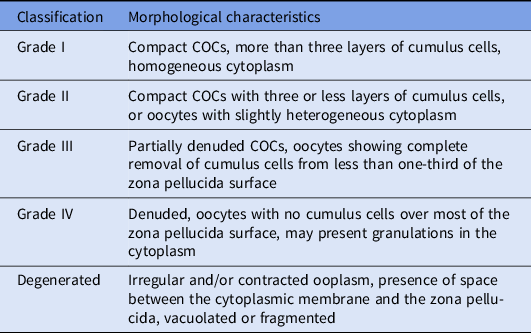
The oocytes collected were transferred to a polystyrene plate and evaluated under a stereomicroscope at ×40 magnification. Once located, the oocytes were washed twice in wash medium (TCM-199 containing 20 mM HEPES and 5 mM sodium bicarbonate buffer) and assessed for cumulus–oocyte complex (COC) morphology, as described by Viana et al. (Reference Viana, de Almeida Camargo, de Moraes Ferreira, de Sa, de Carvalho Fernandes and Junior2004) with modifications (Table 1). Degenerated oocytes were excluded from the study and the remainder were selected for IVM or were stained directly and evaluated for nuclear status without undergoing IVM.
Morphologic classification (Viana et al., Reference Viana, de Almeida Camargo, de Moraes Ferreira, de Sa, de Carvalho Fernandes and Junior2004, with modifications) used for oocytes of Mazama gouazoubira

COC, cumulus–oocyte complex.
In vitro maturation
Once selected, the oocytes were transferred to maturation plates, in 100-µl droplets of maturation medium [TCM-199 supplemented with 10% BSA, 5.0 μg/ml of FSH, 100 μg/ml of hCG, 1.0 μg/ml of estradiol, 0.33 mM of pyruvate, 2 mM of glutamine, 2.2 mg/l of sodium bicarbonate, 16.67 μg/ml of amikacin, as per Siriaroonrat et al. (Reference Siriaroonrat, Comizzoli, Songsasen, Monfort, Wildt and Pukazhenthi2010), modified], containing up to 10 oocytes per drop. The plates were incubated in air with 5% CO2, at a 38.5°C for 24 h.
Evaluation of nuclear maturation
After the maturation time, oocytes were denuded in PBS plus 0.1% hyaluronidase with the aid of an automatic pipette, placed in a drop containing Hoechst 33342 dye (10 μg/ml; Molecular Probes, Inc., Eugene, OR, USA) diluted in TCM-199 HEPES medium and incubated in a semi-controlled humid atmosphere, with 5% CO2 in air, at 38.5°C for 20 min.
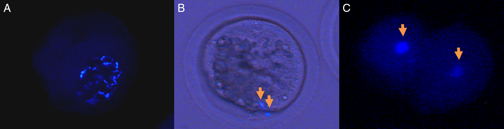
After incubation, the oocytes were washed twice in PBS, placed in a drop of glycerin between a slide and coverslip and observed under an epifluorescence microscope (330–385nm). They were classified as mature (MII stage, visible extrusion of the polar body) or immature oocytes (any other nuclear configuration). Oocytes with no chromatin were classified as degenerated.
Statistical analysis
Descriptive statistics were used for data analysis. Ovarian stimulation data are presented as mean ± standard deviation (SD) of the number of follicles observed. Data on oocyte recovery rate data are expressed as a percentage (%) of the number of oocytes collected in relation to the number of aspirated follicles. The morphological classification of COCs and evaluation of nuclear maturation are reported as percentages (%), as per previously proposed classifications.
Results
Surgical procedures were performed on the superstimulated females, resulting in 94 oocytes, of which 73 oocytes (from 11 laparoscopies) were matured in vitro for later evaluation of the nuclear status, while the remaining 21 oocytes (from three laparoscopies) were only stained, to evaluate the nuclear status of unmatured oocytes in vitro.
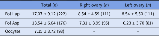
Table 2 presents the mean ± SD of the number of follicles observed by videolaparoscopy, the number of aspirated follicles and the number of oocytes collected during each surgical procedure from each deer. Only those laparoscopies in which the ovaries were observed were included (n = 13). The oocyte recovery rate was 52.8% (7.15 oocytes collected ÷ 13.54 aspirate follicles).
Mean ± standard deviation of the number of follicles observed, aspirated follicles and oocytes from 13 collections by videolaparoscopy in superstimulated females of Mazama gouazoubira

Total, data for both ovaries; Fol Lap, follicles observed by videolaparoscopy; Fol Asp, aspirate follicles.
During the first surgery, adhesions were observed on the ovaries of females 2 and 4 (possibly due to previous surgical procedures). In female 2, these adhesions completely surrounded the ovaries, making it impossible to observe them, therefore this deer was discarded, however during the ‘blind’ punctures performed on this female, one oocyte was collected, matured in vitro and included in the nuclear maturation and morphological evaluation scores. In females 1 and 3, adhesions were observed from the third surgery onward.
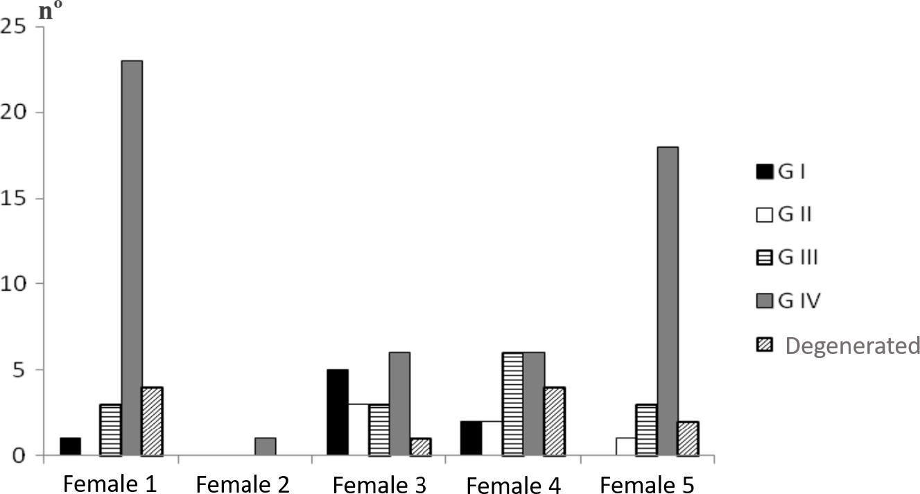
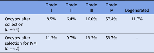
On a few occasions, clots caused the aspiration needle to clog, making it necessary to use the maximum pressure of the device (≈530 mmHg) to clean the aspiration circuit. Figure 1 shows the number of oocytes collected from each female and their morphological classification. Among the oocytes collected, a high rate of denuded oocytes (grade IV) was observed, however the majority presented homogeneous ooplasm. Table 3 shows the values of the morphological classification of the 94 oocytes collected during all 14 video laparoscopies. From 73 oocytes collected during the first 11 procedures, 62 (84.9%) were selected and allocated to IVM. Table 3 also shows the morphological classification of the oocytes submitted to IVM.
Total number of oocytes harvested from each of the overstimulated females of the Mazama gouazoubira species according to the categories of morphological classification.

Morphological classification of oocytes immediately after collection (n = 94) and oocytes selected for IVM (n = 62) from superstimulated females of Mazama gouazoubira

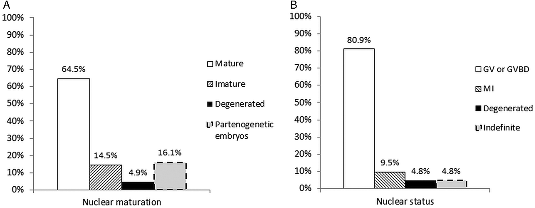
Evaluation of the nuclear maturation of oocytes selected for IVM (n = 62) is presented in Figure 2A. Evaluation of the nuclear status of oocytes collected but not submitted to IVM is presented in Figure 2B. Figure 3 shows representative micrographs of the classification of the nuclear status of M. gouazoubira oocytes.
(A) Representation of the nuclear maturation rate of 62 Mazama gouazoubira oocytes submitted to IVM. (B) Representation of the nuclear status rates of 21 M. gouazoubira oocytes, immediately after collection, not submitted to IVM. GV or GVBD: germinal vesicle or germinal vesicle breakdown; MI: meiosis I. Indefinite, oocytes in which nuclear status could not be defined.

Representative micrographs showing the classification of the nuclear status of Mazama gouazoubira oocytes. (A) Immature oocyte: germinal vesicle breakdown (GVBD). (B) Mature oocyte: meiosis II, visualization of the nucleus and polar body (see arrows). (C) Division activated spontaneously by parthenogenesis, visualization of two nuclei (see arrows).

Discussion
The hormonal protocol used to induce ovarian stimulation in M. gouazoubira was efficient, as the mean number of follicles observed (17.07 ± 9.12) was similar that described in sheep, where the mean ranges between 10.4 and 14.7 follicles per sheep (reviewed by Stangl et al., Reference Stangl, Kühholzer, Besenfelder and Brem1999) under different hormonal protocols. In deer, different hormonal protocols have been tested for superovulation and oocyte collection and have shown broad variation in follicular growth from 3.21 to 18.2 follicles per deer (Argo et al., Reference Argo, Jabbour, Goddard, Webb and Loudon1994; Morrow et al., Reference Morrow, Asher, Berg, Tervit, Pugh, McMillan, Beaumont, Hall and Bell1994; Asher et al., Reference Asher, Jabbour, Thompson, Tervit and Morrow1995; Berg et al., Reference Berg, Thompson, Pugh, Tervit and Asher1995; Bainbridge et al., Reference Bainbridge, Hunter, Chapple, Flint and Jabbour1996; Asher et al., Reference Asher, O’Neill, Scott, Mockett and Fisher2000; Siriaroonrat, Reference Siriaroonrat2006; Cursino et al., Reference Cursino, Salviano, Abril, dos Santos Zanetti and Duarte2014). The best results for ovarian stimulation were achieved in Cervus eldi using a protocol of multiple FSH application (Siriaroonrat, Reference Siriaroonrat2006). For superovulation in M. gouazoubira, hormonal protocols tested by Zanetti and Duarte (Reference Zanetti and Duarte2012) and Zanetti et al. (Reference Zanetti, Munerato, Cursino and Duarte2014) resulted in total follicular stimulus that ranged from 1.4 to 11.5 follicles per animal, depending on the protocol used. A protocol of multiple FSH application resulted in lower total follicular stimulation (10.2) than verified in our study.
In deer, the rate of oocyte recovery rate by laparoscopic oocyte pick-up ranged between 46 and 71% (Bainbridge et al., Reference Bainbridge, Catt, Evans and Jabbour1999; Comizzoli et al., Reference Comizzoli, Mermillod, Cognie, Chai, Legendre and Mauget2001; Locatelli et al., Reference Locatelli, Vallet, Huyghe, Cognie, Legendre and Mermillod2006; Siriaroonrat, Reference Siriaroonrat2006), values that are similar to those obtained in this study (52.8%). The number of oocytes obtained in our experiment (7.15 ± 3.72 oocytes/deer/surgery) was similar to that reported by other authors. In sheep, this number ranged from 4 to 10 oocytes per donor (Alberio et al., Reference Alberio, Olivera, Roche, Alabart and Folch2002; Gibbons et al., Reference Gibbons, Bonnet, Cueto, Catala, Salamone and Gonzalez-Bulnes2007), while in goats, it ranged from 5.6 to 13.4 oocytes per goat (reviewed by Abdullah et al., Reference Abdullah, Liow, Rahman, Chan, Wan-Khadijah and Ng2008). In C. eldi, 13.4 oocytes per female were collected (Siriaroonrat, Reference Siriaroonrat2006), while in C. nippon, nine oocytes per female were reported (Comizzoli et al., Reference Comizzoli, Mermillod, Cognie, Chai, Legendre and Mauget2001). Another study on C. nippon demonstrated variations from 1.7 to 5.6 oocytes per female depending on the season, in a species considered to be seasonal breeders (Locatelli et al., Reference Locatelli, Vallet, Huyghe, Cognie, Legendre and Mermillod2006).
Regarding the occurrence of adhesions after laparoscopic oocyte pick-up, Cordeiro et al. (Reference Cordeiro, Teixeira, Oliveira, Di Filippo, Dias, Beretta, Dória, Feliciano, Coutinho and Vicente2014) reported that, when performing weekly collections in goats, washing the ovaries with collection liquid (PBS plus 10 UI/ml of heparin) helped minimize adherence in 85% of goats, mainly from the fourth surgery onward. However, in our work, washing the ovaries with PBS plus heparin was insufficient to prevent the formation of adhesions, which occurred mainly from the third surgery onward. Therefore, the number of oocytes collected during each surgery may have been influenced by the adhesions that formed in the ovaries, as areas of adhesion can conceal follicles that could be aspirated. This may also affect evaluation of the ovarian stimulation protocol by making the repeatability of the technique for follicular aspiration unfeasible, depending upon the extent of ovarian adhesion.
Studies on oocyte maturation generally do not use denuded oocytes, as they are considered low quality, with reduced ability to complete meiotic maturation (Warriach and Chohan, Reference Warriach and Chohan2004). Despite the large numbers of grade IV (denuded) oocytes in this study, most of them presented homogeneous ooplasm, indicating good oocyte quality. This led us to consider the vacuum pump pressure and/or the diameter of the needle used. The occurrence of needle clogging and the need to increase the vacuum to clean the circuit was observed in this experiment, and could be a contributing factor in the number of denuded oocytes collected.
Pressure (mmHg) and flow (ml of water/min) are important physical characteristics of follicle aspiration with a needle coupled to a vacuum pump and they are directly related to the efficiency of COC recovery (Crocomo et al., Reference Crocomo, Marques Filho, Landim Alvarenga and Bicudo2012). In sheep, Baldassarre et al. (Reference Baldassarre, Wang, Kafidi, Gauthier, Neveu, Lapointe, Sneek, Leduc, Duguay and Zhou2003) and Koeman et al. (Reference Koeman, Keefer, Baldassarre and Downey2003) suggested that pressures between 50 and 70 mmHg were optimal values for the vacuum pump setting, while values between 6.5 and 10 ml/min of water were indicated for flow (Alvarez et al., Reference Alvarez, Anel, Anel, Rodríguez, Kaabi, Boixo, Paz and Olmedo1999; Rodríguez et al., Reference Rodríguez, Anel, Anel, Kaabi, Boixo, De Paz and Olmedo2000; Alberio et al., Reference Alberio, Echegoyen, Olivera, Roche, Alabart, Fernandez-Arias and Folch2001). However, a more recent study suggested that the ideal conditions for aspiration are flow ranging between 10 and 20 ml/min of water, an aspiration system with a diameter of 1–2 mm and the use of short needles, regardless of their calibre (Rodríguez et al., Reference Rodríguez, Anel, Alvarez, Anel, Boixo, Chamorro and Paz2006). Even though we followed the conditions outlined by Rodríguez et al. (Reference Rodríguez, Anel, Alvarez, Anel, Boixo, Chamorro and Paz2006), our study obtained high rates of denuded oocytes. However, if we consider the levels recorded, we used greater pressure than that recommended in the literature, therefore lowering these levels could improve the collection of better quality oocytes.
A study on the nuclear dynamics of M. gouazoubira oocytes demonstrated that a period of IVM of 20–25 h is required for more than 50% of the oocytes to reach metaphase II (Cursino et al., Reference Cursino, Zanetti, Saraiva and Duarte2009). However, this study was limited, as the low number of oocytes per group was low, such that in the group 20–25 h of IVM, only five oocytes were evaluated. In C. elaphus, 24 h of culture were required for 50% of the oocytes to reach metaphase II and 27 h for 75% (Berg et al., Reference Berg, Thompson and Asher2002). Our study corroborated the results cited above, as IVM for 24 h enabled the nuclear maturation of more than 50% of oocytes in vitro.
In deer, IVM for 24 h resulted in rates ranging from 50 to 78% of oocytes at metaphase II (Krogenaes et al., Reference Krogenaes, Ropstad, Thomassen, Pedersen and Forsberg1994; Comizzoli et al., Reference Comizzoli, Mermillod, Cognie, Chai, Legendre and Mauget2001; Berg et al., Reference Berg, Thompson and Asher2002; Siriaroonrat et al., Reference Siriaroonrat, Comizzoli, Songsasen, Monfort, Wildt and Pukazhenthi2010). However, these studies discarded the use of denuded oocytes for IVM. Although the majority of the oocytes submitted to maturation (n = 62) was classified as denuded (59.7%), in this study, the percentage of oocytes that reached metaphase II (64.5%) was within the range described by the literature. Therefore, it is probable that better maturation rates would have been reached whether we only worked with oocytes covered with cumulus cells.
A relatively high percentage of oocytes spontaneously activated by parthenogenesis was verified. Perhaps this is inherent to the species, as occurs in camels (Camelus dromedarius) (reviewed by Abdoon et al., Reference Abdoon, Kandil, Zeng and Cui2011), in which about 33% of oocytes activated spontaneously. Another possibility is that inadequate oocyte maturation occurs, in either the nucleus or the cytoplasm, which increases the occurrence of parthenogenesis (Xu and Greve, Reference Xu and Greve1988). Factors that could lead to inadequate IVM include a prolonged period of culture, which can lead to oocyte ageing and thereby increase their sensitivity to activation stimuli due to handling and in vitro conditions (Albertini et al., Reference Albertini, Sanfins and Combelles2003), and the use of oocytes from small follicles. Regarding the period of maturation, even though IVM for 24 h is optimal for many deer species, this may be too long for M. gouazoubira.
Aged oocytes and/or those collected from small follicles tend to have low H1 kinase activity levels and greater susceptibility to spontaneous activation by parthenogenesis. Basal levels of H1 kinase activity were verified in aged oocytes from pigs after undergoing prolonged IVM (Kikuchi et al., Reference Kikuchi, Izaike, Noguchi, Furukawa, Daen, Naito and Toyoda1995). Moreover, diminished levels of H1 kinase were detected in 25% of bovine oocytes from small follicles (less than 2 mm) (Ledda et al., Reference Ledda, Bogliolo, Calvia, Leoni and Naitana1997; Yang et al., Reference Yang, Kubota, Suzuki, Taneja, Bols and Presicce1998) also verified parthenogenetic activation of 19% of oocytes from prepubertal sheep compared with 2% of oocytes from adult females. This fact was associated with the prevalence of follicles between 1 and 2 mm in diameter in prepubertal females.
In our opinion, identifying the oocyte status immediately after collection is fundamental. Certifying that the oocytes were maturing in vivo is important, because if they were maturing in vivo and are subsequently submitted to in vitro cultivation, the gametes age and this diminishes their potential to produce embryos. In this study, oocyte evaluation allowed us to confirm that in vivo maturation did not occur, as most oocytes were classified as germinal vesicle or germinal vesicle breakdown.
In conclusion, we were able to demonstrate that viable oocytes can be recovered by videolaparoscopy when deer are administered a hormonal protocol based on multiple FSH application. The medium used for IVM was effective for maturing M. gouazoubira oocytes. This study, which used a model species, is a relevant source of knowledge and can be used as the basis for establishing conservation techniques such as in vitro production of embryos and cloning. However, further studies are required to improve the technique to obtain oocytes without denuding them and without the occurrence of adhesion in the ovaries. Moreover, it is essential to conduct research that focuses on the next steps of in vitro production of embryos, including sperm hyperactivation, fertilization, co-cultivation, vitrification and embryo transfer.
Acknowledgements
The authors would like to thank Naiara Z Saraiva for her assistance in laboratory procedures, Ana Paula Gering for her assistance during anaesthesia, Antonio Carlos L Andrade for his assistance during deer handling and the NUPECCE staff for their assistance during surgeries.
Financial support
This work was supported by the São Paulo Research Foundation (FAPESP) under process no. 2010/14785-1 and 2010/50748-3.
Conflicts of interest
None
Ethical standards
This work was evaluated and approved by the Ethics and Animal Welfare Committee (CEBEA) of the Faculty of Agricultural and Veterinary Sciences (FCAV) of São Paulo State University (UNESP), Jaboticabal campus, under protocol no. 009100/11. The study complies with the ethical principles in animal experimentation adopted by the Brazilian College of Animal Experimentation (COBEA).