




No CrossRef data available.
Article contents
Investigating temporal lobe contribution to confrontation naming using MRI quantitative volumetrics
Published online by Cambridge University Press: 01 July 2005
Abstract
Confrontation naming ability entails the operation of distinct cognitive operations and the integrity of a distributed neural network. Previous research has indicated a critical role for the left temporal lobe region in naming ability, but there is less agreement about the relative role of distinct temporal lobe regions. In the current paper, we used quantitative MR volumetrics to investigate the relative contribution of the hippocampus and extrahippocampal temporal lobe (segmented gray and white matter) volumes to confrontation naming performance in 53 patients with temporal lobe epilepsy. Findings showed (1) a stronger relationship for left temporal lobe volume than right temporal lobe volume in predicting naming performance; (2) both left temporal lobe white matter volume and left hippocampus volume contributed a significant amount of unique variance to spontaneous naming performance; and (3) left temporal lobe white matter volume but not left hippocampus volume predicted recognition naming performance. (JINS, 2005, 11, 358–366.)
Information
- Type
- Research Article
- Information
- Journal of the International Neuropsychological Society , Volume 11 , Issue 4 , July 2005 , pp. 358 - 366
- Copyright
- © 2005 The International Neuropsychological Society
INTRODUCTION
Neuropsychological models of confrontation naming ability incorporate a multistage dynamic process involving a distinct set of cognitive operations and the integrity of a distributed neural network (Goodglass & Wingfield, 1997). For example, naming an object to visual presentation entails the integrated activity of visual perceptual processes, concept identification, and lexical retrieval. It is generally acknowledged that naming difficulties can emerge following disruption of any of these cognitive operations, and with lesions to a variety of cortical or subcortical brain regions, or to the connections between these regions.
The study of patients with temporal lobe epilepsy (TLE) has provided a very useful clinical model to investigate the neuroanatomical correlates of confrontation naming ability, and more specifically the impact of temporal lobe damage. Naming difficulties are common in patients with TLE, particularly when the seizure focus is in the (language dominant) left hemisphere (Hermann et al., 1997; Langfitt & Rausch, 1996), and with an early age of recurrent seizure onset or precipitating insult (Saykin et al., 1995).
There is less agreement about the specific contribution of distinct regions within the left temporal lobe for these naming difficulties. One issue concerns the relative contribution of the anterior lateral temporal lobe and hippocampus to the evident naming difficulties in TLE patients. Specifically, two contrasting proposals have been presented. One proposal argues that naming deficits are critically linked to the adequacy of the dominant temporal cortex and the hippocampus does not play a significant role, while a second viewpoint suggests that the dominant hippocampus also plays a role in confrontation naming ability (Davies et al., 1998; Sawrie et al., 2000).
Although naming problems are not discretely localizable and can occur with focal lesions to brain areas within (and outside) the classic language zone, lesions to the left temporal lobe are the most common site (Goodglass & Wingfield, 1997). In non-aphasic populations such as TLE, naming deficits are the most substantial language morbidity (Hermann & Wyler, 1988; Saykin et al., 1995). Electrical stimulation disrupts confrontation naming ability across a wide region of cortical sites within the left (dominant) temporal lobe (Hamberger et al., 2001; Ojemann et al., 1989). In addition, the extent of lateral temporal lobe resection is a significant correlate of confrontation naming ability following left anterior temporal lobectomy (Hermann et al., 1999).
The role of the hippocampus in cognition has typically focused on its critical involvement in anterograde learning ability, or the acquisition and consolidation of new information (Squire & Alvarez, 1995). It is generally assumed that the hippocampus plays a limited role in language processes such as naming ability. Nevertheless, there is a growing body of literature implicating hippocampal integrity for language functions more broadly (MacKay et al., 1998), and to confrontation naming ability more specifically. For example, Sawrie et al. (2000) found that visual confrontation naming ability was significantly correlated with left sided hippocampus measures of H magnetic spectroscopy among a series of 46 patients with focal mesial temporal lobe epilepsy. Indeed, the degree of association of naming ability with hippocampal indices of metabolic integrity was similar to that found between hippocampus metabolic activity and two measures of anterograde episodic memory ability (i.e., WMS logical memory and CVLT total word recall). In addition, pre to post ATL surgery decline in confrontation naming ability is substantial following left sided resection that includes non-pathological (i.e., functional) hippocampus tissue (Davies et al., 1998; Seidenberg et al., 1998) while naming decline is minimal following resection of already sclerotic hippocampal tissue. Conversely, confrontation naming ability before anterior temporal lobectomy is typically more impaired in TLE patients with existing evidence of hippocampal pathology in comparison to those without evidence of significant hippocampal sclerosis (Hermann et al., 1997). These findings are consistent with the possibility of hippocampal participation in the distributed neural system underlying confrontation naming ability.
Clarification of the role and contribution of temporal neocortex and hippocampus for naming ability in TLE has important conceptual and clinical implications. Available data in the pre-post ATL literature has systematically examined hippocampal status and naming ability through direct histopathologic tissue ratings (Davies et al., 1998; Seidenberg et al., 1998), neuronal cell counts (Sass et al., 1992), and MRS ratios (Sawrie et al., 2000). Although some work has been recently reported examining MRI volumes and naming performance in dementia populations (Fama et al., 2000; Grossman et al., 2004), this remains a relatively unexplored area in TLE particularly with a focus on examining the contribution of both hippocampus and lateral temporal cortex to confrontation naming performance.
Quantitative MRI volumetrics provides a methodology from which to consider this issue and entails some advantageous features in studying the neuroanatomical substrates of object naming ability. First, in-vivo MRI permits examination of both the left and right temporal lobes and their contribution to naming performance. Second, it also permits the examination of segmented gray and white tissue volume. The ability to distinctly examine white matter integrity is potentially important since virtually all models of confrontation naming ability invoke a distributed model characterized by multiple stages and operations in the process of naming a visually presented object. One would anticipate that white matter integrity would be critical (particularly within the dominant temporal lobe) for the successful integration and connectivity of this distributed network (Filley, 2001). Of relevance, several recent papers have demonstrated the presence of both hippocampal and extrahippocampal temporal lobe volumetric abnormalities in TLE. Thus, volumetric abnormalities are evident throughout the mesial temporal lobe region and lateral temporal lobe including both gray and white matter volume (Bernasconi et al., 2003; Hermann et al., 2003; Marsh et al., 1997; Moran et al., 2001).
In the current study we used quantitative MRI volumetrics to decompose the role and contribution of the temporal lobe, hippocampus and lateral temporal lobe (gray and white matter), to confrontation naming ability in patients with TLE. In examining this issue, we specifically entertain three questions: (1) what is the relative contribution of left and right temporal lobe volume in predicting confrontation naming performance in TLE? Based on findings from the clinical TLE and ATL surgery literature, we expected left temporal lobe volume (hippocampus and temporal cortex) to be a stronger predictor than right temporal lobe volume in predicting naming performance; (2) within the left temporal lobe, what is the relative contribution of hippocampus volume and temporal lobe gray and white matter volume in predicting naming performance? This analysis permits us to evaluate predictions from the lateral temporal cortex and hippocampal proposals described above. In addition, the relative contribution of temporal lobe gray and white matter volume can also be examined; and (3) what is the contribution of left temporal lobe volume and left hippocampus volume to recognition of names for pictures? This analysis permits us to determine if the relative contribution of lateral temporal lobe volume and hippocampus varied in relation to the lexical retrieval demands of the naming task.
METHODS
Research Participants
The study sample consisted of 53 patients with TLE and 52 healthy controls. Subjects were recruited from the University of Wisconsin Department of Neurology Epilepsy Program. The TLE sample is composed of 28 subjects (11 right, 11 left, 6 bilateral as determined by 24-hr video–EEG monitoring) who were seen for evaluation as potential candidates surgical intervention for relief of intractable seizures, while the remaining 25 subjects were not surgical candidates and were actively treated patients in the Epilepsy program. Individuals were excluded due to mental retardation (IQ < 70) or additional neurological diagnoses. Initial selection criteria for the epilepsy patients included: (1) chronological age from 18 to 60 years and right-handed; (2) complex partial seizures of definite or probable temporal lobe origin; and (3) absence of extrahippocampal lesion on MRI. TLE was determined by a consensus conference that included a board certified neurologist reviewing available data concerning clinical semiology in conjunction with interictal EEGs, neuroimaging findings, and developmental and clinical history. Healthy controls were either friends or family members of the TLE patients. They were also between the ages of 18 and 60 years, with no current substance abuse, medical or acute psychiatric condition, no history of loss of consciousness longer than five minutes, and no history of developmental learning disorder. Controls also received an MR scan.
TLE patients were typically interviewed in the presence of a family member regarding details of their epilepsy history and clinical course. Available medical records concerning previous epilepsy-related hospitalizations and records from physicians who treated the patients' epilepsy were reviewed and abstracted blinded to the MRI and neuropsychological findings.
MR Volumetrics
Images were obtained on a 1.5 Tesla GE Signa MRI scanner. Three different sequences were acquired for each subject: (1) T1-weighted images, using a spoiled grass sequence were acquired with the following parameters: TE = 5 ms, TR = 24 ms, 40° flip angle, number of excitations (NEX) = 2, field of view (FOV) = 26, slice thickness = 1.5 mm, slice plane = coronal, matrix = 256 × 192; (2) proton density (PD), and (3) T2-weighted images acquired with the following parameters: TE = 36 ms (for PD) or 96 ms (for T2), TR = 3000 ms, NEX = 1, FOV = 26, slice thickness = 3 mm, slice plane = coronal, matrix = 256 × 192, with an echo train length = 8.
MRIs were processed using a semi-automated software package, Brain Research: Analysis of Images, Networks, and Systems (BRAINS; Andreasen et al., 1996; Harris et al., 1999; Magnotta et al., 2002). Neuroimaging analyses were conducted blinded to group status, and clinical and sociodemographic characteristics of the subjects. MRI regions of interest for this investigation included segmented temporal lobe tissue volume (gray and white) and hippocampus.
The T1 weighted images were spatially normalized so that the anterior–posterior axis of the brain was realigned parallel to the anterior commissure-posterior commissure line and the interhemispheric fissure was aligned on the other two axes. A 6-point linear transformation was used to warp the standard Talairach atlas space onto the resampled image. Images from the three pulse sequences were then co-registered using a local adaptation of automated image registration software. Following alignment of the image sets, the PD and T2 images were resampled into 1mm cubic voxels following which an automated algorithm classified each voxel into gray matter, white matter, CSF, blood, or “other.” The brains were then “removed” from the skull using a neural network application that had been trained on a set of manual traces. Manual inspection and correction of the output of the neural network tracing was conducted. A stereotaxic method based on the Talairach atlas yields measures of left and right frontal, temporal, parietal and occipital lobes. The BRAINS software and procedures have been shown to be of high inter-rater reliability, intra-rater reliability, and scan–rescan reproducibility, particularly for the MRI indices that are the focus of this study (Harris et al., 1999).
A neural network application was used to trace the hippocampus using guidelines established and psychometrically-validated by the University of Iowa (Pantel et al., 2000) with manual correction of the traces by a qualified technician. These tracings included the pes or head of the hippocampus, the body and the tail. Within the hippocampus, the subiculum, Ammon's horn, and dentate gyrus were included. The white matter structures of the alveus, fimbria, and the fornix were excluded. Within the sagittal orientation, the alveus and the uncal recess marked the anterior border. Posteriorly, the tail of the hippocampus ended at the atrium of the lateral ventricle. Ventrally, the white matter of the parahippocampal gyrus defined the border. Dorsally, the temporal horn of the lateral ventricle served as the border except in the tail of the hippocampus, where the pulvinar of the thalamus was the border.
Naming Measures
We examined two indices of confrontation naming performance derived from the 60-item Boston Naming Test (Kaplan et al., 1983): (1) spontaneous naming score was determined by the total number of items correctly named by the subject within 20 s of presentation of the picture; and (2) name recognition scores reflected the percent of spontaneously unnamed items that subjects were able to correctly select the name (recognize) of the pictured item from a choice of four words (target word and three distractors). No probing using stimulus cue (phonemic or semantic) was used. Instead, all items missed during spontaneous naming were probed for recognition accuracy and credit was given if the correct name was selected. The total naming score was the sum of items spontaneously named plus the number of additional items correctly recognized.
Statistical Analyses
First, group differences in spontaneous and recognition naming performance on the BNT were examined using one-way group analysis of variance. Next, a series of hierarchical multiple regression analyses were conducted within the TLE group to determine the unique and shared contribution of the MRI regions of interest (ROIs) to spontaneous naming and recognition naming indices of the Boston Naming (BNT). Regions of interest included hippocampus, and temporal lobe (segmented into gray and white matter). In all analyses, gender and height were entered first into the equation in order to statistically assess for potential gender and brain size effects. Findings were similar when ICV was entered first into the regression analyses. Results indicate the amount of unique and shared predicted variance of the MR volumes under study in relation to BNT performance. In an attempt to decompose the contribution of temporal lobe volume and hippocampus across the cognitive operations associated with naming performance, we also conducted a regression analysis for naming recognition scores (i.e., percent of items whose names were correctly recognized following a failure to provide the name in the spontaneous probe). Thus, examination of name recognition performance provides a means to contrast the contribution of hippocampus volume and lateral temporal lobe volume when the lexical retrieval demands of naming performance are minimized.
RESULTS
Demographic and Clinical Seizure Variables
There were no significant differences in age, education, or sex distribution between the TLE and controls (ps > .05). The two groups did significantly differ in IQ level (p < .001). Additional information concerning age of recurrent seizure onset and duration of epilepsy are provided in Table 1.
Demographics of TLE and control groups

Confrontation Naming Performance
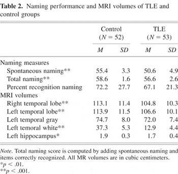
As expected, the TLE group was impaired on confrontation naming ability compared with the control group even when education level was entered as a covariate [F(1,104) = 28.25, p = .001, η2 = .21]. Performance on BNT was not significantly correlated with age of epilepsy onset, r = .23 (p > .05) or duration of epilepsy r = −.21 (p > .05). Overall, the TLE group did not spontaneously name 16% of the items from the BNT compared to 8% for the controls. When provided a recognition name probe for these missed items, the TLE group correctly identified the correct name for 67% of the items not spontaneously named compared to 72% for controls, [Table 2; F(1,104) = 1.33, p > .05].
Naming performance and MRI volumes of TLE and control groups %*p < .01. %**p < .001.

Spontaneous Naming and MR Temporal Lobe Volumes
A hierarchical multiple regression analysis (gender, height, left temporal, right temporal) yielded a significant overall relationship with BNT performance [F(4,52) = 2.51, p = .05], and a multiple correlation value of .42. Thus, these variables together accounted for 17% of the variance in BNT performance. However, only the left temporal volume was a significant predictor of BNT performance (B = .55, t = 2.09, p = .04). Overall, left temporal volume accounted for 7.5% of unique variance in BNT performance compared with less than 1% unique variance attributed to the right temporal lobe. Together, both left and right temporal lobes accounted for nearly 9% of shared variance in BNT performance. Table 3 provides the data contrasting the amount of unique variance on BNT performance that is associated with these MR volumes. Figure 1a provides the scatter-plot of the relationship between left temporal lobe volume and spontaneous naming performance.
Left and right temporal lobe contribution to Boston Naming performance


(a) Relationship between total left temporal lobe volume and spontaneous naming. (b) Relationship between left temporal lobe white matter volume and spontaneous naming. (c) Relationship between left temporal lobe white matter volume and recognition naming.
Spontaneous Naming and Segmented Left Temporal Lobe Volumes
A hierarchical multiple regression analyses (gender, height, left temporal white, left temporal gray, and left hippocampus) examined the relationship between segmented left temporal lobe volume and the left hippocampus volume with BNT performance. There was a significant overall relationship with BNT performance [F(5,52) = 4.48, p = .002], with a multiple correlation index of .57. Thus, this set of variables accounted for 32% of the variance in BNT performance. As shown in Table 4, both left temporal white volume and left hippocampus volume were significant unique predictors of BNT performance. Left temporal white matter volume accounted for 7.8% of unique variance in BNT performance (B = .33, t = 2.32, p = .02), and left hippocampus volume (B = .35, t = 2.75, p = .01), accounted for approximately 11% of unique variance in predicting BNT performance (see Figure 1b).
Left temporal lobe and left hippocampus contribution to Boston Naming performance %**p < .01.

Naming Recognition and Left Temporal Lobe MR Volumes
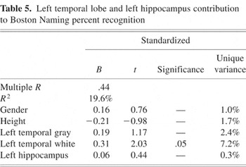
A hierarchical multiple regression (gender, height, left temporal white, left temporal gray (minus hippocampus) and left hippocampus) with BNT recognition naming performance as the dependent variable yielded a multiple correlation index of .44 and accounted for 19.6% of the variance [F(5,51) = 2.24, p = .06]. As shown in Table 5 and figure 1c, left temporal white matter volume accounted for 7.2% of the unique variance (B = .31, t = 2.03, p = .04) while hippocampus volume accounted for only 0.3% of unique variance (B = .06, t = .40, p > .05).
Left temporal lobe and left hippocampus contribution to Boston Naming percent recognition

DISCUSSION
The current study employed quantitative MR volumetrics to investigate the relative contribution of left and right hemisphere temporal lobe tissue volume and hippocampus volume for predicting confrontation naming ability among patients with TLE. Consistent with the clinical lesion literature, we found that left hemisphere temporal lobe volume was a significant predictor of BNT spontaneous naming performance while right hemisphere temporal lobe volume was not. Decomposition of the left hemisphere temporal lobe volume contribution to BNT performance indicated that left temporal temporal lobe (particularly white matter volume) and left hippocampus volume showed a similar degree of association (unique predictive variance) for spontaneous confrontation naming performance. In addition, left temporal lobe white matter volume showed a stronger association with BNT recognition naming performance than left hippocampus volume. Thus, the hippocampus contribution to naming performance may be linked to the demands associated with lexical retrieval demands of visual confrontation naming performance. These findings are discussed in more detail below.
The association of left temporal lobe tissue volume for naming performance is not surprising and is consistent with classic language models (Goodglass & Wingfield, 1997). The findings also serve to highlight the importance of left temporal lobe white matter volume in both spontaneous naming and recognition naming performance. This is consistent with the viewpoint of naming ability as a multistage cognitive task that relies on a distributed neural network for its successful operation. White matter integrity is critical for the communication and integrated activity of discrete regions within a functional neural network, and these data confirm that disruption in this connectivity can have important implications for confrontation naming performance. These findings are consistent with the results from a recent study (Grossman et al., 2004) using MR voxel based morphology to study grey matter cortical atrophy and naming performance in three clinical groups (Alzheimer's dementia, frontotemporal dementia, and corticobasal degeneration). A distributed network of regions was identified to be associated with naming performance, and atrophy of the left lateral cortex was the strongest correlate of naming performance across all three groups.
Consistent with our previous reports, the TLE group had significantly reduced regional volumes compared to controls (Hermann et al., 2003). It is worth noting that the current findings do not appear to be related to extent of volume loss in particular regions. For example, both left hippocampus (11.2%) and right hippocampus volume (14.3%) show a similar degree of volume loss in the TLE group compared to the controls, but the left hippocampus predicted substantially greater variance in the prediction of naming performance (see Table 3). Similarly, left temporal lobe white matter and left hippocampus showed a similar degree of volume loss (12.5% and 11.2%, respectively), and showed different patterns of association with naming performance (spontaneous compared to recognition).
Examination of potential gender influences and laterality of seizure focus also did not alter the current findings. There are no significant differences in naming ability for males and females, and all reported correlations remained significant when gender was entered into the regression equations as a predictor variable. Similarly, while there was trend for a significant difference in naming ability between the subset of left and right TLE groups (t = 1.83, p = .08), there were similar relationships between volumes and naming performance irrespective of seizure focus.
In the current study, the entire extrahippocampal temporal lobe was included in the temporal lobe ROI, segmented into gray and white matter volume, but no further regional parcellation was undertaken. Thus, the current findings cannot determine the relative contribution of specific subregions within the extrahippocampal temporal lobe to BNT performance. Similarly, the region examined here does not overlap with the temporal lobe region of resection in the standard surgical ATL, but rather encompasses a broader area. Thus, the findings here also cannot directly address findings observed following ATL. In addition, the current study examined visual confrontation naming performance and it is quite likely that a different neural architecture would emerge for other naming tasks with different response characteristics such as generative naming (Hamberger et al., 2001).
With these caveats and limitations in mind, several points are worth noting. A recent proposal (Vargha-Khadem et al., 1997) has suggested that the parahippocampal region may play a critical role in the acquisition and retrieval of semantic knowledge and as such can serve to mediate basic language-based functions (such as naming). Similarly, the left temporal pole region, which is also included in the MR ROI measurement of the temporal lobe region, has been identified as a critical component for naming performance of both unique identities (places and people), as well as particular categories of objects (Damasio et al., 1996; Grabowski et al., 2001). Recent volumetric studies have confirmed the presence of significant atrophy in the temporal polar region in TLE (Coste et al., 2002; Moran et al., 2001), and the temporal pole is also typically included within the zone of resection in the standard ATL. It is possible, that stronger associations with naming ability would emerge if specific temporal lobe regions of interest were examined and measured. Nevertheless, the findings observed here clearly implicate a significant contribution of temporal lobe white matter in the neural network underlying confrontation naming.
The positive association between MR left hippocampal volume and naming ability is not consistent with the perspective which specifically delimits its role to the acquisition or formation of new memories (Kensinger et al., 2001; Schmolck et al., 2002). Several possible interpretations can be posed to account for the current findings indicating a significant (and unique) predictive role of hippocampal volume integrity for naming ability. First, the disruption in naming impairment may not reflect the effects of direct hippocampal volume loss but instead reflect the impact of (partial) disconnection (degradation) of the hippocampus from the temporal lobe neocortex and the object identity system. It is also possible that hippocampal volume loss is a marker (co-related) of volume loss in discrete regions of the temporal lobe (or other brain regions) that play critical roles in naming performance. We do note that there is a weak relationship between hippocampal volume and (whole) temporal lobe gray matter volume (r = −.05, p > .10) and a small association between hippocampus volume and temporal lobe white matter volume (r = .31, p < .05). Nevertheless, examination of more discrete regions would be helpful in this regard.
Alternatively, one can consider the possibility that the findings do indeed reflect the participation of hippocampus to naming ability. There is a convergence of both clinical and neuroimaging data which is consistent with this possibility. As reviewed earlier, several studies have shown that TLE patients without significant hippocampal pathology have relatively intact naming performance before surgery and show a substantial decline following ATL compared to those with pre-existing hippocampal pathology (Hermann et al., 1997; Seidenberg et al., 1998). Sawrie et al. (2000) found that MR spectroscopy of left hippocampus showed a significant relationship with Boston Naming Test performance, similar in magnitude to that observed for episodic memory tasks. Two published conference abstracts (Shear et al., 1997; Snyder et al., 1994) using MR volumetrics report findings similar to that reported here; a significant relationship between MRI measured hippocampal volume and confrontation naming in TLE patients.
Because these subjects have chronic epilepsy which often begins early in life, it is possible that the current findings are affected by language reorganization. We think this is a less plausible explanation for the results reported here. First, we only included TLE subjects who showed a strictly right manual motor preference for writing. In addition, the results do not change when age of recurrent epilepsy onset is entered (first) in a hierarchical regression analysis. Finally, similar results concerning hippocampus association with BNT performance was noted in a study of AD patients (Wilson et al., 1996). Specifically, hippocampus volume was significantly associated with both BNT naming ability (r = .33) and delayed story recall (r = .36).
If these findings do indeed implicate a role of the hippocampus in confrontation naming ability, it is germane to consider the nature of this involvement or participation. These findings indicate that the hippocampus participates to a substantially greater degree for spontaneous naming performance than for recognition naming performance. Thus, hippocampal contribution may be more substantial when lexical retrieval demands are present (spontaneous naming). In contrast, when lexical retrieval demands are minimized (name recognition), hippocampal participation is less evident while left lateral temporal lobe continues to make a contribution.
Another issue to consider in relation to the possible participation of hippocampus to naming performance relates to stimulus dimensions. Several studies have implicated age of acquisition as a critical determinant of naming performance. Age of acquisition predicts naming accuracy and naming speed in elderly normals, patients with aphasia, AD, and semantic dementia (Hirsch & Funnell, 1995; Lambon Ralph et al., 1998). In general, items that are acquired at a later age show less accuracy and require more time to name than items typically acquired at an earlier chronological age. In TLE, object names acquired in late childhood are more vulnerable to loss following ATL than items acquired early in childhood (Bell et al., 2000, 2002). Perhaps the hippocampus contribution to spontaneous naming ability varies in relation to age of acquisition with later acquired words showing greater dependency or participation of the hippocampus compared to early acquired words and concepts. Consistent with this possibility, a recent fMRI study found that early and late learned words produced different patterns of neural activation (Fiebach et al., 2003). Further investigation of this issue with structural and functional neuroimaging approaches would be of interest.
In summary, the current findings illustrate the potential utility of quantitative volumetrics in dissecting the operation of the neural network underlying confrontation naming ability. Findings point to a significant contribution by both left temporal lobe white matter and left hippocampus. Additional research utilizing both clinical lesion findings and neuroimaging are needed to further investigate the clinical and theoretical implications of these results.
ACKNOWLEDGMENTS
This work was supported by NIH RO1-37738 and MOI RR03186 to Bruce Hermann and Michael Seidenberg.
References
REFERENCES
Andreasen, N.,
Rajarethinam, R.,
Cizadlo, T.S.A.,
Swayze, V.W.,
Flashman, L.A.,
O'Leary, D.S.,
Ehrhardt, J.C., &
Yuh, W.T.
(1996).
Automated atlas-based volume estimation of human brain regions from
MR images.
Journal of Computer Assisted Tomography,
20,
98–106.Google Scholar
Bell, B.,
Davies, K.,
Hermann, B., &
Walters, G.
(2000).
Confrontation naming after anterior temporal lobectomy is related to
age of acquisition of the object names.
Neuropsychologia,
38,
83–92.Google Scholar
Bell, B.,
Hermann, B.,
Seidenberg, M.,
Davies, K.,
Cariski, D.,
Rosenbek, J.,
Woodard, A.,
Rutecki, P., &
Bishop, M.
(2002).
Ipsilateral reorganization of language in early onset left temporal
lobe epilepsy.
Epilepsy & Behavior,
3,
158–164.Google Scholar
Bernasconi, N.,
Bernasconi, A.,
Caramanos, Z.,
Antel, S.B.,
Andermann, F., &
Arnold, D.
(2003).
Mesial temporal damage in temporal lobe epilepsy: A volumetric MRI
study of the hippocampus, amygdala, and parahippocampal region.
Brain,
126,
462–469.Google Scholar
Coste, S.,
Ryvlin, P.,
Hermier, M.,
Ostrowsky, K.,
Adeleine, P.,
Froment, J.C., &
Mauguiere, F.
(2002).
Temporopolar changes in temporal lobe epilepsy.
Neurology,
59,
855–861.Google Scholar
Damasio, H.,
Grabowski, T.J.,
Tranel, D.,
Hichwa, R.D., &
Damasio, A.R.
(1996).
A neural basis for lexical retrieval.
Nature,
380,
499–505.Google Scholar
Davies, K.,
Bell, B.,
Bush, A.,
Hermann, B.,
Dohan, F.C., &
Jaap, A.
(1998).
Naming decline after left anterior temporal lobectomy correlates
with pathological status of resected hippocampus.
Epilepsia,
39,
407–419.Google Scholar
Fama, R.,
Sullivan, E.V.,
Shear, P.K.,
Cahn-Weiner, D.A.,
Marsh, L.,
Lim, K.O.,
Yesavage, J.A., &
Tinklenberg, J.R.
(2000).
Structural brain correlates of verbal and nonverbal fluency measures
in Alzheimer's disease.
Neuropsychology,
14,
29–40.Google Scholar
Fiebach, C.,
Friederici, A.,
Muller, K.,
Yves von Cramon, D., &
Hernandez, A.
(2003).
Distinct brain representations for early and late learned
words.
Neuroimage,
19,
1627–1637.Google Scholar
Filley, C.
(2001).
The behavioral neurology of white matter.
Oxford, UK:
Oxford University Press.
Goodglass, H. &
Wingfield, A.
(1997).
Anomia: Neuroanatomical and cognitive correlates.
San Diego, CA:
Academic Press.
Grabowski, T.J.,
Damasio, H.,
Tranel, D.,
Ponto, L.L.,
Hichwa, R.D., &
Damasio, A.R.
(2001).
A role for left temporal pole in the retrieval of words for unique
entities.
Human Brain Mapping,
13,
199–212.Google Scholar
Grossman, M.,
McMillian, C.,
Moore, P.,
Ding, L.,
Glosser, G.,
Work, M., &
Gee, J.
(2004).
What's in a name: Voxel-based morphometric analyses of MRI and
naming difficulty in Alzheimer's disease, frontotemporal dementia and
corticobasal degeneration.
Brain,
127,
628–649.Google Scholar
Hamberger, M.,
Goodman, R.,
Perrine, K., &
Tammy, T.
(2001).
Anatomic dissociation of auditory and visual naming in the lateral
temporal cortex.
Neurology,
56,
56–61.Google Scholar
Harris, G.,
Andreasen, N., &
Cizadlo, T.
(1999).
Improving tissue classification in MRI: A three-dimensional
multispectral discriminant analysis method with automated training class
selection.
Journal of Computer Assisted Tomography,
23,
144–154.Google Scholar
Hermann, B.,
Chelune, G.,
Loring, D.,
Trenerry, M.R.,
Perrine, K.,
Barr, W.B.,
Strauss, E., &
Westerveld, M.
(1999).
Visual confrontation naming following left anterior temporal
lobectomy: A comparison of surgical approaches.
Neuropsychology,
13,
3–9.Google Scholar
Hermann, B.,
Seidenberg, M.,
Bell, B.,
Rutecki, P.,
Sheth, R.,
Wendt, G.,
O'Leary, D., &
Magnotta, V.
(2003).
Extratemporal quantitative MR volumetrics and neuropsychological
status in temporal lobe epilepsy.
Journal of the International Neuropsychological Society,
9,
353–362.Google Scholar
Hermann, B.,
Seidenberg, M.,
Schoenfeld, J., &
Davies, K.
(1997).
Neuropsychological characteristics of the syndrome of mesial
temporal lobe epilepsy.
Archives of Neurology,
54,
369–376.Google Scholar
Hermann, B. &
Wyler, A.
(1988).
Effects of anterior temporal lobectomy on language function: A
controlled study.
Annals of Neurology,
23,
585–588.Google Scholar
Hirsch, K. &
Funnell, E.
(1995).
Those old, familiar things: Age of acquisition, familiarity, and
lexical access in progressive aphasia.
Journal of Neurolinguistics,
9,
23–32.Google Scholar
Kaplan, E.,
Goodglass, H., &
Weintraub, S.
(1983).
Boston Naming Test
(2nd ed.).
Philadelphia:
Lea and Febiger.
Kensinger, E.,
Ullman, M., &
Corkin, S.
(2001).
Bilateral medial temporal lobe damage does not affect lexical or
grammatical processing: Evidence from amnesic patient H.M.
Hippocampus,
11,
347–360.Google Scholar
Lambon Ralph, M.,
Graham, K.S.,
Ellis, A.W., &
Hodges, J.R.
(1998).
Naming in semantic dementia-what matters?
Neuropsychologia,
36,
775–784.Google Scholar
Langfitt, J. &
Rausch, R.
(1996).
Word-finding deficits persist after left anterotemporal
lobectomy.
Archives of Neurology,
53,
72–76.Google Scholar
MacKay, D.,
Stewart, R., &
Burke, D.
(1998).
H.M. revisited: Relations between language comprehension, memory,
and the hippocampal system.
Journal of Cognitive Neuroscience,
10,
377–394.Google Scholar
Magnotta, V.,
Harris, G.,
Andreasen, N.,
O'Leary, D.,
Yuh, W.T., &
Heckel, D.
(2002).
Structural MR image processing using the BRAINS2 toolbox.
Computerized Medical Imaging Graphics,
26,
251–264.Google Scholar
Marsh, L.,
Morrell, M., &
Shear, P.
(1997).
Cortical and hippocampal volume deficits in temporal lobe
epilepsy.
Epilepsia,
38,
576–587.Google Scholar
Moran, N.,
Lemieux, L.,
Kitchen, N.D.,
Fish, D.R., &
Shorvon, S.D.
(2001).
Extrahippocampal temporal lobe atrophy in temporal lobe epilepsy and
mesial temporal sclerosis.
Brain,
124,
167–175.Google Scholar
Ojemann, G.A.,
Ojemann, J.,
Lettich, E., &
Berger, M.
(1989).
Cortical language localization in left-dominant hemisphere.
Journal of Neurosurgery,
71,
316–326.Google Scholar
Pantel, J.,
O'Leary, D.,
Crestinger, K.,
Bockholt, H.J.,
Keefe, H.,
Magnotta, V., &
Andreasen, N.
(2000).
A new method for the in vivo volumetric measurement of the human
hippocampus with high neuroanatomical accuracy.
Hippocampus,
10,
752–758.Google Scholar
Sass, K.J.,
Sass, A.,
Weserveld, M.,
Lecncz, T.,
Novelly, R.A.,
Kim, J.H., &
Spencer, D.D.
(1992).
Specificity in the correlation of verbal memory and hippocampal
neuron loss: Dissociation of memory, language, and verbal intellectual
ability.
Journal of Clinical and Experimental Neuropsychology,
14,
662–672.Google Scholar
Sawrie, S.,
Martin, R.,
Gilliam, F.,
Faught, E.,
Maton, B.,
Hugg, J.W.,
Bush, N.,
Sinclair, K., &
Kuzniecky, R.
(2000).
Visual confrontation naming and hippocampal function: A neural
network study using quantitative H magnetic resonance spectroscopy.
Brain,
123,
770–780.Google Scholar
Saykin, A.J.,
Stafiniak, P.,
Robinson, L.,
Flannery, K.,
Gur, R.,
O'Connor, M., &
Sperling, M.
(1995).
Language before and after temporal lobectomy: Specificity of acute
changes and relation to early risk factors.
Epilepsia,
36,
1071–1077.Google Scholar
Schmolck, H.,
Kensinger, E.A.,
Corkin, S., &
Squire, L.R.
(2002).
Semantic knowledge in patient H.M. and other patients with bilateral
medial and lateral temporal lobe lesions.
Hippocampus,
12,
520–533.Google Scholar
Seidenberg, M.,
Hermann, B.,
Wyler, A.,
Davies, K.,
Curtis Dohan, F., &
Leveroni, C.L.
(1998).
Neuropsychological outcome following anterior temporal lobectomy in
patients with and without the syndrome of mesial temporal lobe
epilepsy.
Neuropsychology,
12,
303–316.Google Scholar
Shear, P.,
Sullivan, E.V.,
Marsh, L.,
Morrell, M.J.,
Lim, K.O., &
Pfefferbaum, A.
(1997).
Hippocampal volumes correlate with nonmnemonic abilities in temporal
lobe epilepsy.
Journal of the International Neurological Society,
3,
36.Google Scholar
Snyder, P.,
Barr, W.B.,
Schaul, N., &
Lieberman, J.
(1994).
Quantitative MRI correlates of naming disorders in temporal lobe
epilepsy.
Epilepsia,
35(Suppl. 8),
102.Google Scholar
Squire, L.R. &
Alvarez, P.
(1995).
Retrograde amnesia and memory consolidation: A neurobiological
perspective.
Current Opinion in Neurobiology,
5,
169–177.Google Scholar
Vargha-Khadem, F.,
Gadiaun, D.G.,
Watkins, K.E.,
Connelly, A.,
Van Paesschen, W., &
Mishkin, M.
(1997).
Differential effects of early hippocampal pathology on episodic and
semantic memory.
Science,
277,
376–380.Google Scholar
Wilson, R.,
Sullivan, M.,
DeToledo-Morrell, L.,
Stebbins, G.,
Bennett, D., &
Morrell, F.
(1996).
Association of memory and cognition in Alzheimer's Disease with
volumetric estimates of temporal lobe structures.
Neuropsychology,
10,
459–463.Google Scholar

Demographics of TLE and control groups
Naming performance and MRI volumes of TLE and control groups %*p < .01. %**p < .001.

Left and right temporal lobe contribution to Boston Naming performance

(a) Relationship between total left temporal lobe volume and spontaneous naming. (b) Relationship between left temporal lobe white matter volume and spontaneous naming. (c) Relationship between left temporal lobe white matter volume and recognition naming.

Left temporal lobe and left hippocampus contribution to Boston Naming performance %**p < .01.
Left temporal lobe and left hippocampus contribution to Boston Naming percent recognition
 You have
Access
- 41
- Cited by