




No CrossRef data available.
Article contents
P19: a female and tissue specifically expressed gene in Schistosoma mansoni, regulated by pairing with the male
Published online by Cambridge University Press: 05 December 2003
Abstract
A female-specific sequence was isolated from a cDNA library of Schistosoma mansoni and further characterizedSequences reported here are available from DDBJ/EMBL/GenBank under the Accession number Z27402.. Expression of the corresponding gene (p19) depends on pairing with a male. In situ hybridization and immunohistology experiments revealed exclusive expression of the gene in the cells of the vitellarium, suggesting a function in egg formation. In addition, experimental evidence for cross-linking of the protein under oxidative conditions supports the assumption that the p19 gene may code for an egg-shell precursor protein.
Keywords
Information
- Type
- Research Article
- Information
- Copyright
- 2003 Cambridge University Press
INTRODUCTION
Egg formation in trematodes has been considered to be a major target for chemotherapeutic or immunological approaches to control schistosomiasis, not only because egg production is essential to maintain the life-cycle of the parasite, but also because in schistosomes, eggs are the causative agent for pathogenesis (Capron et al. 2002). The eggs of schistosomes are composed of cells produced by 2 different organs, the vitellarium and the ovary. About 40 vitelline cells and 1 oocyte are combined to a single ovum within the ootype. The vitelline cells synthesize most of the molecules used for egg-shell formation and for nutrition of the embryo (Kunz, 2001). The contribution of the oocyte itself for embryonic development appears to consist mainly in providing the genetic material, some stored mRNA and ribosomes.
Schistosomes are unusual in that the vitelline cell differentiation in females is dependent on pairing with the male worms. Gene expression in the vitellaria of females is dependent on signals from the male sexual partner (Grevelding, Sommer & Kunz, 1997). If the female is separated from the male, gene expression is stopped and egg production ceases. If the female remates with a male, transcription in the vitellaria starts again, and egg production continues. Male-regulated gene expression in schistosomes has been shown for egg-shell proteins, ferritin and mucin (Grevelding et al. 1997). Here, we characterize another gene (p19) that is specifically expressed in the vitellaria of females, and that is regulated by the male.
MATERIALS AND METHODS
Parasites and nucleic acid extraction
Adult worms of the Liberia strain of Schistosoma mansoni were reared in Syrian hamsters and obtained by perfusion of the hepatic portal system (Duvall & DeWitt, 1967). Male and female worms were separated with fine brushes and stored in liquid nitrogen. DNA from female and male worms was obtained by a proteinase K-based extraction method (Grevelding, 1995). Total RNA was isolated using commercially available kits following the instructions of the manufacturer (RNeasy Kit, Qiagen). Poly(A)+ RNA was isolated with 200 ml Dynabeads Oligo(dT)25 solution according to the manufacturer's protocol (Dynal).
In vitro culture
Freshly perfused worm pairs were separated, washed 3 times in culture medium and incubated at 37 °C with 5% CO2 in Petri dishes (Grevelding et al. 1997). After increasing numbers of days, samples of 10 females were used for RNA extraction.
Synthesis of cDNA libraries and differential hybridization
Samples of 5 μg of poly(A)+ RNA from male and female worms were used for cDNA synthesis following the instructions for the ZAP synthesis kit (Stratagene). A subtractive female-specific cDNA library was constructed as described (Menrath, Michel & Kunz, 1995). In total, 600 colonies of this library were replica plated onto duplicate filters. Colony hybridization with radio-isotope labelled cDNA of male and female RNA was done according to Sambrook, Fritsch & Maniatis (1989).
Labelling and blotting of nucleic acids
Radioactive in vitro transcripts were obtained with T7 RNA polymerase using (alpha-32P)dUTP. digoxigenin-labelled in vitro transcripts were synthesized following the manufacturer's protocol (Roche). Northern blotting was carried out as described earlier (Knobloch et al. 2002).
Sequence analysis
Nucleotide sequencing was performed with an ABI 373 DNA sequencer from Applied Biosystems, using the dye terminator cycle sequencing kit with AmpliTaq DNA Polymerase FS (Perkin Elmer). Amino acid analyses were done using the programs FastA, Blast and WinPep.
Expression of fusion protein, purification of antibodies and immunoblotting
The complete insert of clone 3H11 was recloned into the BamHI/SalI sites of the pUR 288 vector. Expression of recombinant beta-galactosidase was carried out as previously described (Köster et al. 1988). Correct cloning was confirmed by Western blotting. Antibodies were prepared from rabbit antisera against female schistosomes essentially according to Whitfield et al. (1988), but immunopurification was carried out on a bacterial lawn, not on single colonies. Preparation of worm extracts and immunoblotting were done as described (Wippersteg et al. 2002).
In situ hybridization and immunohistology
Adult parasites were fixed in Bouin's solution (picric acid/acetic acid/formaldehyde 15[ratio ]1[ratio ]5) and embedded in Paraplast. Sections were treated as described (Köster et al.1988). Dig-transcripts were hybridized and detected as described (Finken et al. 1994). Anti-p19 antibodies (1[ratio ]100) were detected with alkaline phosphatase-conjugated goat anti-rabbit IgG (Dianova) with naphthol-AS-MX-phosphate and Fast Red TR (Sigma) as substrate.
RESULTS
Isolation and characterization of clone 3H11
In order to isolate clones coding for female-specific sequences, a subset of 600 colonies of a subtractive cDNA library was screened with male and female cDNA. Eight clones not reactive with male cDNA were isolated, and the plasmid DNA reprobed with each male or female cDNA. Only 1 of the clones was found to hybridize exclusively with female cDNA. This clone, containing a 839 bp insert, was named 3H11 (accession number Z27402).
There was 1 long open reading frame starting with a methionine at position 59 of the cDNA insert. The deduced translation product of 199 amino acids (Fig. 1) had a calculated molecular mass of 22 kDa. Since the molecular mass on Western blots was found to be 19 kDa (see below), the protein was called p19. Frequent amino acids were Asn (13·3%), Tyr (11·6%) and Gly (9·6%). The base composition of the coding sequence was determined to be 73% A/T. At the 3′ end of the sequence, a poly(A) tail and a polyadenylation signal with the canonical consensus sequence of AATAAA were found.

Fig. 1. cDNA sequence of 3H11 and its open reading frame. The putative start and stop codon as well as the signal sequence are shown in bold.
Expression and regulation of p19 mRNA
We analysed the hybridization of in vitro generated antisense transcripts of 3H11 with total RNA prepared from male as well as female parasites (Fig. 2A). On Northern blots, a hybridization signal at the position of 850 nucleotides was detected only in the lane containing RNA from female worms demonstrating that the p19 gene is expressed exclusively in female schistosomes.

Fig. 2. (A) Northern blot analysis of 3H11. Total RNA from adult female and male schistosomes was separated on a denatured ethidium bromide agarose gel (left), blotted and hybridized with antisense transcripts of clone 3H11 (right). Molecular weight marker in nucleotides (M) is 1 kb DNA ladder (BRL). (B) Expression of p19 in females cultivated in vitro that were separated from males for 1, 2, 3, and 6 days, in comparison with freshly perfused females (0) and those that remained paired for 6 days (6p). Poly (A)+ RNA was probed with antisense transcripts of 3H11 (top) and, as a control, with transcripts of the schistosome protein disulfide isomerase (bottom).
We studied the expression of the p19 gene in female worms that were cultured in vitro. Females separated from males were compared with paired females. RNA from worms of increasing separation times was probed with transcripts of 3H11 and, as an internal control, with transcripts of the schistosome protein disulfide isomerase (PDI), an ER-luminal enzyme responsible for protein folding (Finken et al. 1994). The signal on the Northern blot revealed that the expression of p19 decreased tremendously within the first 2 days after separation and totally disappeared after 3 days, whereas in paired females after 6 days of in vitro culture a signal was still detectable (Fig. 2B). In contrast, PDI mRNA in separated females showed no decrease in expression during culture. This demonstrates that, in comparison with PDI, the expression of p19 in females is dependent on pairing with a male.
Cytological localization and Western blotting
In situ hybridizations were carried out with antisense transcripts of clone 3H11. Only the cells of the vitellaria were labelled (Fig. 3A). The label was clearly restricted to the cytoplasm of the cells. No hybridization signal was visible in the male nor in other tissues of the female, clearly documenting that the vitellaria are the only site of p19 expression. Sense transcripts as a negative control did not show any reaction (data not shown).

Fig. 3. (A) Localization of p19 mRNA in a cross-section of a schistosome female (F) showing greyish blue staining of the cytoplasm of the vitelline cells (V) with BCIP/NBT. The surrounding parenchyma and the tegument (T) are not labelled. The dark staining material in the gut (G) is digested haemoglobin. At the bottom left of the figure, part of a male (M) is shown. (B) Immunohistological localization of the p19 protein, stained with naphthol-AS-MX-phosphate and Fast Red TR in a tissue section documenting selective labelling of the cytoplasm of the vitelline cells (V) in the female. The surrounding parenchyma (P), the gastrodermis (Ga) and the tegument (T) are not labelled. At the upper left side of the figure, part of a male (M) is shown.
To obtain specific anti-p19 antibodies, antisera against protein homogenates of female schistosomes were affinity purified on the 3H11-beta-galactosidase fusion protein and, for further purification, post-adsorbed on beta-galactosidase. The size of the p19 protein predicted from the 3H11 sequence was expected to be 22·5 kDa. On Western blots of male and female proteins, the antibodies detected no proteins in this size range (Fig. 4). Since egg-shell precursor proteins are known to cross-link under oxidative conditions, we added the antioxidant n-propyl gallate (10 mM) during protein extraction (Köster et al. 1988). Under these conditions, the antibodies detected a prominent band of 19 kDa that was present exclusively in females. Some minor bands in the upper range were detected by the antibody in both the male and the female lane, and thus resulted from unspecific cross-reaction. Pre-immune sera, as a negative control, did not show any reaction (data not shown). This result confirmed that the p19 protein predicted from the cDNA is expressed in schistosomes. Computer-assisted analysis indicated that the amino terminus had a hydrophobic region of 18 amino acids that could function as a signal sequence for ER entry. Reduced by this signal sequence of 2 kDa, the calculated value falls into the size range seen on the Western blot.

Fig. 4. Immunoblot of male and female proteins, extracted with (+) and without antioxidant (−), probed with the anti-p19 antibody showing a single band of 19 kDa only in females. Left: amidoblack-stained total protein extracts to document that the female lane is not overloaded (A). High molecular weight marker in kDa (M) (Sigma).
The antibodies were used to localize the p19 protein in tissue sections. The label was clearly restricted to the cytoplasm of the vitelline cells only. No signal was visible in the nuclei (Fig. 3B). No other cells were labelled nor was there any reaction in the male. Again, pre-immune sera as a negative control did not show any reaction (data not shown).
DISCUSSION
Several different genes are known in S. mansoni that are exclusively or preferentially expressed in the vitelline cells of mature females. Some with high probability code for egg-shell precursor proteins (Chen, Rekosh & LoVerde, 1992). Others code for the yolk protein ferritin (Schüssler et al. 1995), for phospholipid hydroperoxide glutathione peroxidase (Roche et al. 1996), or are of unknown function (fs800) (Reis et al. 1989). For the egg-shell precursor genes and for the ferritin gene it has been shown that their expression in the female depends on pairing with a male (Grevelding et al. 1997), whereas for the 2 other genes the situation has not yet been investigated. In this paper, we introduce a new gene (p19) whose activity is restricted exclusively to the vitellarium, and, in addition, it is expressed only in females that are paired with a male.
Since during egg formation in the ootype, the content of the vitelline cells completely merges with the zygote's cytoplasm to form the early embryo, one can assume that most gene products that are synthesized in the vitelline cells, have a function in egg or embryonic differentiation or nutrition.
On the basis of sequence alignments, no convincing information about the function could be obtained. Comparison of the deduced protein sequence with protein data banks showed weak similarity to the egg-shell precursor protein B1 of Fasciola hepatica (Zurita et al. 1987). Permutation analysis (Altschul & Erickson, 1985), however, revealed that there is no similarity in amino acid sequence, but instead a similarity in amino acid composition. This results from the high glycine and tyrosine content that the p19 protein and the B1 protein have in common. Therefore, the conclusion of a common evolutionary origin must not necessarily be justified.
A strong bias towards high glycine/tyrosine content and the frequent occurrence of the Tyr-Gly sequence motif are general characteristics of trematode egg-shell precursor proteins (Rice-Ficht et al. 1992; Bobek, Rekosh & LoVerde, 1991). This suggests that the p19 gene may code for an egg-shell precursor. This assumption is supported by the observation of cross-linking of the monomeric protein under oxidative conditions. Several of the other female-specific genes expressed in vitelline cells, as p14, p48 (Chen et al. 1992), and fs800 (Reis et al. 1989), all show multiple ORFs; this, however, is not the case in the p19 sequence.
The assumption that the p19 is an egg-shell precursor protein needs further substantiation. The most obvious and indispensable set of experiments here would be to analyse monomeric egg proteins isolated from schistosome egg-shells to compare the native protein sequence with the nucleotide sequence of the clone.
Note added after completion
As this work was nearing completion, a sequence of Schistosoma japonicum has been submitted to DDBJ/EMBL/GenBank under the Accession number AAP05897.1 that has 55·9% homology to p19 along its entire amino acid sequence (AliBee-Multiple Alignment, http://www.genebee.msu.su).
The authors thank Dr Ingo Ebersberger for sequence analyses, Dr Christoph Grevelding for advice, Stephan Sroka and Didina David for technical assistance, and the Deutsche Forschungsgemeinschaft (Grant Ku 282/15-1) for financial support. J. K. is grateful to the Düsseldorf Entrepreneurs Foundation for a scholarship.
References
REFERENCES
ALTSCHUL, S. F. &
ERICKSON, B. W.
(
1985).
Significance of nucleotide sequence alignments: a method for random sequence permutation that preserves dinucleotide and codon usage.
Molecular Biology and Evolution 2, 526–538.Google Scholar
BOBEK, L. A.,
REKOSH, D. M. &
LOVERDE, P. T.
(
1991).
Schistosoma japonicum: Analysis of eggshell protein genes, their expression, and comparison with similar genes from other schistosomes.
Experimental Parasitology 72,
381–390.CrossRefGoogle Scholar
CAPRON, A.,
RIVEAU, G. J.,
BARTLEY, P. B. &
McMANUS, D. P.
(
2002).
Prospects for a schistosome vaccine.
Current Drug Targets – Immune Endocrine and Metabolic Disorders 2,
281–290.CrossRefGoogle Scholar
CHEN, L.,
REKOSH, D. M. &
LOVERDE, P. T.
(
1992).
Schistosoma mansoni p48 eggshell protein gene: characterization, developmentally regulated expression and comparison to the p14 eggshell protein gene.
Molecular and Biochemical Parasitology 52,
39–52.CrossRefGoogle Scholar
DUVALL, R. H. &
DEWITT, W. D.
(
1967).
An improved perfusion technique for recovering adult schistosomes from laboratory animals.
American Journal of Tropical Medicine and Hygiene 16,
483–486.CrossRefGoogle Scholar
FINKEN, M.,
SOBEK, A.,
SYMMONS, P. &
KUNZ, W.
(1994).
Characterization of the complete protein disulfide isomerase gene of Schistosoma mansoni and identification of the tissues of its expression.
Molecular and Biochemical Parasitology 64,
135–144.CrossRefGoogle Scholar
GREVELDING, C. G.
(
1995).
The female-specific W1 sequence of the Puerto Rican strain of Schistosoma mansoni occurs in both genders of a Liberian strain.
Molecular and Biochemical Parasitology 71,
269–272.CrossRefGoogle Scholar
GREVELDING, C. G.,
SOMMER, G. &
KUNZ, W.
(
1997).
Female-specific gene expression in Schistosoma mansoni is regulated by pairing.
Parasitology 115,
635–640.CrossRefGoogle Scholar
KNOBLOCH, J.,
WINNEN, R.,
QUACK, M.,
KUNZ, W. &
GREVELDING, C. G.
(
2002).
A novel Syk-family tyrosine kinase from Schistosoma mansoni which is preferentially transcribed in reproductive organs.
Gene 294,
87–97.CrossRefGoogle Scholar
KÖSTER, B.,
DARGATZ, H.,
SCHRÖDER, J.,
HIRZMANN, J.,
HAARMANN, C.,
SYMMONS, P. &
KUNZ, W.
(
1988).
Identification and localisation of the products of a putative eggshell precursor gene in the vitellarium of Schistosoma mansoni.
Molecular and Biochemical Parasitology 31,
183–198.CrossRefGoogle Scholar
KUNZ, W.
(
2001).
Schistosome male–female interaction: induction of germ-cell differentiation.
Trends in Parasitology 17,
227–231.CrossRefGoogle Scholar
MENRATH, M.,
MICHEL, A. &
KUNZ, W.
(
1995).
A female-specific sequence of Schistosoma mansoni encoding a mucin-like protein that is expressed in the epithelial cells of the reproductive duct.
Parasitology 111,
477–483.CrossRefGoogle Scholar
REIS, M. G.,
KUHNS, J.,
BLANTON, R. &
DAVIS, A. H.
(
1989).
Localization and pattern of expression of a female specific mRNA in Schistosoma mansoni.
Molecular and Biochemical Parasitology 32,
113–120.CrossRefGoogle Scholar
RICE-FICHT, A. C.,
DUSEK, K. A.,
KOCHEVAR, G. J. &
WAITE, J. H.
(
1992).
Eggshell precursor proteins of Fasciola hepatica, I. Structure and expression of vitelline protein B.
Molecular and Biochemical Parasitology 54,
129–142.CrossRefGoogle Scholar
ROCHE, C.,
LIU, J. L.,
LEPRESLE, T.,
CAPRON, A. &
PIERCE, R. J.
(
1996).
Tissue localization and stage-specific expression of the phospholipid hydroperoxide glutathione peroxidase of Schistosoma mansoni.
Molecular and Biochemical Parasitology 75,
187–195.CrossRefGoogle Scholar
SAMBROOK, J.,
FRITSCH, E. F. &
MANIATIS, T.
(
1989).
Molecular Cloning. A Laboratory Manual, 2nd Edn,
Cold Spring Harbor Laboratory Press,
Cold Spring Harbor, NY.
SCHÜSSLER, P.,
PÖTTERS, E.,
WINNEN, R.,
BOTTKE, W. &
KUNZ, W.
(
1995).
An isoform of ferritin as a component of protein yolk platelets in Schistosoma mansoni.
Molecular Reproduction and Development 41,
325–330.CrossRefGoogle Scholar
WHITFIELD, W. G.,
MILLAR, S. E.,
SAUMWEBER, H.,
FRASCH, M. &
GLOVER, D. M.
(
1988).
Cloning of a gene encoding an antigen associated with the centrosome in Drosophila.
Journal of Cell Science 89,
467–480.Google Scholar
WIPPERSTEG, V.,
KAPP, K.,
KUNZ, W.,
JACKSTADT, P.,
ZAHNER, H. &
GREVELDING, C. G.
(
2002).
HSP70-controlled GFP expression in transiently transformed schistosomes.
Molecular and Biochemical Parasitology 120,
141–150.CrossRefGoogle Scholar
ZURITA, M.,
BIEBER, D.,
RINGOLD, G. &
MANSOUR, T. E.
(
1987).
Cloning and characterization of a female genital complex cDNA from the liver fluke Fasciola hepatica.
Proceedings of the National Academy of Sciences of the USA.
84,
2340–2344.CrossRefGoogle Scholar
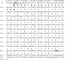
Fig. 1. cDNA sequence of 3H11 and its open reading frame. The putative start and stop codon as well as the signal sequence are shown in bold.
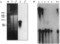
Fig. 2. (A) Northern blot analysis of 3H11. Total RNA from adult female and male schistosomes was separated on a denatured ethidium bromide agarose gel (left), blotted and hybridized with antisense transcripts of clone 3H11 (right). Molecular weight marker in nucleotides (M) is 1 kb DNA ladder (BRL). (B) Expression of p19 in females cultivated in vitro that were separated from males for 1, 2, 3, and 6 days, in comparison with freshly perfused females (0) and those that remained paired for 6 days (6p). Poly (A)+ RNA was probed with antisense transcripts of 3H11 (top) and, as a control, with transcripts of the schistosome protein disulfide isomerase (bottom).
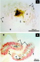
Fig. 3. (A) Localization of p19 mRNA in a cross-section of a schistosome female (F) showing greyish blue staining of the cytoplasm of the vitelline cells (V) with BCIP/NBT. The surrounding parenchyma and the tegument (T) are not labelled. The dark staining material in the gut (G) is digested haemoglobin. At the bottom left of the figure, part of a male (M) is shown. (B) Immunohistological localization of the p19 protein, stained with naphthol-AS-MX-phosphate and Fast Red TR in a tissue section documenting selective labelling of the cytoplasm of the vitelline cells (V) in the female. The surrounding parenchyma (P), the gastrodermis (Ga) and the tegument (T) are not labelled. At the upper left side of the figure, part of a male (M) is shown.

Fig. 4. Immunoblot of male and female proteins, extracted with (+) and without antioxidant (−), probed with the anti-p19 antibody showing a single band of 19 kDa only in females. Left: amidoblack-stained total protein extracts to document that the female lane is not overloaded (A). High molecular weight marker in kDa (M) (Sigma).
 You have
Access
- 12
- Cited by