Introduction
Magnetic nanoparticles (MNPs) are a new type of materials, which have increasingly extensive applications in many fields such as biomedicine [Reference Sun, Zhang, Pang, Hyun, Yang and Xia1, Reference Ei-Boubbou2, Reference Luo, Peng, Hou, Wu, Shen and Wang3], magnetic fluid, catalysis, magnetic resonance imaging (MRI) [Reference Ma, Xie, Zhang, Chen, Tang, Lei, Wu, Zhang and Hu4], environmental protection [Reference Lingamdinne, Chang, Yang, Singh, Choi, Shiratani, Koduru and Attri5], etc. Magnetic iron oxide nanoparticles (MIONPs) are one kind of MNPs, which have superparamagnetic behavior, good biocompatibility, and low toxicity [Reference Bull, Madani, Sheth, Seifalian, Green and Seifalian6]. There are many methods to synthesize MIONPs, including physical synthesis methods [Reference Feijoo, Gonzalez-Garca, Moldes-Diz, Vazquez-Vazquez, Feijoo and Moreira7], chemical synthesis methods [Reference Sari, Fadli and Amri8], and biosynthesis methods [Reference Sulaiman, Tawfeeq and Naji9]. Compared with physical methods and biosynthesis methods, chemical synthesis methods are more often used because they are simpler and efficient, and the generated MIONPs have high purity, small particle size, and good morphology. Various chemical synthesis strategies including coprecipitation, hydrothermal treatment, high temperature decomposition, sol–gel method, cathodic electrochemical deposition (CED), and pulsed laser ablation (PLA) had been reported.
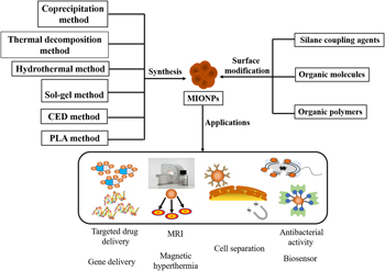
However, since MIONPs have a large surface-to-volume ratio and exhibit high surface energies, they tend to aggregate together to minimize this kind of energy. Furthermore, the bare MIONPs have high chemical activity, which makes them easily to be oxidized in air [Reference Wu, He and Jiang10]. So, the surface modification of MIONPs is very necessary. Inorganic metal oxides [such as silica (SiO2) and noble metal], organic small molecules [such as citric acid and oleic acid (OA)], or organic polymer [such as chitosan (CS), dextran, polyethyleneimine (PEI), and polyethylene glycol (PEG)] (Fig. 1) were used to modify MIONPs. Surface modification not only increases the stability of MIONPs mainly via preventing them from agglomerating and being oxidized but also provides the possibility for further functionalization [Reference Zhu, Ji, Yu, Niu, Farooq, Akram, Udego, Li and Niu11]. Pušnik et al. [Reference Pušnik, Goršak, Drofenik and Makovec12] found that the aspartic-acid-absorbed MIONP could be dispersed to form relatively concentrated aqueous suspensions displaying a good colloidal stability at an increased pH. Byoun et al. [Reference Byoun, Jang and Yoo13] modified MIONP with silica to generate a kind of multifunctional nanoparticles—highly fluorescent multiple Fe3O4 nanoparticles core-silica shell nanoparticles (FL multi-Fe3O4@SiO2 NPs) which possessed fluorescence and magnetism, these unique nanoparticles could widely be applied in nanomedicine. However, some modifications will result in certain side effects, e.g., the size of complex will be increased after MIONP was modified with PEI, which was not conducive to be endocytosed by cell. Even some functionalized MIONPs discharged into the environment can adsorb harmful substances (such as benzenes and phenols), and the combinations are harmful to the environment and our body after being evaporated into the air. It is urgently necessary to optimize the strategies for surface modification of MIONPs. Therefore, we summarized the chemical synthesis approaches and surface modification strategies of MIONPs, application fields, and the faced challenges of MIONPs or functionalized MIONPs in this review to promote the exploitation and applications of them.
The preparation strategy of functionalized MIONPs.

Synthesis method of MIONPs
Coprecipitation method
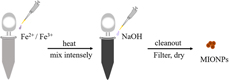
Coprecipitation method is also called multicomponent cationic method, which involves in Fe2+ salt, Fe3+ salt (usually chloride, nitrate, or sulfate), and excess precipitator (e.g., sodium hydroxide or ammonia water) [Reference Suh, Yuet, Hwang, Bong, Doyle and Hatton14], as shown in Fig. 2. It is one of the most commonly used methods for the rapid preparation of large quantities of MIONPs [Reference Tamer, Gündoğdu, Boyacıi and Pekmez15]. Businova et al. [Reference Businova, Chomoucka, Prasek, Hrdy, Drbohlavova, Sedlacek and Hubalek16] successfully obtained the black MIONPs from FeCl2·4H2O and FeCl3·6H2O after those raw materials were reacted with concentrated ammonia at 80 °C for 2 h [Reference Mahdavi, Ahmad, Haron, Namvar, Nadi, Rahman and Amin17]. Wahab et al. [Reference Wahab, Imran, Ikram, Naz, Aqeel, Rafiq, Majeed and Ali18] synthesized iron oxide (Fe3O4) nanoparticles doped with cobalt (Co) and manganese (Mn) using coprecipitation, and 16% Co-doped nanoparticles shown the highest saturation magnetization, they would be a promising candidate acted as nanocatalyst to improve conventional wastewater management methods.
Schematic showing the process of coprecipitation synthesis for MIONPs.

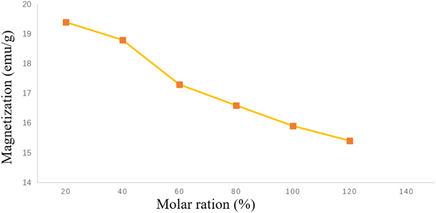
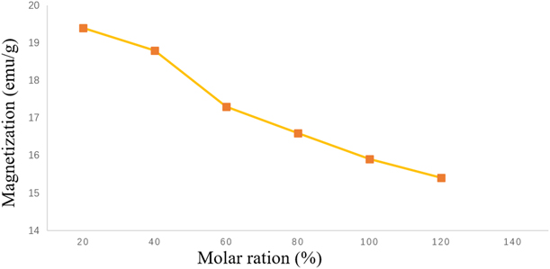
The preparation of MIONPs with coprecipitation method has many advantages such as easy to operate, low requirement for equipment, low price of iron salt and precipitant, short process, relatively high yield, and easy to expand to industrial production. The generated MIONPs are similar to sphere, which is convenient to be modified on their surface. However, severe agglomeration, a wide range of particle-size distribution (PSD), and irregular shape of those MIONPs are also observed because the reaction occurred in this method is rapid which makes it difficult to separate the process of explosive nucleation from the subsequent process of slowly crystal growth, so it is hard to obtain single dispersed MNP. In order to reduce the agglomeration and increase the stability of MIONPs, substances with high dielectric constant including the mixture of ethanol and water as solvent or surfactants (such as sodium oleate and sodium citrate), and so on should be used [Reference Liu, Wang, Lu, Du, Peng, Du, Zhang and Yao19]. However, the magnetization decreases gradually with the increasing concentration of citrate ions as shown in Fig. 3. In addition, the generated MIONPs have a low crystallinity and is easily oxidized to the γ-Fe2O3 [Reference Aghazadeh and Karimzadeh20], so the preparing process usually requires the protection of inert gas such as N2 [Reference Alp and Aydogan21].
Effects of molar ration on saturation magnetization of γ-Fe2O3 nanoparticles [Reference Liu, Wang, Lu, Du, Peng, Du, Zhang and Yao19].
Hydrothermal method
In the hydrothermal synthesis method, Fe3+ (source of iron), aqueous solution (reactive medium), ethylene glycol (reactive solvent), ammonium acetate, ammonium bicarbonate, and sodium citrate are mixed and transferred to a special stainless-steel autoclave with a lining made up of polytetrafluoroethylene. After those raw materials are reacted and recrystallized at 200 °C for 8–24 h, the generated MIONPs have the characteristics of high crystallinity, purity, dispersibility, and controlled morphology [Reference Li, Zhang, Tang, Qiu, Wang and Zhou22]. Walton pointed out that this method was the most advanced method for the synthesis of a series of MIONPs with different shapes and complex structures [Reference Walton23].
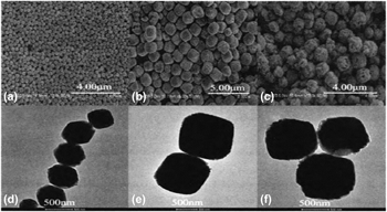
Tian et al. [Reference Tian, Yu, Li and Li24] prepared 4, 5, and 6 nm ultra-small monodisperse MIONPs by varying the volume rations of n-octylamine to n-octanol. Du et al. [Reference Du, Cai, Zhu and Geng25] found that when the reactive time was extended from less than 12–24 h, the particle size and dispersity of the products were increased, and their appearance changed from the broad bean-like to cubic. However, if the reactive time was up to 48 h, the particle size of the samples was decreased rather than increased and their surface became rougher, as shown in Fig. 4. Therefore, the reactive time of contains in the stainless-steel autoclave has great influences on the morphology and size of the MIONPs. In addition, high temperature in closed reactive vessels can increase the magnetism of MIONPs, and relatively high pressure is of great benefit to avoid the volatilization of components and improve the purity of the products. Torres-Gómez et al. [Reference Torres-Gómez, Nava, Argueta-Figueroa, García-Contreras, Baeza-Barrera and Vilchis-Nestor26] developed a simple and efficient method for preparing nanoparticles with good saturation magnetization and without cytotoxicity via only modifying the syntheses temperature of hydrothermal method.
Scanning electron microscope (SEM) and TEM images of MIONPs prepared at different times [Reference Du, Cai, Zhu and Geng25]. (a-c) SEM images of MIONPs prepared at different times. (a) 12h. (b) 24h. (c) 48h. (d-f) TEM images of MIONPs prepared at different times. (d) 12h. (e) 24h. (f) 48h.

However, compared with the coprecipitation method, the hydrothermal method requires special equipment, the production cost is relatively higher, and there is a serious oxidation problem in the reactive process, so this method is difficult to be applied to industrial production of MIONPs.
Thermal decomposition method
Reactive precursors including iron acetylacetonate, Fe(CO)5, or iron oleate are added to a mixture containing surfactant at high temperature in the thermal decomposition method. Different sizes of MIONPs are produced through regulating the reactive temperature and time [Reference Hyeon, Lee, Park, Chung and Na27]. Surfactants can slow the nucleating process of nanocrystals and affect the adsorption of additives on the reactive core, further inhibiting the growth of synthetical particles, and finally making the MIONPs smaller [Reference Wu, Wu, Yu, Jiang and Kim28]. By means of this method, MIONPs with single dispersion, narrow PSD, and high crystallinity are synthesized.
Chen et al. [Reference Chen29] reported that the particle size of MIONPs was 12.5, 17.6, and 22.5 nm when the reflux solvents were heated to different boiling points including 320 °C (1-octadecene), 360 °C (eicosane), or 380 °C (docosane) and maintained for 30 min, respectively. MIONPs with a diameter of 12.5 and 17.6 nm were sphere-like, while those with a diameter of 22.5 nm appeared cube-like morphology. So temperature dependence was existed during the nucleation and growth of monodisperse nanoparticles, reactive temperature at more than 320 °C contributed to the growth of MIONPs. Hufschmid et al. [Reference Hufschmid and Arami30] found that the different iron-containing chemicals would affect the synthesis of MIONPs, iron pentacarbonyl was particularly suitable for synthesizing small-sized MIONPs (<10 nm), iron oleate and iron oxyhydroxide were often used to synthesize MIONPs with larger size (10–30 nm). Therefore, the reflux temperature, reflux solvent, and organic materials are important parameters for the preparation of MIONPs with high temperature decomposition method. However, those synthesized MIONPs are mostly hydrophobic, which greatly limits their applications, and researchers need to further improve the strategy to obtain water-soluble MIONPs.
Sol–gel method
The sol–gel method is based on the hydrolysis and condensation of precursors in a colloidal solution to form a network of metal oxides. Particles with smaller size will be got if the hydrolysis rate is lower and vice versa. In addition, the size of product is also closely associated with the solvent concentration, pH, reactive temperature, etc. [Reference Fu and Ravindra31]. When the size of MIONPs is smaller than a certain value, the magnetic orientation force produced by external magnetic field is too small to resist thermal motion of superparamagnetic iron oxide nanoparticles (SPIONs) as their size is small. Once the magnetic field is removed, the magnetization of nanoparticle closes to zero, there will be no residual magnetism [Reference Kandasamy and Maity32]. Those MIONPs which are more sensitive to external magnetic field and only show magnetism in the presence of exogenous magnetic field are known as SPIONs. Hu et al. [Reference Hu, Chang, Chen, Deng, Li, Zuo, Kang, Yang, Hostetter and Volinsky33] found that the highly pure, well-crystallized, spherical, and monodispersed SPIONs could be obtained with the sol–gel explosion-assisted method when the ratio of dry gel and explosive agent was 12:1, and the hysteresis behavior of them would be weakened with the increase of temperature.
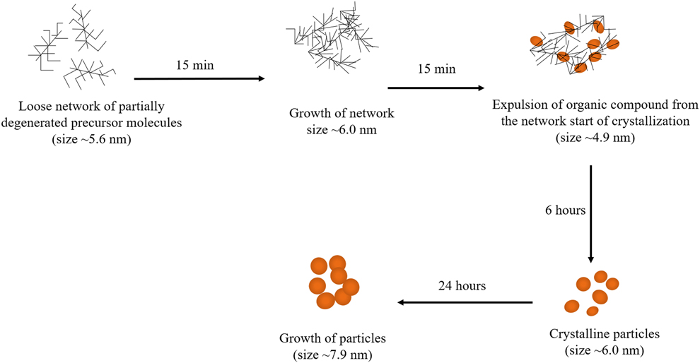
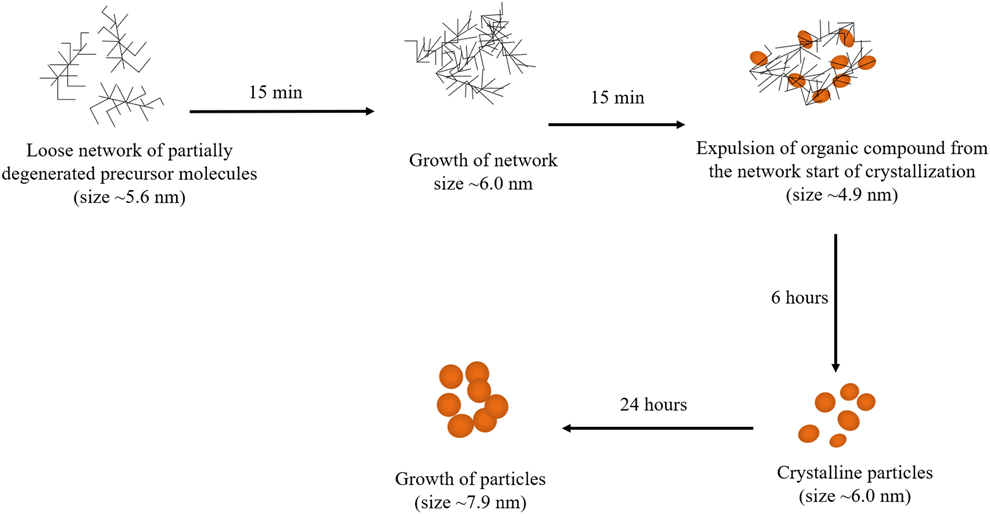
Using the sol–gel method, Richard et al. [Reference Richard, Eder, Caputo, Journe, Ou, Bolley, Louedec, Guenin, Motte, Pinna and Lalatonne34] prepared SPIONs that were controlled from 3 to 9 nm in size and could acted as contrast agents for MRI. Masthoff et al. [Reference Masthoff, Kraken, Menzel, Litterst and Garnweitner35] also obtained MIONPs with small particle size, good dispersion, and high saturated magnetism via this method as shown in Fig. 5. Lu et al. [Reference Lu, Yin, Mayers and Xia36] proved that the thickness of SiO2 coating could be controlled by changing the concentration of the sol–gel solution. In addition, 7-dimethylamino-4-methylcoumarin-3-isothiocyanate (DACITC) and tetramethylrhodamine-5-isothiocyanate (5-TRITC) had also been incorporated into the SiO2 shells to obtain multifunctional MIONPs, which could be simultaneously manipulated with external applied magnetic field and characterized in situ using conventional fluorescence microcopy. These multifunctional nanoparticles have far-reaching application potentials.
Schema of the particle growth process during the nonaqueous sol–gel synthesis in triethylene glycol (TEG) [Reference Masthoff, Kraken, Menzel, Litterst and Garnweitner35].
The sol–gel method can avoid some disadvantages of the coprecipitation method (such as severe agglomeration, a wide range of PSD, and irregular shape of the MIONPs) and produce high quality MIONPs. However, the particle size and morphology of the products could not be completely controlled in this method, and the suitable concentration of solvent and reactive time are required to be further improved.
CED method
CED is a process of depositing the metal ions in electrolyte onto the cathode surface via electrochemical principles. The MIONPs synthesized with this method have high magnetization value and negligible coercivity [Reference Karimzadeh, Aghazadeh, Doroudi, Ganjali and Kolivand37], which makes them to be widely applied in many fields such as drug delivery and magnetic hyperthermia for tumor. Karimzadeh et al. [Reference Karimzadeh, Aghazadeh, Dalvand, Doroudi, Kolivand, Ganjali and Norouzi38] synthesized SPIONs in an aqueous solution with CED method, and these nanoparticles were spherical with a high level of homogeneity and a narrow size distribution, after being coated with polyvinyl alcohol, and they exhibited high drug loading, which has the potential to be used in target drug delivery. The purity, crystallinity, and size of MIONPs generated using the CED method are closely associated with the electric current, electric potential, electrolyte composition, pH, and concentration. Starowicz et al. [Reference Starowicz, Starowicz, Zukrowski, Przewoznik, Lemanski, Kapusta and Banas39] found that the electrolyte concentration was inversely proportional to the particle size of MIONPs.
The surface of MIONPs prepared with CED method is easy to be coated with organic polymers, which gives MIONPs suitable surface properties and crystal structure; hence, this method used for the preparation of MIONPs has been rapidly developed in recent years. Aghazadeh et al. [Reference Aghazadeh and Karimzadeh20] used FeCl2 and Fe(NO3)3 with a molar ratio of 1:2 to obtain bare MIONPs which were characterized by nanosized (about 10 nm diameter), well-dispersed superparamagnetism. Surface of MIONPs synthesized with CED method could be modified with PEG and dextran in situ. Those double-coated MIONPs had an average diameter of approximately 15 nm. Karimzadeh et al. [Reference Karimzadeh, Aghazadeh, Doroudi, Ganjali and Kolivand37] used the same method to synthesize MIONPs and coated them with PEG and PEI, and the particle sizes of bare MIONPs and double-coated MIONPs were 20 and 58 nm, respectively. In addition, both of them had morphology with spherical shape and no obvious aggregation, but the modified MIONPs had relatively better dispersion.
PLA method
The PLA method is environmentally friendly since this strategy can minimize the chemical waste and the potential safety issue, and it has become an increasingly important approach for generating MIONPs. The morphologically stable MIONPs with a narrow size distribution could be formed through PLA of iron pellet in sodium dodecyl sulfate (SDS) and dimethylformamide (DMF) solutions [Reference Ismail, Sulaiman and Abdulrahman40]. Ismail et al. [Reference Ismail, Sulaiman, Abdulrahman and Marzoog41] prepared the MIONPs (50–100 nm) with antibacterial activity from iron target through PLA method in the same solutions. The antibacterial activities of them were depended on their size, concentration, and oxidation state. Once microorganisms contacted to the surface of MIONPs, the ions would be released from MIONPs, then those ions attached to the bacterial cell wall with negative charge via electrostatic adsorption and ruptured it, resulting in protein denaturation and death of bacterium [Reference Fazio, Santoro, Lentini, Franco, Guglielmino and Neri42]. The process of generating MIONPs with PLA method is simple and low cost, few chemical species are required, and the ablation atmosphere is highly controlled without action of any catalyst. However, those generated MIONPs are easily aggregated due to insufficiency of laser energy. Therefore, the laser energy and suitable concentration of solvent are required to be further improved.
Characteristics of MIONPs synthesized with several commonly used methods are summarized in Table I.
Several methods commonly used to synthesize MIONPs.

Note: + represents the degree of each evaluate indicators.
Surface modification of MIONPs
Although previous reports have been suggested that the MIONPs were one of the most basic materials in biomedicine, they still need to be further modified to obtain high-quality MIONPs. In order to avoid aggregation and improve their stability and functions, various strategies for surface modification had been developed. Surface-active groups of MIONPs can couple with bioactive substances such as DNA, protein, or antibody, which will widen their application scopes [Reference Park, Mcconnell, Boddohi, Kipper and Johnson43].
Inorganic small molecules
SiO2
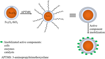
SiO2, an acidic oxide with stable chemical properties, biocompatibility, low toxicity, acid resistance, and high temperature resistance, is one of the preferred materials to modify MIONPs; MIONPs coated with SiO2, the core–shell structure, which also named as MIONPs@SiO2 nanoparticles. SiO2 not only can shield the electrostatic repulsion of MIONPs but also is rich in hydroxyl groups [Reference Hui, Shen, Tian, Bao, Ding, Tian, Shi and Gao44]. Those hydroxyl groups take part in the formation of covalent bridges between MIONPs and proteins, antibodies, DNA, or a series of bioactive molecules [Reference Sonmez, Georgescu, Alexandrescu, Gurau, Ficai, Ficai and Andronescu45] (Fig. 6). Compared with commercial extraction kit, MIONPs@SiO2 with unique characteristics including well-dispersion, high saturation magnetization, good surface topography, and excellent acid resistance could provide better adsorption, elution, and purity for DNA extraction [Reference Fan, Guan, Zhang, Xu, Yang and Guo46]. In recent years, the improved methods including Stöber method and sol–gel method [Reference Shahabadi, Khorshidi, Zhaleh and Kashanian47] have been used to synthesize MIONPs@SiO2 nanoparticles.
Silica shell functionalization of Fe3O4/SiO2 via covalent route [Reference Shahabadi, Khorshidi, Zhaleh and Kashanian47].

Stöber method
Stöber method is a strategy for synthesizing monodisperse SiO2 nanoparticles, which normally involves tetraethyl orthosilicate and the mixture solvent of ethanol and ammonia water [Reference Stöber, Fink and Bohn48]. Kulkarni et al. [Reference Kulkarni, Sawadh and Palei49] coated MIONPs with SiO2 by hydrolysis of tetraethylorthosilicate (TEOS). The saturation magnetization of those MIONPs decreased drastically with the increase of coated SiO2 and they had good monodispersity and stability.
Sol–gel method
Mitra et al. [Reference Mitra, Shahtahmassebi, Roknabadi and Ghows50] prepared the MIONPs with coprecipitation method and then treated them with trisodium citrate, which is one kind of surfactants to improve the dispersibility of MIONPs. Puscasu et al. [Reference Puscasu, Sacarescu, Lupu, Grigoras, Oanca, Balasoiu and Creanga51] found that the SiO2 could be abundant in silanol groups at the surface of SiO2-coated MIONPs and easily interact with cations, which the diameter of MIONPs with or without SiO2 shell was about 20 nm and 15 nm, respectively. Subsequently treated MIONPs were coated by SiO2 shell via sol–gel method, those generated MIONPs@SiO2 nanoparticles had electrostatically stability and superparamagnetic property at room temperature. The thickness of SiO2 shell was about 5 nm, which was related to the reactive time, TEOS concentration, and other parameters [Reference Lu, Yin, Mayers and Xia36]. The prospect of MIONPs@SiO2 in biomedicine will be widen because of their biocompatibility, low toxicity, superparamagnetic properties, monodispersity, and stability.
Noble metal
Gold (Au) and silver (Ag) are common noble metals, which have stable chemical properties, biocompatibility, resistant to oxidation, and corrosion, so they were usually used to modify MIONPs [Reference Li, Yu, Zhao and Xue52]. After being modified with Au, not only the stability of MIONPs under physiological conditions and their binding ability to ligand would be enhanced but also the formation of harmful free radicals could be prevented [Reference Lee, Leu, Lin, Tseng, Lin and Yang53].
Miao et al. [Reference Miao, Tang and Wang54] treated MIONPs with HAuCl4 for generating Fe3O4@Au MNPs. The prepared MIONPs were uniform spherical particles with the mean diameter about 150 nm and coated with bright Au. Electrochemical biosensor based on DNA modified Fe3O4@Au nanoparticles had the ability to successfully detect the trace of heavy metal irons in water because the stable structures of cytosine-Ag+-cytosine and thymine-Hg2+-thymine formed in the presence of Ag+ and Hg2+ could assist the hybridization of DNA probes. Zhao et al. [Reference Zhao, Tu, Liu, Qin, Wang, Qi and Shi55] synthesized dually surfaced dumbbell-like aptamers-functionalized Au–Fe3O4 nanoparticles (Apt-Au–Fe3O4). Modified nanoparticles could act as a smart photo-controlled drug carrier for the unique biological properties, and the aptamers could be released upon plasmon-resonant light (605 nm) radiation. Therefore, the dumbbell-like Fe3O4@Au MNPs have great potential as drug-carriers for highly sensitive diagnostics and therapy of diseases.
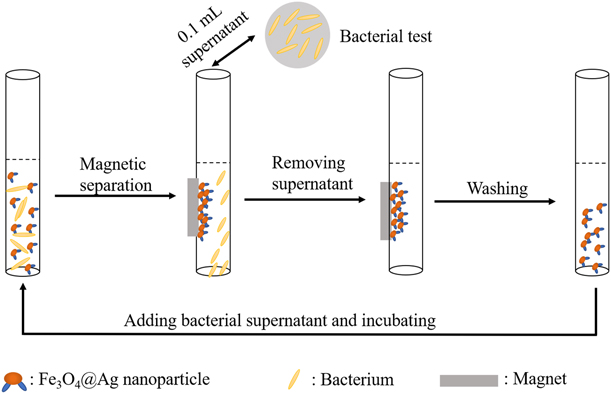
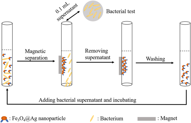
Chen et al. [Reference Chen, Liu, Yanyun, Xiong and Dong56] in situ synthesized the Ag@Fe3O4 nanoparticles by germination of Ag nanocrystals on the surface of Fe3O4 nanoparticles, and Ag loaded on the nanoparticles exhibited good antibacterial performance to Staphylococcus and Escherichia coli (Fig. 7). The size of Ag@Fe3O4 nanoparticles was about 20 nm and they showed superparamagnetic behavior at room temperature. Ochratoxin A (OTA) is an abundant and representative food-contaminating mycotoxin, which has a detrimental effect on our health. Shao et al. [Reference Shao, Ma, Zhao, Lv, Hun, Wang and Wang57] found that the Au@Au–Ag nanogapped nanostructures could couple with MIONPs by OTA aptamer and its complementary DNA sequence. The generated nanoparticles might be used to quickly detect OTA in food based on the change of Raman intensity. Therefore, MIONPs coated with Au and/or Ag have suitable properties such as good antibacterial activity and superparamagnetic behavior, which contributes to their applications in microbiology and food security [Reference Stefan, Leostean, Pana, Soran, Suciu, Gautron and Chauvet58].
Schematic illustration of bacterial test of Fe3O4@Ag nanoparticles [Reference Chen, Liu, Yanyun, Xiong and Dong56].
Organic small molecules
Surfactant
OA
OA is a popular surfactant because of their nontoxic in blood and high soluble in organic solvents such as ethanol, ether, and chloroform. MIONPs modified with OA exhibit the biocompatibility, dispersibility, and stability as a result of reduced surface energy caused by OA [Reference Portet, Denizot, Rump, Lejeune and Jallet59]. Mahdavi et al. [Reference Mahdavi, Ahmad, Haron, Namvar, Nadi, Rahman and Amin17] found that pH, the initial temperature, and stirring rate were important parameters for preparing the monodisperse OA-coated MIONPs. Since the chemical bond was formed between the carboxyl group of OA and the surface of MIONP [Reference Soares, Laia, Carvalho, Pereira, Coutinho, Ferreira, Novo and Borges60], OA and MIONP could be firmly combined together and the formed complex has better dispersibility. Zhang et al. [Reference Zhang, He and Gu61] indicated that the cover density of OA attached to MIONP could significantly affect the size of MNP. The saturated magnetization and superparamagnetic behavior of the modified MIONP would be enhanced with the increase of particle size. Although OA is slightly soluble in water, MNPs coated with double OA could be dissolved in inorganic solvents at the optimum pH, which is convenient for their practical applications especially in biomedical fields.
Citric acid
Similar to OA, the citric acid is another surfactant with three carboxylic acid groups, those groups can be covalently bound with iron ions of the MIONPs. Electrostatic repulsive forces among citric acids prevent MIONPs from aggregating each other, which results in the significantly increased dispersibility of modified MIONPs. The citric acid-modified MIONPs can be stably stored in an aqueous solution, so they are suitable to the application of MRI. Andreas et al. [Reference Andreas, Georgieva, Ladwig, Mueller, Notter, Sittinger and Ringe62] found that the magnetic labeling of human mesenchymal stem cells with citrate-modified MIONPs did not affect their proliferation and differentiation, especially after being injected intramuscularly in rats, those transplanted MIONPs-labeled stem cells were visualized in vivo with MRI. Therefore, citrate-modified MIONPs can be used to track the stem cells in vivo highly efficiently. Saraswathy et al. [Reference Saraswathy, Nazeer, Jeevan, Nimi, Arumugam, Harikrishnan, Varma and Jayasreea63] synthesized the citric acid-modified MIONPs via coprecipitation method. Monodispersity and stability of them could be maintained in water for 6 months. Their optimized parameters including particle size and magnetic property were contributed to produce good contrast for MRI.
Other surfactants
Except for OA and citric acid, some other surfactants could also be used to modify MIONPs. Ardelean et al. [Reference Ardelean, Stoencea, Ficai, Ficai, Trusca, Vasile, Nechifor and Andronescu64] found that MIONPs modified with alicylic acid, glutamic acid, and trichloroacetic acid could prevent the aggregation of them and obtain stable nanostructure even in the biological environment. After being bound to MIONPs, those surfactants still had some free groups, which could be used to further bind molecules onto the surface of MIONPs. Therefore, MIONPs modified with surfactants can reduce their surface energy and form covalent bond, which resulted in the modified MIONPs with better stability and dispersibility.
Silane coupling agent
RSiX3 is a molecular formula of silane coupling agent. R represents an active group, and X represents an alkoxy group with the capability of being hydrolyzed. MIONPs modified with the silane-coupling agent exhibit the hydrophilic and lipophilic properties because this agent contains organic and inorganic groups, which can be chemically bonded with MIONPs. First, the alkoxy group is hydrolyzed to form silanol, then silanol reacts with the hydroxyl group on the surface of MIONPs to form –Si–O–MIONPs via covalent bond.
Amino silane
3-Aminopropyltriethoxysilane (APTES), an amino silane coupling agent, can aminate MIONPs to generate functional MIONPs, which show well-dispersibility, superparamagnetic behavior, and strong magnetization at room temperature [Reference Yamaura, Camilo, Sampaio, Macedo, Nakamura and Toma65]. Jia et al. [Reference Jia, Gao and Cui66] found that SiO2@MIONPs and bare MIONPs were modified with amino silane, respectively, and the former had more amino groups than the latter. Teo et al. [Reference Teo, Wang, Chen, Zhang, Yang, Huang and Tang67] drew conclusion that the surface of APTES-MIONP was enriched in hydroxyl groups and the alkoxy groups, so they could deliver genes with high transfected efficiency without obvious cytotoxicity. Hep G2 cells expressed more genes mediated by APTES-SPIONs when they were exposed to alternating current magnetic field only once.
Hydrogels with specific and even reversible responses to environmental stimuli have been used as excellent candidates for drug delivery. Khosroshahi et al. [Reference Khosroshahi and Asemani68] embedded APTES-SPIONs covalently connected fluorescein isothiocyanate (FITC) in the hydrogel scaffold, and the optical and elastomagnetic properties of those modified SPIONs could be used for bioimaging and tracking the changes of hydrogel scaffold in equilibrium or swelling states using argon laser-induced fluorescence. Fluorescent-based ferroscaffold can be used to study the mechanism on drug release. In addition, polyethersulfone (PES) nanocomposite membranes had been synthesized with IONPs and APTES, which could efficiently remove arsenate ion from drinking water or industrial waste [Reference Rowley and Abu-Zahra69].
Vinylsilane
CH2=CHSi(OCH3)3 is a formula of the vinyltriethoxysilane (VTEO). It can be hydrolyzed in aqueous solution and form a condensation compound of vinylsilicotriol. Sun et al. [Reference Sun and Li70] found that MIONPs coated with VTEO exhibited the superparamagnetism and high magnetic saturation. The dispersibility of modified MIONPs was increased due to the steric hindrance of VTEO, and their dispersibility was related to the ratio of VTEO to MIONPs. In order to reduce the agglomeration of Fe3O4@SiO2 nanoparticles, Ahangaran et al. [Reference Ahangaran, Hassanzadeh and Nouri71] modified SiO2 shell on the surface of Fe3O4 with VTEO and were covalently coupled together to form Fe3O4@SiO2–Si–CH=CH2 nanoparticles, which showed well biocompatibility, dispersibility, and stability.
Methacryloxysilane
Similar to vinylsilane, γ-methacryloxypropyltrimethoxy-silane (γ-MPS) is hydrophilic and lipophilic because of their propenyl groups and methoxy groups. It can be bound to the surface of MIONPs by Fe–O–Si, which is formed via dehydration between hydroxyl of MIONPs and γ-MPS [Reference Chen, Zhao, Zhao, Du and Xie72]. Xiong et al. [Reference Xiong, Li and Xia73] found that the stability of the MIONPs modified with γ-MPS was good not only in aqueous media at different pH but also in mixed solvents of water and hydrophilic organic solvents such as methanol, ethanol, or dimethyl sulfoxide. The compatibility between γ-MPS-MIONPs and organic solvent was mainly affected by the concentration of organic solvent and γ-MPS [Reference Cui, Shen and Lin74]. Compared with MIONPs coated with other silane coupling agents, MIONPs modified with methacryloxysilane have the strongest activity and their heat resistance was improved [Reference Gu, Hou, Gao and Deng75].
Organic polymer
CS and its derivatives
CS is compounded by chemical connection between a β-1,4-glucoside and N-acetyl-D-glucosamine monomers, it is a linear cationic polymer due to a free amino group on it. Moreover, the amino group at the C2 position of the polymer is higher active than the hydroxyl group. Therefore, CS is easy to be reacted under mild conditions. CS exhibits many advantageous properties including nontoxicity, hydrophilicity, biocompatibility, and biodegradability, and it is widely used in the fields of waste treatment [Reference Radwan, Rashad, Sadek and Elazab76], health care, and food hygiene [Reference Shahidi and Abuzaytoun77]. After MIONPs are modified with CS, the number of amino groups will increase, which promotes MIONPs to be attached with more bioactive molecules and expands their potential applications. In order to obtain the MIONPs with active groups, Borowska et al. [Reference Ziegler-Borowska, Chełminiak and Kaczmarek78] double coated MIONPs with CS and amphiphilic polyquaternium, the generated MIONPs carried positive charge, which greatly enhances their ability to bind with biologically active molecules. CS-coated MIONPs could carry chlorhexidine (CHX) to effective against bacterial and fungal planktonic cells, and this strategy had the potential to prevent or treat the biofilm-associated oral diseases [Reference Vieira, Arias, Neto, Kubo, Lima, Camargo, Pessan, Delbem and Monteiro79].
Safee et al. [Reference Safee, Abdullah and Othman80] modified the MIONPs with carboxymethyl chitosan (CCS). The particle size of CCS-MIONP ranged from 9.7 to 12.5 nm, and they had a great potential to remove metal ions from drinking water. In addition, their properties such as superparamagnetism, strong magnetic response, high stability, and biocompatibility were the essential requirements for MRI. Song et al. [Reference Song, Luo, Zhang, Zhu, Ji and Yan81] synthesized CS-coated MIONPs with coprecipitation method and then modified them with PEG and lactobionic acid. Those generated particles showed excellent properties such as nontoxicity, well-dispersibility, and extremely high sensitivity for liver MRI. Therefore, MIONPs modified with CS and its derivatives can be widely applied to environmental improvement, magnetic imaging, and so on.
Dextran
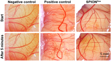
MIONPs could be coated with dextran, which not only avoided the aggregation but also had good biocompatibility [Reference Boustani, Shayesteh, Salouti, Jafari and Shal82]. Unterweǵer et al. [Reference Unterweǵer, László, Matuszak, Janko, Poettler, Jordan, Bäuerle, Szebeni, Fey, Boccaccini, Alexiou and Cicha83] confirmed that the particle size of MIONPs and dextran-coated MIONPs was about 30 nm and 50–80 nm, respectively. The stimulation to cells was independent of the size of dextran-coated SPIONs, and there was no irritative potentiality in a chick chorioallantoic membrane assay. Moreover, ultra-small (30 nm) dextran-coated SPIONs could be endocytosed by macrophages and there were no hypersensitive reactions (Fig. 8). These nanoparticles could be used as contrast agents for MRI. In addition, dextran-coated MIONPs conjugated with doxorubicin (Cet) and cetuximab (Dox) could significantly suppress the proliferation of A549 cells, and they may serve as a promising candidate for targeted therapy of non-small cell lung cancer (NSCLC) [Reference Zhang, Liu, Du, Vermorken, Cui, Zhang, Guo, Ma and Chen84].
The chick chorioallantoic membrane assay with SPIONDex30: vascularization before (top row) and 5 min after application (bottom row) was shown. Phosphate buffer saline (PBS) was used as a negative control and 0.5 wt% SDS as a positive control [Reference Unterweǵer, László, Matuszak, Janko, Poettler, Jordan, Bäuerle, Szebeni, Fey, Boccaccini, Alexiou and Cicha83].
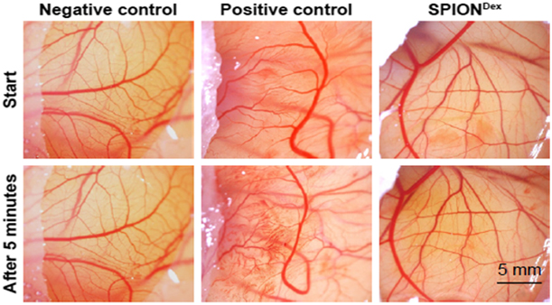
After dextran-coated MIONPs were intravenously injected, those nanoparticles were phagocytosed by resident Kupffer cells in normal reticuloendothelial system within the liver. If there were tumors in the liver, nanoparticles were not phagocytosed and not retained in tumor tissue because the Kupffer cells were absented [Reference Qin, Xu and Yang85]. Consequently, there were significant differences in thermoacoustic signal intensity between normal liver and liver with tumor. Therefore, dextran-coated MIONPs have exceptional safety and the potential to become a novel approach for clinical diagnosis of tumor in the future.
PEI
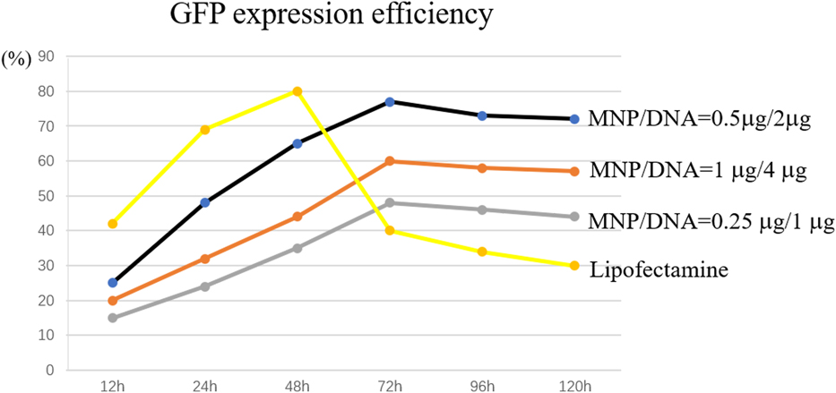
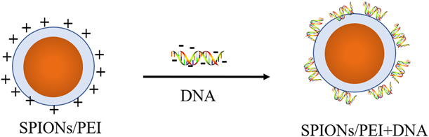
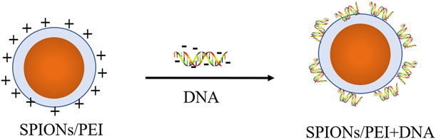
PEI, a water-soluble cationic polymer with amino and vinyl groups, is a common nonviral vector for gene delivery because it can electrostatically bind to DNA with negative change to form the PEI/DNA complex (Fig. 9). In addition, PEI-coated MIONPs can be used for targeted drug delivery, magnetic transfection, magnetic hyperthermia, and so on [Reference Zhao, Cui, Chen, Wang, Cui, Sun, Meng and Liu86].
GFP expression efficiency of magnetofection and liposome transfection in porcine kidney PK15 cells with time [Reference Zhao, Cui, Chen, Wang, Cui, Sun, Meng and Liu86].
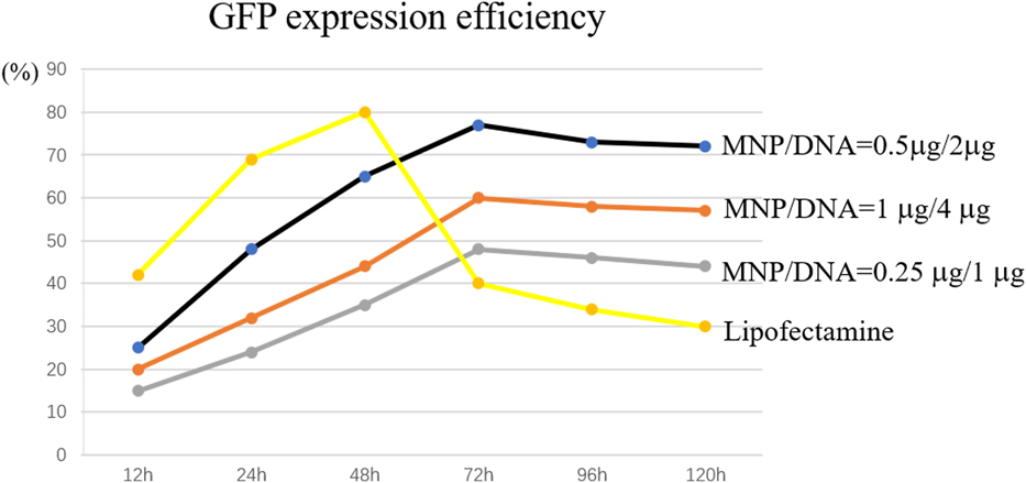
Karimzadeh et al. [Reference Karimzadeh, Aghazadeh, Ganjali, Doroudi and Kolivand87] simultaneously in situ coated MIONPs with PEI and polyvinylpyrrolidone (PVP) with CED method. Double modified MIONPs had positive charge, so DNA could be attached on them. PEI-coated SPIONs could stably exist even in environment with salt at high concentration and in solution at wide range of pH. After plasmids DNA containing green fluorescent protein (GFP) gene were bound to PEI-coated SPIONs (Fig. 10), this complex could be endocytosed into cells, and the transfected efficiency would be increased by being exposed in foreign magnetic fields. The nanoparticles with a size of 50 nm were uptook by lung carcinoma cell 3 times to 4 times faster than 100 nm particles, and 8 times to 10 times faster than 200–500 nm particles [Reference Steitz, Hofmann, Kamau, Hassa, Hottiger, Rechenberg, Amtenbrink and Fink88]. When PEI-coated SPIONs were endocytosed into the cells, the efficacy of escaping lysosomal entrapment for the cationic polymers such as PEI was related to their extensive ability to buffer acidic pH in lysosome through amine protonation; so change of pH would result in destabilization of the lysosome vesicles, followed by release of vectors and plasmids into cytoplasm and delivery of DNA into the nucleus [Reference Arsianti, Lim, Marquis and Amal89].
Structure of PEI-coated SPIONs binding with DNA [Reference Arsianti, Lim, Marquis and Amal89].
In order to track the localization of nanoparticles in cells, Topel et al. [Reference Topel, Topel, Bostancıoğlu and Koparal90] synthesized the PEI-coated MIONPs, which were covalently coupled with a fluorophore molecule, Bodipy-5. The imaging ability of the Bodipy-PEI-coated MIONPs in human lung adenocarcinoma epithelial cells was acquired. The Bodipy-PEI-coated MIONPs had no cytotoxic effects on Ishikawa cells even at higher concentrations, whereas had little toxicity to human lung adenocarcinoma epithelial cells. Therefore, the Bodipy-PEI-coated MIONPs had potential application in biolabeling but they were slightly aggregate. However, Tutuianu et al. [Reference Tutuianu, Popescu, Preda, Rosca, Piticescu and Burlacu91] evaluated the binding capacity of PEI-coated MIONPs to anti-tumor drugs and found that the aggregate phenomenon of MIONPs in culture medium was exploited for further entrapment of other active molecules. After active molecules bound to PEI-coated MIONPs directly contact cells in vivo or in vitro, they were delivered into cells and the biological activities of molecules were preserved. Thus, those reports support the value of PEI-coated MIONPs as tools for drug delivery. PEI-coated MIONPs also have great potential to treat the complicated industrial wastewater matrices. They could efficiently separate and recycle diesel oil or olive oil droplets stabilized by SDS from aqueous media [Reference Lü, Qi, Zhang, Zhang and Zhao92].
PEG
PEG is a hydrophilic, nontoxic, and long chain polymer. PEG-coated MIONPs have been synthesized mainly with sol–gel method, thermal decomposition method, and hydrothermal synthesis method. But these methods require rigorously experimental conditions or higher temperature. Therefore, it is necessary to develop a simpler and more efficient synthetic method for generating PEG-coated MIONPs. Yang et al. [Reference Yang, Ping, Yang, Cao, Sun, Han, Yang, Wang, Chen, Wang and Kong93] synthesized PEG-coated MIONPs via coprecipitation method at 60 °C. Well-dispersed PEG-coated MIONPs were obtained when 4g PEG1000 were added into the sealing beaker, and the average particle size of them was about 15 nm. The reason might be that the ammonia and water vapor were difficult to evaporate under the condition of seal, then the pressure and generated hydroxy were increased.
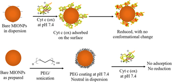
MIONPs modified with phosphonic acid (PA)- and hydroxamic acid (HA)-terminated PEG were colloidally stable and dispersible in water and cell culture medium, especially they were no cytotoxic to primary human peripheral blood cells, which provided great potential for their applications in diagnosis and therapy of cancer [Reference Patsula, Tulinska, Trachtová, Kuricova, Liskova, Španová, Ciampor, Vavra, Rittich, Ursinyova, Dusinska, Ilavska, Horvathova, Masanova, Uhnakova and Horák94]. There is the risk of forming reactive oxygen and developing cancer when excess irons exist in the human body [Reference Zwart, Morgan and Smith95]. Mukhopadhyay et al. [Reference Mukhopadhyay, Joshi, Chattopadhyay and De96] found that the redox state of cytochrome C (Cyt C) was altered by bare MIONPs, which resulted in the decrease of hemoglobin. Therefore, MIONPs must be reasonably modified to carry the drugs in clinical researches. The colloidal stability and half-life of MIONPs in blood were increased after being modified with PEG. Furthermore, PEG-coated MIONPs did not disrupt the redox state of Cyt C (Fig. 11).
Schematic representation of the interaction of Cyt C with the bare and PEG coated Fe3O4 nanoparticles. In the case of bare nanoparticles, the protein is adsorbed onto the surface of nanoparticles by electrostatic interactions, followed by immediate reduction of the heme group. The presence of PEG coating resists the adsorption of Cyt C on its surface, and hence, the reduction is not observed [Reference Mukhopadhyay, Joshi, Chattopadhyay and De96].

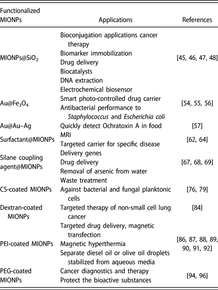
Therefore, MIONPs with different surface modifications can be used in different fields. Choosing a reasonable agent for surface modification according to our needs will have a multiplier effect. The applications of functionalized MIONPs are summarized in Table II.
Applications of functionalized MIONPs.
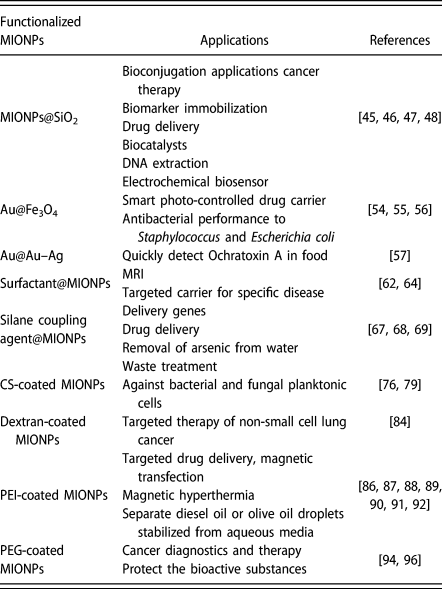
Applications of MIONPs
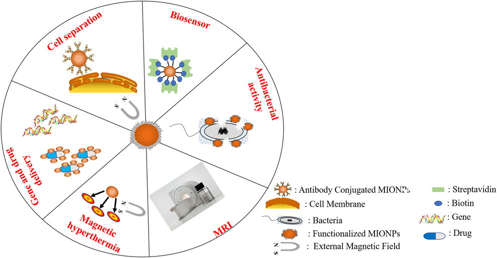
MIONPs have gained enthusiastic attention from academia in biomedicine fields including gene transfer, targeted drug delivery, magnetic hyperthermia, and so on (Fig. 12) due to their unique properties, such as biocompatibility, superparamagnetic behavior, and transfer electrons.
Applications of functionalized MIONPs in biomedicine.
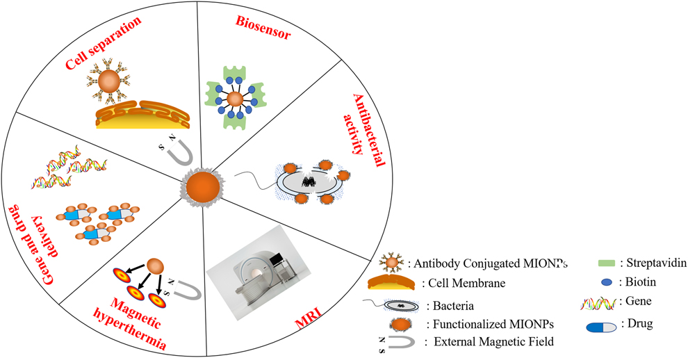
Biosensor
Biosensor is an instrument, which is sensitive to biological substances and can convert their concentrations into electrical signals for detection. MIONPs are particularly attractive in biosensor because of their superparamagnetism and high surface energies. Surface-active groups of functionalized MIONPs can couple with bioactive substances such as specific antibodies that recognize the specific protein. Blyakhman et al. [Reference Blyakhman, Buznikov, Sklyar, Safronov, Golubeva, Svalov, Sokolov, Melnikov, Orue and Kurlyandskaya97] synthesized the multifunctional biosensor with MIONPs and ferrogels, and the biosensor with sensitive elements was deposited by magnetron sputtering onto glass or polymer substrates, which was adapted for ferrogel studies. Dolci et al. [Reference Dolci, Bryche, Leuvrey, Zafeiratos, Gree, Begin-Colin, Barbillon and Pichon98] created an original nanostructured surface plasmon resonance (SPR) biosensor, the assembly of MIONPs modulated the properties of SPR, leading to the huge enhancement of sensitivity prior to streptavidin detection. Therefore, MIONPs can improve the sensitivity and targeting of biosensors.
Antibacterial activity
Recently, the emergence of bacterial strains with resistance to antibiotics has posed a serious threat to public health. Searching for materials which can replace antibiotics has received a considerable attention. MIONPs were found to be good inhibitors for many bacterial strains. Shah et al. [Reference Shah, Yehye, Saad, Simarani, Chowdhury, Alhadi and Al-Ani99] indicated that MIONPs functionalized with GA (MIONPs@GA) could act as water-soluble antioxidants, which had favorable therapeutic and industrial applications for their advanced biocompatibility, hydrophilicity, and further synergistic property to organic–inorganic hybrid antioxidants. In addition, since the large surface area of nanoparticles could be tightly adsorbed on the surface of bacteria, disruption of membrane would lead to the leakage of intracellular components, then the bacteria would be killed. Ismail et al. [Reference Ismail, Sulaiman, Abdulrahman and Marzoog41] found that the smaller particle size of nanoparticles would result in the increase of membrane permeability and bacterial destruction. Therefore, research on the synthesis mechanism of MIONPs is beneficial to improve their antimicrobial activity.
Cell separation
Due to the unique properties (such as superparamagnetic behavior and biocompatibility) of MIONPs, applications of them in cell separation are currently under investigation. However, uncoated MIONPs have some disadvantages including low binding capacity to specific antibody, easily being aggregated for high surface energy, and easily being oxidized in air, which limit them to be used to separate cells. MIONPs with appropriate surface modification could separate target cells from complex sample matrices. Lu et al. [Reference Lu, Ling, Jia, Huang, Li and Yan100] synthesized the PEI-coated MIONPs (about 12.7 nm) to separate and enrich in lung cancer cells from sputum for cytopathology analysis. Tumor cells could be isolated from the fresh whole blood through MIONPs conjugated with special antibody [Reference Xu, Aguilar, Yang, Kuang, Duan, Xiong, Wei and Wang101]. MIONPs modified with molecularly imprinted polymers (MIP) were used to remove steroid from biological samples, which had an abundance of steroid [Reference Zengin, Yildirim, Tamer and Caykara102].
Gene delivery
Gene delivery vectors are divided into virus-mediated and non-viral-mediated systems [Reference Yang, Xu, Jiang, Gao and Gu103]. However, virus-mediated gene delivery systems have certain flaws including tumorgenicity, immunogenicity, and virality [Reference Lu104]. MIONPs with suitable surface modification can carry the exogenous genes including plasmid DNA, microRNA (miRNA), and small interfering RNA (siRNA) into cells via endocytosis, especially the transfection efficiency will be significantly increased under the action of external magnetic field because MIONPs present superparamagnetic behavior. Therefore, MIONPs are a promising vector system with low cost, low toxicity, and high transfection efficiency [Reference Wang, Xu and Ow105].
Targeted drug delivery
In traditional drug delivery system, drugs will be spread throughout the body via blood circulatory system after being orally administrated or being intravenously injected, and therefore, only a small quantity of drugs can act on the target organ; furthermore, side effects may be generated in some healthy organs. Targeted drug delivery is a method of local or selective delivery for drugs to the target organ through the blood circulation, which could obviously increase the efficacy of drugs and significantly reduce their side effects [Reference Riahi, Tamayol, Shaegh, Ghaemmaghami, Dokmeci and Khademhosseini106]. When the size of MIONPs is less than 10 nm, those nanoparticles will quickly pass through the kidney tissue and be cleared away, so they are not able to play a role in the target organ. The particle sizes ranged from 10 to 100 nm are optimal for nanoparticles acted as vector to deliver drugs to the target organ via intravenous injection because those complexes can maintain longer time in blood circulation [Reference Laurent, Forge, Port, Roch, Robic, Elst and Robic107]. Surface modification offers the opportunities for remaining stability of MIONPs during transportation in blood circulation and enhancing the accumulation in the target organ via special antigen–antibody interaction or exogenous magnetic field, improving the efficiency of delivering drugs. PEG-coated MIONPs can be used as a vector for targeted drug delivery because of their superparamagnetic behavior and stability [Reference Chen, Dai, He, Zhang, Wang, Wang and Zhang108]. The intracellular delivery of MIONPs is also one of the key steps in drug delivery, which is closely related to their particle size and adhesion to cell membrane, endocytosis ability of cells, and so on [Reference Kader, Rose, Suhaimi and Manickam109].
Magnetic hyperthermia
MIONPs are one of the ferromagnetic materials, which can be heated in an alternating magnetic field [Reference Fan, Gao, Chen, Fan and Li110, Reference Jang, Park, Kang, Kim and Kim111]. MIONPs injected into the tumor focus could kill tumor cells after being warmed to 41–47 °C under alternating magnetic field [Reference Thomas, Dekker and Kallumadil112], which mainly resulted from the change of growth, differentiation, structures of many proteins, and enzymes in tumor cells at this specified temperature environment [Reference Moroz, Jones and Gray113]. This method generally does not cause damage to normal cells. In order to speed up the death of tumor cells, the treated temperature may be increased to higher than 47 °C in the clinical therapy. Therefore, MIONPs have attracted great attentions in the field of tumor magnetotherapy. However, it is still difficult to maintain the normal physiological state and functions of nontumor cells in magnetic hyperthermia [Reference Kita, Oda, Kayano, Sato, Minagawa, Yanagihara, Kishimoto, Mitsumata, Hashimoto, Yamada and Ohkohchi114]. The treatments of tumors in brain, kidney, and other fine tissues using this strategy are still medicinal challenges.
MRI
MRI, a new medical imaging technology, can produce high-quality image based on the principle of nuclear magnetic resonance under the action of static magnetic field and radio frequency magnetic field [Reference Liu, Chang, Cao and Meng115]. MIONPs with biocompatibility and superparamagnetic behavior can be sent to target site with the aid of external magnetic field. The location of lesion in vivo can be determined according to the difference in magnetic response of MIONPs between pathological and normal tissue. MIONPs have become an important magnetic resonance contrast agent in clinical cancer therapy [Reference Chen, Tao, Xiong, Zhu, Zhang, Ding and Ge116]. However, it is difficult to deliver MIONPs to the exquisite tissues such as brain in the clinical applications.
Conclusion and future perspectives
The most recent advances in methods for synthesis and modification of MIONPs, the advantages and disadvantages of versatile synthesis strategies, and biomedical applications of functionalized MIONPs were summarized in this review. As a new kind of material, MIONPs have gained much attention among researchers due to their distinctive characteristics. Although some recent progresses on MIONPs were excellent, several challenges still exist: (i) The particle shape and size distribution of MIONPs are still not be absolutely controlled; (ii) MIONPs have a tendency to agglomerate and be oxidized; (iii) in vivo and in vitro targeting efficiency and the long-term stability of functionalized MIONPs are low; (iv) most toxicity tests of MIONPs were performed in vitro and data on toxicity in vivo were insufficient; (v) although the particle size of MIONPs is small, some of them will still accumulate in biological sanctuaries where they cannot be diffused even affect health; and (vi) MIONPs trapped in the environment can absorb harmful substances, which causes the air pollution that is difficult to reverse. Therefore, we should focus on the toxicity and degradation mechanisms of MIONPs in vivo and develop the green synthetic procedures with reducing the environmental pollution as much as possible. In addition, the surface modified strategies and the security of functionalized MIONPs need to be further improved for diagnosis and treatment of diseases. It is believed that researches of multifunctional MIONPs are sure to remain a frontier of biomedical science as well as improve the quality of our lives.
Acknowledgments
We thank the National Natural Science Foundation of China (Grant No. 31572488), the Based and Advanced Research Projects of Chongqing (Grant No. cstc2017jcyjAX 0477) for the support of this work.