Introduction
Haemangiopericytomas are tumours of vascular origin, and are rarely seen in the nose and paranasal sinuses. They were initially described by Stout and Murray as tumours primarily composed of pericytic cells.Reference Stout and Murray1 They can occur throughout the body, and have been noted to have a low risk of malignant transformation. The most common sites of occurrence in the head and neck are the sinonasal cavities. Haemangiopericytomas at these sites do not appear to have the same risk of malignancy and metastasis as haemangiopericytomas elsewhere in the body.Reference Compagno and Hyams2
There are histological and biological differences between the sinonasal haemangiopericytoma and its soft tissue counterpart, and the designation ‘haemangiopericytoma-like’ implies that these tumours are related to, yet distinct from, soft tissue haemangiopericytomas.Reference Compagno and Hyams2 A similarity to glomus tumours has also been suggested.Reference Tse and Chan3, Reference Wilson, Hellquist, Ray and Pickles4
From a therapeutic point of view, the mainstay of treatment is wide surgical excision with clear resection margins, as these tumours are relatively radioresistant.Reference Gillman and Pavlovich5 Nowadays, given the amazing development of endoscopic techniques, sinonasal haemangiopericytoma can be managed endonasally, with very few exceptions.Reference Schlosser, Woodworth, Gillespie and Day6 Despite this, these lesions do seem to present a potential risk for malignancy, and for this reason they should be classified according to one of several available classification systems. We used the UICC (2002) tumour–node–metastasis (TNM) classification system in the current study.
We undertook a retrospective evaluation in order to present our 12-year experience with endoscopic, endonasal management of sinonasal haemangiopericytoma.
Materials and methods
We retrospectively reviewed the medical records of 10 patients who had undergone endoscopic, endonasal surgery for haemangiopericytoma of the nose and paranasal sinuses between 1997 and 2008. Data regarding patients' demographics, clinical presentation, tumour localisation and size, treatment, complications, and follow up were collected and analysed.
Our local ethics committee approved this retrospective evaluation.
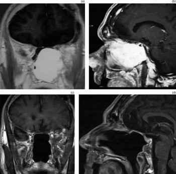
All the patients underwent computed tomography and magnetic resonance imaging (MRI) (figure 1). Angiography was not performed on a routine basis. The choice of whether or not to perform angiography was based on the MRI results: cases with clearly visible feeding vessels from the maxillary artery were selected for pre-operative angiography. If angiography confirmed the presence of these vessels, then pre-operative embolisation was performed 24 hours prior to surgery.
Pre-operative (a) coronal and (b) sagittal magnetic resonance imaging (MRI) scans showing a huge tumour involving the left infratemporal fossa. Post-operative (c) coronal and (d) sagittal MRI scans show the extent of clearance.

Endoscopic resection was carried out following a centripetal strategy, as previously described.Reference Castelnuovo, Battaglia, Locatelli, Delù, Sberze and Bignami7 Once the pedicle of the neoplasm was identified, a wide cuff of macroscopically normal mucosa was excised together with it. The underlying bone was then drilled down to ensure safe margins of resection. Frozen section analysis was always performed to check for the absence of neoplastic cells at the margins of the surgical resection. For tumours pedicled at the cribriform plate or ethmoidal roof, the bone was drilled down and then a skull base plasty performed, regardless of evidence of intra-operative cerebrospinal fluid (CSF) leakage.
Results
Our series included five men and five women, with a mean age at surgery of 59 years. The most common presenting symptoms were nasal obstruction (six patients), rhinorrhoea (four), epistaxis (three) and headache (three), while the most involved site was the ethmoid sinus. None of our patients presented with loco-regional or distant metastasis, either at diagnosis or during follow up.
Three out of 10 patients had previously been treated for the same disease before being referred to our department. In one of these cases, the disease seemed to originate extranasally, at the level of the lacrimal sac. This patient had been treated four times by an ophthalmologist before referral to us. This patient's history has already been described in detail in a previous paper.Reference Castelnuovo, Pagella, Delù, Benazzo and Cerniglia8 Another patient had previously been treated by a neurosurgeon, with removal of the intracranial, intradural portion of the lesion located lateral to and above Meckel's cave. In this patient, in agreement with the neurosurgeon, a staged resection was planned in order to reduce the risk of intracranial bleeding and to facilitate the extradural, transnasal removal of the remaining part. All patients underwent endoscopic, endonasal resection of the lesion. One patient underwent pre-operative embolisation of the tumour, due to the presence of major feeding vessels from the maxillary artery. Three patients required endoscopic skull base plasty after tumour resection.
Only one major post-operative complication was observed (stroke); for this reason, this patient was discharged after 14 days, with neurological sequelae. No other significant complications were observed. Patients were discharged after a mean hospitalisation duration of 6.2 days.
Recurrence of disease was noted in one patient (10 per cent) after 73 months of follow up; this patient subsequently underwent revision surgery (combined approach). Despite this, local recurrence manifested again, and the patient underwent a third revision procedure including orbital exenteration. In both these revision procedures, the intra-operative margin check was negative. Finally, this patient died of cerebrovascular disease, with the presence of local recurrence but no regional or systemic metastasis.
No adjuvant radiotherapy was given to any patient.
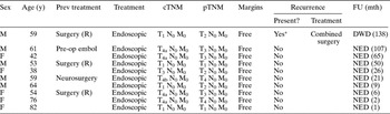
All patients but one (as detailed above) were alive and well after a mean follow up of 42.5 months (range one to 138 months). Detailed clinical data are given in Table I.
Patient data

*At 73 and 92 months. Y = years; prev = previous; cTNM = UICC (2002) tumour–node–metastasis; pTNM = UICC (2002) tumour–node–metastasis; FU = follow up findings; mth = months; M = male; F = female; R = recurrence; DWD = died with disease; NED = no evidence of disease; pre-op embol = pre-operative embolisation
Discussion
Approximately 15 per cent of all soft tissue haemangiopericytomas occur in the head and neck.Reference Barnes and Barnes9 In this region, haemangiopericytomas tend to prefer the nasal cavity and paranasal sinuses. These lesions are most frequently reported in the seventh decade of life, and the usual presentation includes nasal obstruction and epistaxis.Reference Thompson, Miettinen and Wenig10
Our patients' findings were essentially similar, although the mean age at diagnosis was 59 years. Despite the series being small, no difference regarding the incidence was observed between men and women, in agreement with other authors.Reference Kuo, Lin, Eng and Huang11
The standard diagnostic protocol for sinonasal haemangiopericytoma comprises endoscopic evaluation together with neuroradiological investigation. When necessary, additional investigations are conducted; angiography (and embolisation if necessary) is a valuable tool especially in extended cases.
From a histological viewpoint, an accepted definition and classification of haemangiopericytoma simply does not exist. While haemangiopericytomas share many immunohistochemical features with glomus tumours, and some authors even consider them to be a biological variant, others refer to the former as ‘haemangiopericytoma-like’ tumours.Reference Tse and Chan3, Reference Wilson, Hellquist, Ray and Pickles4, Reference Thompson, Miettinen and Wenig10 This designation implies that these tumours are related to, yet still distinct from, soft tissue haemangiopericytomas. Surprisingly, there is limited knowledge about the biological behaviour and natural history of haemangiopericytomas.
Furthermore, a distinction between benign and malignant soft tissue haemangiopericytomas cannot be made in all cases on the basis of histological findings alone.Reference Enzinger and Smith12, Reference Diaz-Flores, Gutierrez, Varela, Rancel and Valladares13 In this sense, from a prognostic point of view only severe nuclear pleomorphism and bone invasion seem to correlate with recurrence development.Reference Thompson, Miettinen and Wenig10 Notwithstanding this, these lesions do not seem to produce distant metastases; in patients who have died, it is not clear whether the cause of death was the disease itself or other, concomitant factors.Reference Thompson, Miettinen and Wenig10 Only exceptional cases have been reported to metastasise.Reference Gorestein, Facer and Weiland14
Our experience seems to confirm these observations. We observed only one death in our series, which was unlikely to be related to the presence of haemangiopericytoma. In this case, the unexpected element was the extreme tendency of the disease to recur, although our surgical procedures invariably produced a clean surgical field and histologically negative margins. In this patient, the biological behaviour of the disease was probably the critical factor leading to the development of a recurrence. This patient died with haemangiopericytoma, but probably not of haemangiopericytoma. In this sense, we support other authors' assertions that the malignancy of sinonasal haemangiopericytoma is probably only local.Reference Thompson, Miettinen and Wenig10 Therefore, until the development of effective molecular therapies, surgery should be considered the treatment of choice for sinonasal haemangiopericytoma, with a wide excision with tumour-free margins being the advisable option.Reference Compagno and Hyams2, Reference Catalano, Brandwein, Shah, Urken, Lawson and Biller15–Reference Billings, Fu, Calcaterra and Sercarz18
Clinically and biologically, haemangiopericytomas tend to have a benign course, although limited malignant potential has been reported.Reference Thompson, Miettinen and Wenig10 Therefore, we tried to stage our patients' lesions according to the UICC (2002) TNM staging system. However, in our series the T stage did not influence the surgical strategy or the outcome. All patients underwent surgery using an endoscopic technique, while the combined (external and endoscopic) approach was used for recurrent disease. On an anecdotal level, the poor prognostic significance of the T stage was confirmed by the fact that our only failure was a lesion staged initially as T2.
In our series, three patients had been previously treated and referred to us with recurrent disease. One of these patients, also described elsewhere, had been treated four times by an ophthalmologist.Reference Castelnuovo, Pagella, Delù, Benazzo and Cerniglia8 In this case, the disease seemed to originate from the lacrimal sac, and the patient was referred to us due to the presence of an intranasal recurrence. Unfortunately, this patient also suffered a recurrence after the initial revision surgery, and thus required a second revision procedure including orbital exenteration. In this case, despite the presence of negative surgical margins at the end of each procedure, the disease showed an extreme tendency to recur. It has been reported that infiltration of the surgical margins is the most important predictor of haemangiopericytoma recurrence; therefore, we always performed frozen section analysis during surgical resection.
Another of our patients had extensive disease and had previously been treated transcranially by the neurosurgeons to remove the intradural portion of the lesion; he presented to us with persistent disease in the nose and paranasal cavities. A staged procedure was planned in order to reduce the risk of intracranial bleeding, and to facilitate and make safer the endoscopic, transnasal resection of the extradural portion.
• Sinonasal haemangiopericytomas are rare, vascular lesions that arise in the nose and paranasal sinuses
• Wide surgical excision with free resection margins is the mainstay of treatment
• An endoscopic, endonasal approach is preferred for this tumour
Catalano et al. advocated a staging system akin to the Kadish staging system for olfactory neuroblastomas, as they found that the prognosis of sinonasal haemangiopericytoma patients strongly depended upon tumour stage and the completeness of the primary resection.Reference Catalano, Brandwein, Shah, Urken, Lawson and Biller15 We strongly agree with the need for a complete primary resection; in our experience, the outcome seems to be unrelated to T stage once the tumour has been removed with free margins of resection. We consider this last condition to be absolutely mandatory, although our fatal case poses several questions regarding the natural course and biological behaviour of these lesions. Molecular investigations, rather than histopathological ones, may perhaps provide answers to these questions. Nevertheless, we emphasise the critical importance of a safe ‘oncological plane’; for vascular lesions with a benign clinical course, this requires the removal of underlying bone by extensive drilling. Our patients' low incidence of recurrence, and the fact that all but one were alive and well at study completion, lead us to believe that sinonasal haemangiopericytoma has a benign clinical course in most cases. Therefore, a classification system based on T stage seems to be of poor prognostic value, and is inadvisable in our opinion. In fact, in most cases recurrence seems to be related directly to incomplete resection (although this was not properly confirmed in our series).
This means that, in skilled hands, a purely endoscopic resection can be safely proposed even for large lesions. On the basis of the ‘oncological plane’ concept, tumours which appear pedicled to the cribriform plate require mandatory drilling of the lamina cribra. In such cases, a skull base plasty is useful regardless of evidence of intra-operative CSF leakage. This situation was present in three patients in our series.
Pre-operative embolisation seems not to be a critical factor in sinonasal haemangiopericytoma management, especially in skilled hands. However, we advise pre-operative embolisation at least in cases of very large lesions or those with intracranial extension, in order to reduce surgical time and blood loss. A staged procedure can be proposed in cases with intracranial, intradural extension.
Conclusion
Sinonasal haemangiopericytomas are vascular lesions that arise in the paranasal region. They are biologically and clinically benign in most cases, but sometimes recur. Adequate surgery with free resection margins is the treatment of choice. Nowadays, with very few exceptions, the resection should be conducted endoscopically. Patient outcome is generally good, and the risk of recurrence seems to be related to the completeness of resection.