I. INTRODUCTION
Dopamine (DA) is a monoamine neurotransmitter found in mammalian brain and is essential for the normal functioning of the central nervous system.Reference Mao, Bao, Gan, Li and Niu1 It is one of the most important neurotransmitters and plays a significant role in the functioning of central nervous system, as also the renal and hormonal systems. Abnormal levels of DA causes neurological disorders such as Parkinson’s disease, schizophrenia, Alzheimer’s diseaseReference Grace2–Reference Weignberger and Lipska4 and Huntington’s chorea.Reference Wilson and Johnson5 DA is widely applied to the treatment of circulatory collapse syndrome, which is a medical emergency, caused by myocardial infarction, trauma, renal failure, recent cardiac surgery, or congestive cardiac failure.Reference Phillips, Stuber, Heien, Wightman and Carelli6,Reference Wightman, May and Michael7 Therefore, it is important to develop a sensor for the detection of DA. DA is an electroactive compound but its electroactivity suffers on conventional electrodes. Moreover, interferences from various other metabolic analytes like ascorbic acid (AA), which is essential for skin, connective tissues and immune system and uric acid (UA), which is the final oxidation product of the urine metabolism, present in real samples impose further limitations on the effectiveness of electrodes. Hence, there is a need to develop modified electrodes to improve the speed of electron transfer, sensitivity, detection limit and to lessen the electrocatalytic oxidation/reduction potential.
Various reports are available detailing the ongoing attempts for modification of electrode with materials having good electroactivity like metal nanoparticles,Reference Heinz, Przuntek, Winterer and Pietzcker8,Reference Yin, Shang, Meng and Ai9 conductive polymers,Reference Luczak and Brzezinska10–Reference Li, Chen and Zhang13 carbon nanotubes.Reference Tiwari and Singh14,Reference Tiwari, Singh, Gupta and Aggarwal15 Among these, carbon nanotubes (CNTs) have attracted much attention due to their high chemical stability, high surface area, unique electronic properties, high aspect ratio and relatively high mechanical properties.Reference Tiwari and Singh14 As electrode materials, CNTs can be used for promoting electron-transfer between the electroactive species and electrode and provide a novel method for fabricating chemical sensor or biosensor.Reference Tsai, Li and Liao16 CNTs promote electron transfer between electroactive species and electrode. However, CNT is not soluble in all solvents. It could be made soluble by noncovalent functionalizationReference Star, Stoddart, Streuerman, Diehl, Boukai, Wong, Yang, Chung, Choi and Heath17 or covalent modification.Reference Liu, Rinzler, Dai, Hafuer, Bradley, Boul, Lu, Iverson, Shelimov, Huffman, Macias, Shona, Lee, Colbart and Smalley18 In particular, wrapping of CNTs in polymeric chains has been shown to be useful for improving their solubility without impairing their physical properties.Reference Tiwari, Singh, Singh and Banks19 Nafion, a perfluorinated polymer is an electrochemistry-promoting polymeric binder. The inclusion of Nafion as a constituent of the nanocomposite for modifying the electrode leads to stable dispersion of multiwalled carbon nanotubes (MWCNTs).
Polyaniline (PANI) and its derivatives have attracted much attention in the field of conductive polymers due to their excellent electrical property and extensive potential applications. Phenylenediamines can be oxidized in two steps to the semiquinonediimine radicals and the quinonediimines as reported.Reference Siegfried, Baltruschata, Hönesb and Lungub20 They play a central role in hair dyingReference Dolinsky, Wilson, Wisneski and Emers21 and color photography,Reference Theys and Sosnovsky22 are an antiozonant for natural and synthetic elastomers, act as a stabilizer of polymers and as a monomer polymerization inhibitor and are also used as antioxidants e.g., in rubber or in fuels like gasoline or kerosene.Reference Huntink, Datta and Noordermeer23 Phenylenediamines are very susceptible to oxidative polymerization via oxidation of one or both amino groups to give linear azopolymers, polyaminoanilines, ladder polyphenazines, and phenazine/iminoquinonoid-unit containing polymers. Phenylenediamine derivatives are extensively used in electrochemical polymerization aloneReference Sayyah, Rehim, Deeb, Kamal and Azooz24 and along with other polymers like PANI etc.Reference Haldorai, Lyoo and Shim25 for the preparation of chemically modified electrodes. However, there are very less reports available in literature to use phenylenediamine derivatives as sensor.Reference Ravlchandran and Baldwin26,Reference Thompsona, Lawrencea, Davisb, Jiangc, Jonesc and Compton27 p-phenylenediamine, derivates of aniline are conductive material which play an important role in electron transfer system in biological reaction. The oxidation of p-phenylenediamine was first studied by BandrowskiReference Bandrowski28 and Erdmann.Reference Erdmann29 To the best of our knowledge, no reports are available that might have used N-(1,3-dimethylbutyl)-N′-phenyl-p-phenylenediamine in sensor.
In this report, we have prepared and characterized the N-(1,3-dimethylbutyl)-N′-phenyl-p-phenylenediamine (p-PDA) -functionalized MWCNTs via electrostatic interaction of amine groups of N-(1,3-dimethylbutyl)-N′-phenyl-p-phenylenediamine and sulfonic group of Nafion/MWCNTs composite. This prepared nanocomposite has been applied as an electrochemical sensor for the determination of neurotransmitter DA.
II. EXPERIMENTAL
A. Reagents and materials
MWCNTs were obtained from Aldrich (dia. 110–170 nm, +90% purity), Nafion (5 wt%) and N-(1,3-dimethylbutyl)-N′-phenyl-p-phenylenediamine were purchased from Aldrich (Sigma-Aldrich, Inc., St. Louis, MO). All other chemicals used were of analytical grade. All the solutions were prepared with triple distilled water. All the experiments were performed in 0.1 M phosphate buffer solution (pH 7.0).
B. Instruments
The electrochemical measurements were performed with CHI 630 C series electrochemical system. Three-electrode system was used for recording cyclic voltammetry (CV) where modified glassy carbon electrode (GCE) was used as working electrode, an Ag/AgCl filled with 3 M KCl as reference electrode and platinum (Pt) wire as counter electrode. All electrochemical measurements were carried out in 4 mL 0.1 M phosphate buffer solution (PBS), pH 7.0 and deaerated by bubbling nitrogen for 15 min prior to the experiments. SEM of the nanocomposite film was performed with JEOL840A; Japan. Transmission electron microscopy (TEM) measurements of MWCNTs and N-(1,3-dimethylbutyl)-N′-phenyl-p-phenylenediamine (p-PDA)/Nafion/MWCNTs nanocomposite were performed on a Tecnai G2 instrument. Ultraviolet-visible experiment was performed on (UV-Vis; Hitachi 3900, Techcomp Ltd., Hong Kong). Infra red spectroscopy study was performed on Varian 3100.
C. Preparation of N-(1,3-dimethylbutyl)-N′-phenyl-p-phenylenediamine (p-PDA)/Nafion/MWCNTs nanocomposite
MWCNTs were purified by hydrochloric acid treatment for 1 h before using. Nanocomposite was prepared by simple ultrasonication. First, 0.2 mg of purified MWCNTs were dispersed in a 1 mL of 1% w/v Nafion with ultrasonication at room temperature for 20 min. Subsequently, 3 mg of p-PDA were added in MWCNTs/Nafion composite and ultrasonicated 15 min till a well dispersed homogeneous suspension of p-PDA/Nafion/MWCNTs composite has been formed.
D. Modification of GCEs with p-PDA/Nafion/MWCNTs nanocomposite
GCEs were polished with alumina slurry and then ultrasonicated in distilled water and allowed to dry at room temperature. Five microliters of p-PDA/Nafion/MWCNTs solution was dropped on the pretreated GCE and allowed to air-dry at room temperature. The prepared electrode was washed with triple distilled water twice to remove nonadsorbed or loosely bound p-PDA molecules.
III. RESULTS AND DISCUSSION
A. Interaction of the MWCNTs with p-PDA
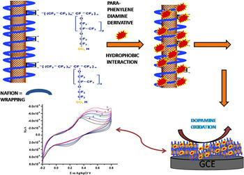
Although CNTs have very exciting properties as discussed earlier, the wall of carbon nanotubes are highly hydrophobic in nature, hence are not soluble in different solvents. To overcome this difficulty various approaches have been taken as reported in the literatureReference Star, Stoddart, Streuerman, Diehl, Boukai, Wong, Yang, Chung, Choi and Heath17,Reference Liu, Rinzler, Dai, Hafuer, Bradley, Boul, Lu, Iverson, Shelimov, Huffman, Macias, Shona, Lee, Colbart and Smalley18 and the interaction of Nafion for the solubilization of MWCNTs is one of them.Reference Tiwari, Singh, Singh and Banks19 Nafion is an ionomer consisting of hydrophobic perfluoroalkyl chain and a hydrophilic sulfonic group. In the formation of nanocomposite, it is proposed that the perfluoroalkyl chain of Nafion interact hydrophobically with the hydrophobic wall of MWCNTs resulting in the wrapping of Nafion to MWCNTs which further interact with phenyl rings hydrophobically and electrostatically with positively charged ammonium ion of p-PDA, resulting in the formation of nanocomposite as presented in Scheme 1.
B. Physical characterization of p-PDA/Nafion/MWCNTs nanocomposite
1. Morphological characterization of p-PDA/Nafion/MWCNTs nanocomposite
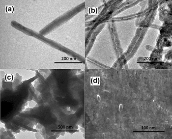
TEM results show that while the pure MWCNTs do not possess any type of adsorption [see Fig. 1(a)], a film-like structure can be seen on the wall of MWCNTs after modification with Nafion [see Fig. 1(b)] indicating that the Nafion molecules are chemically adsorbed on the MWCNTs and prevent their reaggregation. The Fig. 1(c) shows TEM micrograph of prepared p-PDA/Nafion/MWCNTs nanocomposite in Nafion matrix. Some dark spots could be observed which are probably due to interaction of positively charged p-PDA on negatively charged Nafion-MWCNTs. The scanning electron microscopy (SEM) images of the prepared p-PDA/Nafion/MWCNTs nanocomposite [see Fig. 1(d)] shows some rough yet homogeneous and well-dispersed patterns at the electrode surface.
(a) TEM image of pure MWCNTs (b) TEM image of nafion modified MWCNTs (c) TEM micrograph of prepared p-PDA/Nafion/MWCNTs nanocomposite in nafion matrix (d) Scanning Electron Micrograph of p-PDA/Nafion/MWCNTs nanocomposite film.

2. Infrared spectroscopy studies of p-PDA/Nafion/MWCNTs nanocomposite
The adsorption of p-PDA to the wall of MWCNTs is also confirmed by comparing the IR spectra of p-PDA, Nafion, MWCNTs/Nafion and p-PDA/Nafion/MWCNTs nanocomposites. Fig. S1 shows the absorption IR spectrum of p-PDA (see Supplementary material I) which contains peaks in the region of 3186–3375 cm−1 which is probably due to substituted amine group; 3008 and 1516 cm−1 which is attributed to C–H stretching and bending vibration of alkyl group of PDA respectively. The peak at 1637 cm−1 is attributed to aromatic C=C str of PDA while the peak at 1264 cm−1 is most probably due to C–N str vibration of p-PDA.
The absorption bands of Nafion contain peaks at 3426 and 1241 cm−1 attributed to –OH stretching and –CF2 stretching respectively. The peak at 1630 cm−1 is assigned to –OH bending whereas the peaks at 1052, 971, and 637 cm−1 are due to –SO3, COC and C–S stretching vibrations respectively (Supplementary material II, curve a). Upon addition of MWCNTs to Nafion (Supplementary material II, curve b); the peaks at 971 and 1052 cm−1 due to –SO3 stretching and COC stretching respectively shifted to 984 and 1029 cm−1 while the peak due to C–F stretching shifted to 1230 cm−1 (1241 cm−1 in Nafion). The shift in C–F stretching vibration shows the interaction of hydrophobic per fluorocarbon chain to MWCNTs. The sharp peak at 1634 cm−1 is due to combined effect of O–H bending and C=C of MWCNTs in the spectra of MWCNTs/Nafion. The peak at 637 cm−1 due to C–S stretching vibration in Nafion shifted to 529 cm−1 after interaction with MWCNTs.
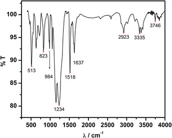
Upon the addition of p-PDA to Nafion coated MWCNTs (see Fig. 2); the peak in the region of 3184–3375 cm−1 of p-PDA due to –NH stretching is shifted to 3335–3443 cm−1 which may be due to electrostatic interaction of positively charged amine group of p-PDA and sulfonic group of MWCNTs/Nafion. Further, shift in the peaks position of C–N str vibration of p-PDA (1264 cm−1 shifted to 1234 cm−1) is attributed to the combined effect of C–F stretching and C–N stretching in p-PDA, also confirms the interaction of MWCNTs/Nafion to p-PDA. Hence, the data obtained supports the formation of p-PDA/Nafion/MWCNTs nanocomposite.
Infrared spectra of p-PDA/Nafion/MWCNTs nanocomposite.

3. UV-Visible spectroscopy studies of p-PDA/ Nafion /MWCNTs nanocomposite
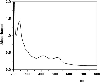
The formation of p-PDA/Nafion/MWCNTs nanocomposite was further studied by UV-Vis spectroscopy in the range of 200–800 nm. The UV–Vis spectra were also recorded for the p-PDA aqueous solution (see Supplementary material III), MWCNTs aqueous solution (data not shown), p-PDA/Nafion/MWCNTs aqueous solution (see Fig. 3). Figure S3 shows the UV–Vis spectrum of free p-PDA aqueous solution where the major bands attributed to π-π* transition of phenylene group of PDA is found to be at 247 nm with a shoulder at 283 nm which is characteristic of p-phenylendiamine derivatives. The peak at 512 nm is proposed to be due to n-π* transition in p-PDA structure.
After interaction with Nafion/MWCNTs (see Fig. 3) the peak at 247 shifted to 241 nm and the shoulder at 243 nm diminishes. Moreover, the peak at 512 nm shifted to 518 nm and a new peak appears at 416 nm, which may be attributed to transition between π bond of MWCNTs and p-PDA. The decrease in intensity of peaks observed is due to adsorption of p-PDA to the Nafion wrapped MWCNTs, which is due the decrease in free p-PDA complex in aqueous solution. Hence, the above results further corroborate the attachment of p-PDA molecule on the MWCNTs template and the growth of tubular nanocomposite.
UV-Visible spectroscopy of p-PDA/Nafion/MWCNTs nanocomposite aqueous solution.

4. Electrochemical characterization of p-PDA Nafion /MWCNTs nanocomposite
Phenylenediamine derivatives are redox-active, conducting compounds and have ability to electropolymerize at electrode surface and hence have been utilized in the formation of chemically modified electrodes. Among these o-PDA and m-PDA do not possess reduction wave in the electrochemistry of o-PDA and m-PDA because the oxidized intermediate species is not sufficiently stable to enable the detection of corresponding current wave for the reduction of this oxidized intermediate species. While in the case of p-PDA both oxidation and reduction peaks appear because the stability of oxidized species enables to generate reduction peak by the reduction of this oxidized intermediate. This behavior of p-PDA is observed because during electrochemical oxidation of p-PDA polymerization process is inhibited by the blocking of paraposition,Reference Heineman, Wieck and Yacynych30–Reference Ye, Zhang and Zhou32 which is not possible in the other two cases. This property of p-PDA makes it suitable redox mediator to be utilized in the formation of nanocomposite material along with carbon nanotube in its monomer state.
Figure S4 (Supplementary material IV) shows the cyclic voltammograms of the p-PDA/Nafion/MWCNTs nanocomposite modified GCE in the potential range of −0.4 to 0.8 V at different scan rates (10, 20, 50, 60, 100, 150, and 200 mV/s) in 0.1 M phosphate buffer solution having pH 7.0, while no faradic response was observed at bare GCE, Nafion modified GCE and MWCNTs/Nafion modified GCE (data not shown). To evaluate the contribution of MWCNTs, the control experiment was also performed but due to unstable electrode material as high leaching of p-PDA occurred from Nafion film the CV could not be recorded.
The cyclic voltammogram of p-PDA/Nafion/MWCNTs nanocomposite modified electrode displays one redox peak with anodic peak at 275 mV and cathodic peak at 201 mV versus Ag/AgCl reference electrode at the scan rate of 10 mV/s. The peak separation was observed to be 74 mV at the scan rate of 10 mV/s (versus Ag/AgCl). The formal potential was calculated by the midpoint of cathodic and anodic peaks versus Ag/AgCl (Epa + Epc/2) at the scan rate 10mV/s and found to be 238 mV. The peak-to-peak separation increases upon increasing scan rates and the peak current ration ipa/ipc has found to be more than 1. The effect of variation of scan rate (ν) and peak currents (ipa and ipc) was investigated in the range of 10–200 mV/s. Inset I to Fig. S4 displays the Randles-sevick plot which shows a linear relationship between peak currents (i.e., ia and ic) with square root scan rates indicating the electrochemical process is diffusion controlled in nature. The surface coverage area (Γ aq) for the electroactive species was estimated by: Γ aq= Q/nFA,Reference Bard and Faulkner33 where Q is the charge obtained by integrating the anodic peak at low scan rate (10 mV/s), n is the number of electrons involved per p-PDA molecules (n = 1) and A is the geometric electrode area. In the present case, the value of Γ aq was determined to be 4.25 × 10⁻¹⁰ mol/cm2.
5. Electrocatalytic oxidation of DA on the p-PDA/Nafion/MWCNTs nanocomposite-modified electrode
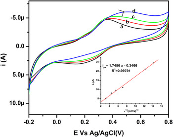
Figure 4 shows the electrochemistry of p-PDA/Nafion/MWCNTs nanocomposite modified GCE in the absence (curve a) and presence of 125, 250, and 500 µM (curve b, c, d) DA in phosphate buffer solution (pH 7.0) at the scan rate of 10 mV/s. The oxidation of DA starts at 0.34 V and attains maxima at 0.50 V. It can be seen that DA oxidation shows enhanced anodic peak as expected and already reported.Reference Pandey, Chauhan and Singh34 The oxidation of DA involves two electron transfer and generation of orthoquinone; then orthoquinone derivative undergoes cyclization to indole which is not able to reduce completely in reverse scan. Hence, the oxidation peak enhances and corresponding reduction peak decreases continuously upon addition of DA.
Cyclic voltammetric response of dopamine : curve (a) in the absence and curve (b) (c) (d) in the presence of 125,250,500 μM concentration of dopamine at the p-PDA/Nafion/MWCNTs nanocomposite modified GCE in 0.1 M phosphate buffer solution (pH 7.0).

The role of MWCNTs in DA oxidation was ascertained by obtaining response also on p-PDA/Nafion-modified GCE in the presence of 250 µM DA (Inset II to Fig. S4). From the results obtained on the composite modified electrode in the presence of 250 µM DA (see Fig. 4, curve c), it is clear that the presence of both p-PDA and MWCNTs results in the improvement of the response and the current density of DA oxidation increases. Moreover, the presence of MWCNTs improves the electrochemical applicability of p-PDA. Taking all these precedence into account it can be inferred that the interaction between p-PDA and MWCNTs might be the decisive factor for the good response of the sensor; acting as electron mediators, improving the electron transfer, fixing the film, avoiding the leaching out of individual material from the electrode surface and increasing the stability of the electrode as reported earlier.Reference Tiwari, Singh, Gupta and Aggarwal15 Inset to Fig. 4 shows the variation of peak current versus square root of scan rate in the presence of 125 µM DA which shows that the oxidation of DA at p-PDA/Nafion/MWCNTs modified electrode is diffusion controlled in nature.
C. Optimization of experimental conditions
1. Calibration plot
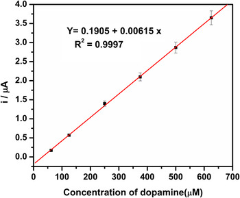
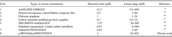
Figure 5 shows the linear calibration curve in the range 62–625 µM of DA in 0.1 M PBS at pH 7 with correlation coefficient of 0.9997 (n = 6) and the sensitivity of composite modified electrode was observed to be 0.006 µA/µM. The detection limit is around 5 µM at signal to noise ratio of 3 which is better than other reported worksReference Yin, Shang, Meng and Ai9,Reference Zheng, Ye, Zhou, Zhang and Yu35–Reference Thiagarajan and Chen39 as indicated in Table I.
Calibration plot of dopamine detection at p-PDA/Nafion/MWCNTs nanocomposite modified GCE in 0.1 M phosphate buffer solution (pH 7.0).

Comparison of linear range and detection limit of some modified electrode and p-PDA/Nafion/MWCNTs nanocomposite modified GCE for the oxidation of DA.

2. Effect of interferences
Selectivity of the sensor was evaluated by performing interference study. DA oxidation was done in the presence of hydrogen peroxide (H2O2), urea, glucose, ascorbic acid, oxalic acid, phenol as interfering substances. It is found that the tested substances did not significantly interfere with the determination of DA oxidation. The current obtained for each interfering substance was recorded and compared it with the current obtained in the presence of DA. It could be easily concluded that tested substances did not significantly interfere with the determination of DA.
3. Effect of p-PDA and MWCNTs concentration on sensor performance
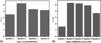
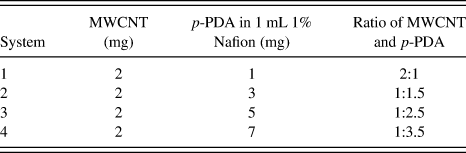
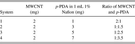
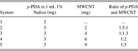
We also studied the effect of varying the concentration of p-PDA and MWCNTs in p-PDA/Nafion/MWCNTs nanocomposite. For studying this effect, p-PDA and MWCNTs have been taken in different ratios for preparing nanocomposite as indicated in Table II. The results obtained for the oxidation of 250 µM DA at subsequent electrodes [Fig. 6(a) for p-PDA and (b) for MWCNTs] indicates that the optimized ratio used for the preparation of p-PDA/Nafion/MWCNTs nanocomposite in the present work is most suitable for effective working of the electrode.
Bar graph (a) Detection of 250 μM concentration of dopamine by varying different concentration of p-PDA and keeping MWCNTs constant, (b) Detection of 250 μM concentration of dopamine by varying different concentration of MWCNTs and keeping p-PDA constant.

Schematic representation of interaction among N-(1,3-dimethylbutyl)-N'-phenyl-p-phenylenediamine, nafion and multiwalled carbon nanotubes for the formation of nanocomposite.

Preparation of nanocomposite using p-PDA and MWCNTs taken in different ratios.
[A] preparation of nanocomposite by varying amount of p-PDA and keeping amount of MWCNTs constant.
[B] Preparation of nanocomposite by varying amount of MWCNTs and keeping amount of p-PDA constant
4. Performance of sensor
The stability of electrode was studied by the effect of observable changes obtained by cycling 20 repeated cycles in CV of p-PDA/Nafion/MWCNTs nanocomposite modified GCE. The result justifies the stability of p-PDA nanocomposite incorporated in Nafion film along with MWCNTs. When 20 continuous cycles scan was carried out at 10 mV/s scan rate, only 4.5% decrease of the initial response at 125 µM was recorded. The RSD of 2.5% was observed for three successive measurements of one nanocomposite modified electrode at 125 µM DA indicating good reproducible behavior of the proposed sensor. The long-term stability of the proposed sensor was also studied. Lifetime is more than 4 months when the electrode was kept at ambient conditions, when not in use.
IV. CONCLUSION
We have utilized a simple method to prepare p-PDA/Nafion/MWCNTs nanocomposite. The new material exhibits excellent electrocatalytic properties due to good electrical conductivity of MWCNTs combined with excellent film-forming ability. Direct coating of the prepared material was used as sensor of DA, which showed fast charge transfer rate, good stability and high sensitivity.
ACKNOWLEDGMENTS
The present work has been performed by funds sanctioned by DBT, India. The authors are grateful for this financial support. The author is also highly grateful to Dr. Madhu Yashpal, Institute of Medical Science, BHU, Varanasi for providing TEM facility.