In his original exposition, ShoneReference Shone, Sellers, Anderson, Adams, Lillehei and Edwards 1 described in 1963 a constellation of left heart lesions characterised by mitral valve stenosis, most often supra-annular mitral ring, with associated subaortic stenosis with or without coarctation of the aorta. Thereafter known as Shone’s complex or Shone’s syndrome, the clinical setting is now commonly characterised by a congenital mitral valve anomaly resulting in stenosis, with resultant downstream left heart underdevelopment including subvalvar aortic stenosis, valvar aortic stenosis, bicuspid aortic valve, and/or coarctation of the aorta.Reference Tchervenkov, Tahta, Jutras and Beland 2 As in the original description, many of these patients have other cardiac lesions including atrial and ventricular septal defects.Reference Bolling, Iannettoni, Dick, Rosenthal and Bove 3 Most reports recently have focussed on the surgical outcomes in small series of infants and children with Shone’s complex.Reference Tchervenkov, Tahta, Jutras and Beland 2 – Reference Brown, Ruzmetov and Vijay 6 These reports illustrate that these patients often require repeat surgical and catheter-based interventions. The late outcomes in this population are not well described, as single-centre experiences with long-term follow-up have not been reported. On the other hand, a number of surgical series indicate that the 5-year survival may be as low as 65–80%.Reference Brauner, Laks, Drinkwater, Scholl and McCaffery 5 , Reference Malhotra, Lacour-Gayet and Campbell 7 , Reference Ikemba, Eidem and Fraley 8 Survival has been postulated to be linked to eventual growth of left heart structures including the mitral valve, the left ventricle itself, as well as the aortic valve annulus. Postnatal left heart growth is seen in a number of other CHD lesions including neonatal aortic valve stenosis and coarctation of the aorta.Reference Petit, Ing, Mattamal, Pignatelli, Mullins and Justino 9 – Reference Page and Levine 12 The degree of catch-up growth in left heart structures in Shone’s complex patients is unknown. Unfortunately, in the management of children with Shone’s complex, many questions persist. The role of the atrial septal defect may be hazardous, although it may allay left atrial hypertension.Reference Petit, Justino and Fraser 13 Therapy for pulmonary hypertension in the setting of Shone’s complex is also unclear. We sought to characterise the outcomes, survival, and the need for repeat interventions in our institution over a 20-year period, as well as to evaluate the role of the atrial septal defect in the potential for left heart structural growth in children with Shone’s complex.
Materials and methods
Patients were considered to have Shone’s complex if they had congenital anatomic mitral valve stenosis in addition to at least one of the following: (1) subvalvular aortic stenosis, (2) valvar aortic stenosis, (3) supravalvar aortic stenosis, (4) aortic arch hypoplasia, or (5) coarctation of the aorta. Patients with mitral and aortic stenosis who were palliated with single-ventricle strategy were considered to have hypoplastic left heart syndrome and were excluded from this study. Furthermore, patients with complete atrioventricular canal defect, double-outlet right ventricle, or surgical intervention before presentation to our institution were excluded.
Mitral valve anatomy was classified as follows: (1) typical congenital mitral stenosis, consisting of thickened mitral valve leaflets with diminished diastolic excursion; (2) parachute mitral valve, consisting of a single papillary muscle; (3) mitral valve arcade, consisting of shortened chordae with absent/diminished inter-chordal space; and (4) supra-annular mitral valve ring. Some patients were noted to have more than one type of mitral valve disease. Associated cardiac lesions that existed at presentation, including ventricular septal defect and atrial septal defect, were noted. Patient age and weight at initial presentation were noted, and the duration of follow-up was noted. For patients lost to follow-up, the last encounter at our centre was considered the last date of follow-up for statistical analysis.
The cardiology database was reviewed for each surgical and catheter-based intervention, and attention was focussed on (1) mitral valve surgical repair/balloon valvuloplasty, (2) mitral valve replacement, and (3) total number of interventions. Operative reports were reviewed for details, including surgical closure of the atrial septal defect. Date and age at death or heart transplantation were also noted.
The initial echocardiographic images at the time of diagnosis were reviewed for each patient. Measurement of left heart structures was performed by two independent reviewers (R.P. and C.D.), and z-scores were calculated on the basis of the Colan z-score data for the following: (1) mitral valve annulus dimension, (2) left ventricular end-diastolic dimension, (3) left ventricular end-systolic dimension, and (4) aortic valve annulus dimension.Reference Sluysmans and Colan 14 Final echocardiograms were also reviewed, and the same measurements were assessed for each patient.
Presence of an atrial septal defect was noted in the baseline echocardiogram and in the postoperative echocardiogram for patients who underwent surgical intervention. Residual atrial-level shunting after surgery (yes or no) was noted. Doppler assessment of the mean trans-mitral gradient was also noted. Right ventricular pressure estimate based on the tricuspid regurgitation jet was also reviewed. Patients with ventricular septal flattening on the parasternal short-axis view or with a tricuspid regurgitant jet >3 m/second were considered to have pulmonary hypertension, in the absence of pulmonary arterial anomaly.
For the subset of patients who underwent cardiac catheterisation, haemodynamic data were reviewed. Patients with right ventricular systolic pressure >30 mmHg or with pulmonary vascular resistance >4 Wood units were considered to have pulmonary hypertension. The left atrial pressure, trans-mitral valve gradient, and left ventricle end-diastolic pressure were also recorded. Intervention type, when applicable, was categorised as mitral valvuloplasty, aortic valvuloplasty, coarctation dilation, angioplasty and/or stenting, and atrial septostomy.
Statistical analyses were performed using SAS 9.4 (SAS Institute, Cary, North Carolina, United States of America). Statistical significance was assessed at the 0.05 level unless otherwise noted. Descriptive statistics were calculated for all variables of interest and included means and standard deviations, medians and interquartile ranges, and counts and percentages, when appropriate. Normality of continuous variables was assessed using histograms, normal probability plots, and the Anderson–Darling test for normality. Continuous data were compared between groups of interest using two-sample t-tests when normally distributed. Non-normal data were compared using Wilcoxon’s ranked-sum test. Comparisons between categorical variables were performed using the χ2 test. When expected cell counts were small (<5), an exact test was used in place of the χ2 test. Change in left heart structural dimensions was compared using Wilcoxon’s signed-rank tests. Univariate analysis of factors associated with left heart structural growth was performed using linear regression models. Factors included mitral valve classification type, the presence of initial atrial septal defect, and the presence of pulmonary hypertension. Relationships between continuous measurements were further quantified using Pearson’s correlation coefficient and associated 95% confidence intervals. Kaplan–Meier survival curves were generated to estimate freedom from death or heart transplantation and freedom from repeat intervention. The log-rank test was used to compare survival distributions between risk factor groups. Our institution’s review board approved this study.
Results
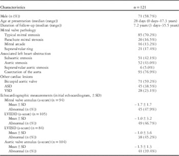
Between May, 1978 and September, 2010, 121 patients with Shone’s syndrome presented to our institution and were followed-up for a median of 7.2 (0.01–35.5) years. Patient characteristics at presentation are shown in Table 1. The median age at presentation was 28 days (0 days–17.3 years). An atrial septal defect was present in 45 patients (38.5%) preoperatively. Typical mitral valve stenosis was the most common category of mitral stenosis, which was seen in 85 (70.2%) patients.
Patient characteristics.

ASD=atrial septal defect; LVEDD=left ventricular end-diastolic dimension; LVESD=left ventricular end-systolic dimension; VSD=ventricular septal defect
An “abnormal” z-score is classified as a z-score of <−2.0
Preoperative catheterisation data were available for 60 patients (Table 2). Patients who had preoperative atrial septal defects had similar left ventricular end-diastolic pressure and mean pulmonary artery pressures compared with patients without an atrial septal defect; however, the mean left atrial pressure was lower in patients with an atrial communication (12.9±6.8 mmHg) compared with patients without atrial septal defects (19.5±6.9) (p=0.03). In addition, pulmonary vascular resistance was lower in those with an atrial septal defect (p=0.03).
Preoperative catheterisation characteristics.

Bold value indicates statistical significance
ASD=atrial septal defect (defined as any left-to-right atrial shunt); LVEDP=left ventricle end-diastolic pressure; PVR=pulmonary vascular resistance; Qp:Qs, ratio of pulmonary-to-systemic blood flow
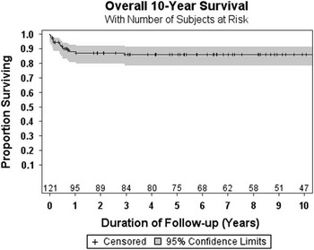
There were 17 deaths among the 121 patients (14.0%). The median age at death was 4.9 months (3 days–15.5 years); seven of the deaths were of infants before any intervention. The 10-year survival for the entire cohort was 86.1% (95% confidence interval: [78.2–91.2%]), and 14 of the 17 deaths occurred within the first 10 months of life (Fig 1). In our study population, two patients underwent orthotopic heart transplantation at 5 months and 9 months of age, respectively. Both of them died shortly after their transplant. Therefore, the 10-year, transplant-free survival remains the same as the overall survival at 86.1%.
The overall cohort survival. Kaplan–Meier survival curve of the entire Shone’s complex cohort demonstrates that at 10 years the survival is 86%. Most of the mortality occurs within the 1st year of life. Shaded region is the 95% confidence interval.

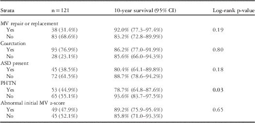
The cohort of patients who underwent mitral valve repair or replacement had similar survival (92.0%; 95% confidence interval: [77.3–97.4%]) compared with the remainder of the cohort (83.2%; 95% confidence interval: [72.8–89.9%]). Pulmonary hypertension was common in our cohort with 53 patients (44.9%) meeting the predefined diagnostic criteria. The 10-year survival for those with pulmonary hypertension was significantly lower (78.7%; 95% confidence interval: [64.8–87.6%]) than those without pulmonary hypertension (93.6%; 95% confidence interval: [83.7–97.5%]; p=0.03). The presence of an atrial septal defect preoperatively and an abnormal initial mitral valve annulus z-score were not associated with death or heart transplantation on survival analysis (Table 3).
Ten-year survival by strata.

Bold value indicates statistical significance
ASD=atrial septal defect; CI=confidence interval; MV=mitral valve; PHTN=pulmonary hypertension
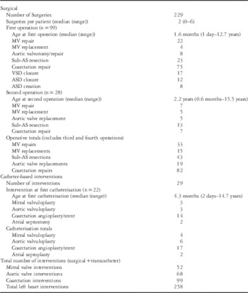
The 121 patients underwent a median of 2 (0–6) surgical or catheter-based procedures per patient, and in total there were 258 procedures including 229 operations and 29 interventional catheterisations (Table 4). Among all, 17 patients (14.0%) underwent no interventions, whereas 33 (27.3%) patients underwent mitral valve surgical repair, and only 15 (12.4%) patients underwent mitral valve replacement. The median age at the first surgical intervention was 1.6 months (1 day–12.7 years), whereas the median age at first catheterisation was 4.3 months (2 days–14.7 years). The most common interventions at initial surgical intervention included surgical coarctation repair (n=75), subaortic resection (n=23), and mitral valve repair (n=22). Of the 99 patients who underwent initial surgical intervention, 28 (28.3%) underwent at least one additional surgical intervention.
Surgical and catheter interventions.

AS=aortic stenosis; ASD=atrial septal defect; MV=mitral valve; VSD=ventricular septal defect
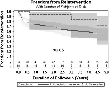
Catheter-based interventions included coarctation dilation/stent (n=17), balloon aortic valvuloplasty (n=6), mitral valvuloplasty (n=4), and atrial septoplasty (n=2). Table 4 illustrates the number and types of interventions on the left heart. Coarctation of the aorta was associated with increased risk of re-intervention (p=0.05) (Fig 2).
The freedom from re-intervention. Kaplan–Meier curve demonstrating freedom from repeat left heart intervention reveals that patients with coarctation of the aorta are at a greater risk for repeat intervention. Shaded region is the 95% confidence interval.

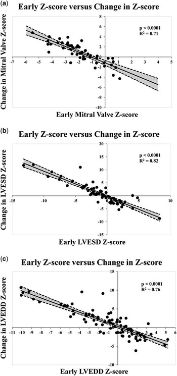
Although the range of baseline left heart structure dimensions was large, there was normalisation over time noted in mitral valve annulus z-scores, in left ventricular end-diastolic dimension z-scores, and in aortic valve annulus z-scores (Table 5). In addition, with the exception of the aortic valve annulus z-score, regression analysis revealed that higher baseline left ventricular end-diastolic dimension z-scores, left ventricular end-systolic dimension z-scores, and mitral valve z-scores were associated with diminution of z-scores over time, with the opposite being true of lower baseline z-scores for mitral valve, left ventricular end-diastolic dimension, and left ventricular end-systolic dimension (Fig 3). This suggests normalisation of left heart dimensions over time. Patients who were left with a residual atrial communication after initial surgery demonstrated a lesser increase in the z-score of the left ventricular end-diastolic dimension (p<0.01) and left ventricular end-systolic dimension (p<0.01) over time. Finally, there was an inverse relationship between the age at presentation and the change in aortic valve z-score (r=−0.27; 95% confidence interval: (−0.49 to 0.00); p=0.05), mitral valve annulus z-score (r=−0.44; 95% confidence interval: (−0.64 to −0.18); p<0.01), left ventricular end-diastolic dimension z-score (r=−0.49; 95% confidence interval: (−0.63 to −0.30); p<0.01), and left ventricular end-systolic dimension z-score (r=−0.36; 95% confidence interval: (−0.56 to −0.11); p=0.01).
Left heart dimensions over time. Normalisation of the mitral valve annulus ( a ), left ventricular end-systolic dimension (LVESD) ( b ), and left ventricular end-diastolic dimension (LVEDD) ( c ) over time.

Matched echocardiogram z-scores indicating interval growth and normalisation of left heart structures over time.

Bold value indicates statistical significance
Ao valve=aortic valve; LVEDD=left ventricular end-diastolic dimension; LVESD=left ventricular end-systolic dimension; MV=mitral valve
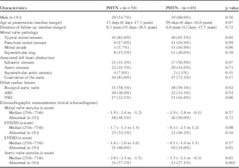
Pulmonary hypertension was common among the cohort, with 53 patients (43.8%) having pulmonary hypertension based on catheterisation or echocardiographic data. Patients with pulmonary hypertension had a lower 10-year survival (78.7%; 95% confidence interval: [64.8–87.6%]) compared with those without (93.6%; 95% confidence interval: [83.7–97.5%]; p=0.03). Although typical mitral valve stenosis was associated with pulmonary hypertension in our cohort (p=0.01), there were no other left heart obstructive or other cardiac lesions associated with pulmonary hypertension (Table 6). The subgroup of patients with pulmonary hypertension had lower left ventricular end-systolic dimension (p=0.03) and aortic valve annulus (p=0.03) z-scores. There was no statistical relationship between having pulmonary hypertension and having an atrial septal defect (p=0.54).
Demographics and echocardiogram measurements by pulmonary hypertension status.

ASD=atrial septal defect; LVEDD=left ventricular end-diastolic dimension; LVESD=left ventricular end-systolic dimension; PHTN=pulmonary hypertension; VSD=ventricular septal defect
An “abnormal” z-score is classified as a z-score of <−2.0
Discussion
This study comprises the largest reported institutional experience of children with Shone’s complex. The medical and surgical challenges are augmented by inter-patient anatomical variations that fall under the general classification of Shone’s complex. Our study demonstrates that children with Shone’s complex are at risk of multiple interventions, but that mortality is generally limited to the period of infancy. These patients bear a high burden of repeat procedural intervention. Children with Shone’s complex appear to experience significant growth of left heart structures over time.
Comparison of outcomes among reports of Shone’s complex relies upon common criteria for diagnosis. Although the original report by ShoneReference Shone, Sellers, Anderson, Adams, Lillehei and Edwards 1 described supravalvular mitral ring and subaortic stenosis in all patients – with coarctation and/or parachute mitral valve in four of the eight cases – most series have defined the complex as mitral valve disease of varying category plus at least one additional level of left heart obstruction.Reference Bolling, Iannettoni, Dick, Rosenthal and Bove 3 , Reference Brauner, Laks, Drinkwater, Scholl and McCaffery 5 – Reference Ikemba, Eidem and Fraley 8 In accordance, we defined our cohort similarly.
Our cohort appears to have a significantly better long-term survival compared with other published series. MalhotraReference Malhotra, Lacour-Gayet and Campbell 7 reported that in a series of 43 patients with Shone’s complex, the 10-year survival was only 61.3% for those who underwent cardiac transplantation as compared with 83.1% among those who underwent staged repair. It is important to note that 30% of patients in that series were managed with cardiac transplantation. The authors warn against primary transplant as the therapeutic option because of a donor supply shortage and the extended time on the waiting list predisposes patients to potentially irreversible congestive heart failure and pulmonary hypertension. This highlights the variable nature and severity of the Shone’s complex lesion, in addition to the variability in practice among differing institutions. Likewise, other smaller published series with similar exclusion criteria have noted poor midterm survival. A midterm study by Brauner et alReference Brauner, Laks, Drinkwater, Scholl and McCaffery 5 reported that among 19 patients with Shone’s complex, the 7-year survival was 73%, despite having an older age at initial operation. Interestingly, they did not find an association between pulmonary hypertension and mortality, which contradicts the findings of the present study; however, they have noted a reversal in the degree of pulmonary hypertension in long-term survivors, which they partially attribute to improvement in perioperative care. Although previous reports have linked mortality to the need for mitral valve surgery, we did not find such a relation.Reference Bolling, Iannettoni, Dick, Rosenthal and Bove 3 , Reference Ikemba, Eidem and Fraley 8 Although not all of our patients underwent surgical intervention for mitral valve pathology, those who did underwent repeated interventions on the mitral valve. This provides further evidence that an aggressive approach to repairing obstructive lesions may improve long-term survival.
The cause for variation in survival among centres is unclear. As there are differing reports on the association between mortality and need for mitral valve intervention, the anatomical aetiology of mitral valve obstruction should be questioned. In previous reports, parachute mitral valve and supramitral ring are the most common types of mitral stenosis; however, in the present study, typical mitral stenosis was found in the majority of patients. In addition, it may relate to the surgical era, as multiple previously published series include patients from the 1960s through the 1980s.Reference Bolling, Iannettoni, Dick, Rosenthal and Bove 3 , Reference Brauner, Laks, Drinkwater, Scholl and McCaffery 5 , Reference Brown, Ruzmetov and Vijay 6 In contrast, 115 out of the 122 patients in our cohort presented between 1990 and 2010 – an era of improved understanding of anatomical variation, intraoperative diagnostic testing, and surgical refinement.
Traditionally, the criteria followed at each centre for pursuing two-ventricle physiology versus single-ventricle palliation have not been uniform. In addition, multiple previously published series are from a time when the Norwood-type palliation was not commonly offered at most centres.Reference Bolling, Iannettoni, Dick, Rosenthal and Bove 3 , Reference Brauner, Laks, Drinkwater, Scholl and McCaffery 5 , Reference Brown, Ruzmetov and Vijay 6 As a result, patients with more significant mitral stenosis and/or hypoplasia of the left ventricle may have undergone promotion to two-ventricle circulation; however, if deemed a single-ventricle candidate, they were excluded from the present analysis, similar to previously published reports.Reference Brauner, Laks, Drinkwater, Scholl and McCaffery 5 , Reference Brown, Ruzmetov and Vijay 6 Previous studies have demonstrated that the smaller left ventricle and the smaller mitral valve are independently associated with failure of biventricular circulation.Reference Rhodes, Colan, Perry, Jonas and Sanders 15 , Reference Schwartz, Gauvreau and Geva 16
Another possibility is that many centres are reluctant to attempt two-ventricle circulation when left heart underdevelopment is present. Some centres may elect to rely on the atrial septal defect as a “pop-off” to relieve left atrial hypertension. This may be effective in reducing left atrial pressures, but may lead to important overcirculation.Reference Petit, Justino and Fraser 13 In addition, it has been shown that there is a rapid decline in left atrial pressure postoperatively, which previous authors have used as justification for pushing towards biventricular circulation in a borderline hypoplastic left heart that would have been previously deemed a single-ventricle candidate.Reference Tchervenkov, Tahta, Jutras and Beland 2 Our data demonstrate that closure of the atrial septal defect is associated with improved left heart growth. The majority of patients in our cohort had no atrial-level shunt after the first surgical intervention in order to promote left heart growth. These patients with no atrial septal defect did experience significant left heart growth and were at no greater risk of pulmonary hypertension.
A significant proportion of our cohort (43.8%) had some degree of pulmonary hypertension. Previous studies have linked the development of pulmonary hypertension with mortality in Shone’s complex patients.Reference Tchervenkov, Tahta, Jutras and Beland 2 , Reference Bolling, Iannettoni, Dick, Rosenthal and Bove 3 In our large cohort, we also demonstrated an association with mortality. Longitudinal data to assess pulmonary artery pressures in this cohort were not available; however, the prevalence and severity of secondary pulmonary hypertension in this population warrant further investigation. Further unclear is the potential role of selective pulmonary vasodilator therapy in this population. Data from smaller published series indicate that pulmonary hypertension may dissipate following abolishment of mitral valve disease.Reference Brauner, Laks, Drinkwater, Scholl and McCaffery 5
We have found that these patients are subject, often, to multiple left heart surgical and catheter-based interventions. In particular, the presence of coarctation of the aorta is associated with an increased likelihood for repeat intervention. Despite the many challenges predisposed in this disease complex, survival appears to be more favourable than what earlier smaller studies have indicated. Significant left heart growth will occur in the population, particularly among patients with the smallest left heart structures at diagnosis. Our data demonstrate that the absence of an atrial septal defect, although associated with an increased left atrial pressure, is in fact not associated with an increased risk of mortality or pulmonary hypertension.
Limitations
This is a retrospective study with its inherent limitations. In addition, a number of patients were lost to follow-up because of relocation or transfer of care to other centres. A subset of patients did not have sufficient baseline or follow-up echocardiographic data available to accurately measure left heart dimensions and calculate normative z-scores. Nonetheless, a large proportion of the cohort was captured for the long-term, follow-up study.
Acknowledgement
None.
Financial Support
This research received no specific grant from any funding agency, commercial, or not-for-profit sectors.
Conflicts of Interest
None.
Ethical Standards
This study was approved by the Ethical Committee of the institution.