Implications
The multipotent developmental characteristics of neural stem (NS) cells and amniotic fluid-derived stem (AFS) cells may increase porcine cloning efficiency, if the undifferentiated state of the donor cells affects the success rate. However, there were no reports on production of cloned embryos using porcine NS and AFS cells. This study was carried out to assess the developmental ability of cloned embryos derived from porcine NS and AFS cells and compared the developmental ability with that of embryos cloned from other donor cells such as the differentiated cells from NS and AFS cells, fetal fibroblast cells, adult fibroblast cells and mammary gland epithelial cells.
Introduction
The development of nuclear transfer (NT) techniques has enabled researchers to generate cloned pig (Betthauser et al., Reference Betthauser, Forsberg, Augenstein, Childs, Eilertsen, Enos, Forsythe, Golueke, Jurgella, Koppang, Lesmeister, Mallon, Mell, Misica, Pace, Pfister-Genskow, Strelchenko, Voelker, Watt, Thompson and Bishop2000; Polejaeva et al., Reference Polejaeva, Chen, Vaught, Page, Mullins, Ball, Dai, Boone, Walker, Ayares, Colman and Campbell2000; Yin et al., Reference Yin, Tani, Yonemura, Kawakami, Miyamoto, Hasegawa, Kato and Tsunoda2002; Lee et al., Reference Lee, Wu, Tian, Barber, Hoagland, Riesen, Lee, Tu, Cheng and Yang2003; Lagutina et al., Reference Lagutina, Lazzari and Galli2006; Du et al., Reference Du, Kragh, Zhang, Li, Schmidt, Bøgh, Zhang, Purup, Jørgensen, Pedersen, Villemoes, Yang, Bolund and Vajta2007). However, the efficiency of somatic cloning technology in pigs remains still lower than in other livestock species (Samiec et al., Reference Samiec, Skrzyszowska and Smorag2003). Although some believe that embryonic stem (ES) cells are not much better as the donor cell for NT (Oback and Wells Reference Oback and Wells2002), the success rate for producing cloned mice from several ES cell lines appears to be higher than the rates for other differentiated donor cells (Wakayama et al., Reference Wakayama, Rodriguez, Perry, Yanagimachi and Mombaerts1999; Rideout et al., Reference Rideout, Wakayama, Wutz, Eggan, Jackson-Grusby, Dausman, Yanagimachi and Jaenisch2000). The results suggest that the undifferentiated state of the donor cells may increase the birth rate of cloned animals. Yamazaki et al. (Reference Yamazaki, Makino, Hamaguchi-Hamada, Hamada, Sugino, Kawase, Miyata, Ogawa, Yanagimachi and Yagi2001) also reported that cloned embryos develop to term with high efficiency (6%) in freshly isolated fetal immature neural cells transferred into enucleated oocytes. Thus, the undifferentiated state of the donor cell, such as tissue-specific stem cells, may increase the animal cloning efficiency even if the cell is derived from a somatic cell lineage. Investigating other somatic stem cells used in NT is necessary.
NS cells (Gage, Reference Gage2000; Jiang et al., Reference Jiang, Vaessen, Lenvik, Blackstad, Reyes and Verfaillie2002) can be cultured over the long term and are able to self-renew and differentiate into neurons, astrocytes and oligodendrocytes when exposed to cytokines in vitro (Reynolds and Weiss, Reference Reynolds and Weiss1992 and Reference Reynolds and Weiss1996; Schwartz et al., Reference Schwartz, Nethercott, Kirov, Ziaeian, Young and Klassen2005). Thus, the NS cell is one type of well-defined somatic stem cell that can proliferate and differentiate in vitro. Amniotic fluid is known to contain multiple cell types derived from the developing fetus (Priest et al., Reference Priest, Marimuthu and Priest1978; Polgár et al., Reference Polgár, Adány, Abel, Kappelmayer, Muszbek and Papp1989). Cells within this heterogeneous population can give rise to diverse differentiated cells including those of adipose, muscle, bone and neuronal lineages (In ‘t Anker et al., Reference In ‘t Anker, Scherjon, Kleijburg-Van Der, Noort, Claas Frans, Willemze, Fibbe and Kanhal Humphrey2003; Prusa et al., Reference Prusa, Marton, Rosner, Bettelheim, Lubec, Pollack, Bernaschek and Hengstschläger2004; Tsai et al., Reference Tsai, Lee, Chang and Hwang2004). Human AFS cells can give rise to neurogenic, adipogenic, osteogenic, myogenic and endothelial lineages, inclusive of all embryonic germ layers (De Coppi et al., Reference De Coppi, Bartsch, Siddiqui, Xu, Santos, Perin, Mostoslavsky, Serre, Snyder, Yoo, Furth, Soker and Atala2007). The multipotent developmental characteristics of NS cells and AFS cells may increase porcine cloning efficiency, if the undifferentiated state of the donor cells affects the success rate, as observed with ES cell donors. The proliferative ability of NS and AFS cells may provide an opportunity to apply the cloning technique to the agricultural field or for basic research.
However, there were no reports on production of cloned embryos using porcine NS and AFS cells. This study was carried out to assess the developmental ability of cloned embryos derived from porcine NS and AFS cells and compared the developmental ability with that of embryos cloned from other donor cells such as the differentiated cells from NS and AFS cells, fetal fibroblast (FF) cells, adult fibroblast (AF) cells and mammary gland epithelial (MGE) cells.
Material and methods
Unless otherwise mentioned, all chemicals used in this study were purchased from Sigma Chemical Co. (St. Louis, MO, USA).
Animals
Porcine fetuses were obtained off-site from a pregnant sow placed under general anesthesia and terminated before waking, according to a protocol approved by the Institutional Animal Care and Use Committee. All tissues were acquired in compliance with National Institutes of Health and institutional guidelines.
Isolation of NS, AFS, FF, AF and MGE cells
The isolation of porcine NS cells followed the following protocol. A embryonic day 30 fetal pig was obtained off-site from a pregnant sow placed under general anesthesia and terminated before waking. The subventricular zone of the brain tissue were cut up into smaller pieces, and treated with enzyme solution and inhibitory solution. Enzyme solution was composed of dissociation medium (20 ml), cysteine (6.4 mg) and papain (200 μl). Inhibitory solution was composed of dissociation medium (6 ml), bovine serum albumin (BSA; 60 mg) and Trypsin inhibitor (60 mg). Dissociation medium was composed of Na2SO4 (98 mmol/l), K2SO4 (30 mmol/l), MgCl2 (5.8 mmol/l), CaCl2 (0.25 mmol/l), Hepes (1 mmol/l), glucose (20 mmol/l), Phenol red (0.001%) and NaOH (0.125 mmol/l). After the tissue chunks were broken up, the appropriate amount of cells (1 × 10⁶ cells/6-cm plate or 2 × 10⁶ cells/10-cm plate) were added into a non-coated bacterial plate containing NS cells complete medium. The cells were incubated in 37°C, 5% CO2 incubator. NS cells complete medium was composed of 10 ml NS cells basal medium, 10 μl EGF (epidermal growth factor; 100 ng/μl) and 10 μl bFGF (basic fibroblast growth factor; 10 ng/μl, Gibco). NS cells basal medium was composed of 48.5 ml of NS cells medium, 1 ml B27 supplement and 0.5 ml N2 supplement (Gibco). NS cells medium was composed of 500 ml neurobasal medium (Gibco), 5 ml × 100 penicillin/streptomycin and 5 ml × 100 l-glutamine. When the spheres had formed and grown larger, NS cells were passaged through enzymatic dissociation method as same as that described above for NS cells isolation from brain tissue. Neurospheres were subpassaged as a single cell suspension.
The isolation of porcine AFS cells followed the following protocol. Amniotic fluid was collected from embryonic day 30 porcine amniotic fluid and the recovered cells were cultured in 24-well dishes. After expansion to confluence, a single-cell suspension was prepared by gentle trypsinization, and cells were harvested by trypsinization. AFS cells were grown in α-minimum essential medium (MEM) medium (Gibco, Invitrogen) containing 15% ES-fetal bovine serum (FBS), 1% glutamine and 1% penicillin/streptomycin (Gibco), supplemented with 18% Chang B and 2% Chang C (Irvine Scientific) at 37°C with 5% CO2 atmosphere. AFS cells were subcultured routinely at a dilution of 1 : 4 to 1 : 8 and not permitted to expand beyond 70% of confluence. Clonal AFS cell lines were generated by the limiting dilution method in 96-well plates. Replace the medium every 3 days.
The techniques for isolation porcine FF cells, AF and MGE cells used in this study were similar as described previously (Zheng et al., Reference Zheng, An, Peng, Shi and Zhang2006).
Differentiation of NS and AFS cells in culture
The protocol for differentiation of porcine NS and AFS cells used in this study were similar as described by De Coppi et al. (Reference De Coppi, Bartsch, Siddiqui, Xu, Santos, Perin, Mostoslavsky, Serre, Snyder, Yoo, Furth, Soker and Atala2007).
RNA extraction and reverse transcription-PCR
Analysis of mRNA expression by reverse transcription (RT)-PCR was carried out using standard protocols. Briefly, total RNA was extracted from NS cells, AFS cells and their differentiated cells, respectively, using Purescript RNA Isolation Kit (Gentra Systems Inc., Minneapolis, MN, USA) according to the manufacturer’s protocol. Any residual genomic DNA was eliminated by treatment with DNase (DNA-free™; Ambion, Inc., Austin, TX, USA). First-strand cDNA was synthesized using AffinityScript Multiple Temperature cDNA Synthesis Kit (La Jolla, CA, USA). PCR was carried out in a final volume of 50 μl with 3 μl of cDNA template, 0.75 μl of forward and reverse primers (0.5 μg/μl; Qiagen, Valencia, CA, USA), and 1.25 units of DNA Taqpolymerase (Amersham, Piscataway, NJ, USA) on a Genius thermocycler (Techne, Minneapolis, MN, USA). Initial denaturation for 5 min at 95°C was followed by 40 cycles of 30 s at 94°C, 15 s at the corresponding annealing temperature and 45 s at 72°C. The final step consisted of 5 min of extension at 72°C. PCR products were run on 2% agarose gels and visualized with ethidium bromide against a 50-bp or 100-bp ladder.
In vitro maturation of pig oocytes
Ovaries were retrieved from prepubertal gilts at a local slaughterhouse and transported to the laboratory in physiological saline at 30°C to 35°C within 2 h. Oocytes were aspirated from antral follicles (3 to 7 mm in diameter) and cultured in a 100 μl droplet of maturation medium (BSA-free NCSU23 with 10% porcine follicular fluid, 0.1 mg/ml cysteine, 1% MEM nonessential amino acid and 0.2 mM pyruvate) with hormonal supplementation (2 g/ml Follitropin-V, Vetrepharm, Ontario, Canada) in NCSU23 medium at 38.5°C under 5% CO2 in air for 44 h.
Preparation of donor cells
NS and AFS cells, their differentiated cells, FF, AF and MGE cells were used for nuclear transfer, respectively. Immediately before whole-cell injection, donor cells were trypsinized, washed by centrifugation and resuspended in injection medium of TL-Hepes and 10% polyvinylpyrrolidone solution at 1 : 1.
Enucleation and whole-cell injection
Recipient oocytes were prepared by centrifugation for 10 min in an eppendorf centrifuge at 12 000 × g in 200 μl TL-Hepes medium to allow detection of the first polar body. Only oocytes with excellent morphology and a visible polar body were selected for this experiment. For enucleation, groups of oocytes were transferred into droplets of TL-Hepes containing 5 μg/ml cytochalasin B (CB), which had previously been placed in the operation chamber on the microscope stage. In the initial experiments, enucleation was accomplished by aspiration of the first polar body and the Metaphase II plate in a small amount (<15% of the oocyte volume) of cytoplasm. Successful enucleation was confirmed by examination after staining with 5 μg/ml Hoechst33342. Successful enucleation was confirmed by staining the isolated cytoplasm. Whole-cell injection was conducted according to the following protocol. Briefly, donor cells were transferred to TL-Hepes containing 10% (w/v) polyvinylpyrrolidone and kept at room temperature. A microdrop (10 μl) of injection medium under light mineral oil was placed in the lid of a 60-mm sterile culture dish, which was positioned on an inverted microscope (Olympus) equipped with micromanipulators (Nikon). Individual donor cells was aspirated into the injection pipette with a sharp, beveled tip (inner diameter 10 to 12 μm, 15 to 18 μm, and 20 to 25 μm) and injected into an enucleated oocyte via the slit that was already made during enucleation process. The cell expelled into the cytoplasm of the oocyte.
Activation of oocytes
Reconstructed embryos were washed and preincubated 20 s in activation medium (0.25 M mannitol solution supplemented with 0.01% polyvinyl alcohol, 0.5 mM Hepes, 0.1 mM CaCl2-H2O and 0.1 mM MgCl2-6H2O with pH 7.2) at room temperature. Electrical stimulation was delivered with a BTX Electro Cell Manipulator (Biotechnologies and Experimental Research, Inc., San Diego, CA, USA) to a chamber with two parallel platinum wire electrodes (200 μm outer diameter) spaced 1 mm apart overlaid with activation medium. The reconstructed oocytes were exposed to an electrical pulse for 10 s at 5 V alternating current followed by a 1 × 30 μs pulse at 2.2 kV/cm direct current at room temperature. Nonmanipulated, but ultraviolet (UV)-exposed, oocytes were activated 3 h after UV exposure as a control. Following somatic cell injections, oocytes were either immediately activated and then cultured in NCSU23 medium containing 10 μg/ml CB and cycloheximide for 5 h or left in NCSU23 medium at 38.5°C under 5% CO2 in air for 1.5, 3 and 6 h before electrical activation treatment.
In vitro culture of cloned embryos
After activation treatments, surviving cloned embryos were thoroughly washed and cultured in 50-μl drops of NCSU23 supplemented with 1% MEM nonessential amino acid and 0.4 mg/ml BSA for 7 days at 38.5°C in 5% CO2 in air without a medium change. After 72 to 96 h of in vitro culture, cleavage-stage embryos were selected. Twenty to forty cleaved embryos were cultured together in a 50-μl drop of NCSU-23 medium supplemented with 10% FBS at 38.5°C in an incubator with 100% humidity and 5% CO2 in air for additional 72 h. At the end of the in vitro culture period (days 6 to 7), embryos were evaluated morphologically for blastocyst formation. The two-cell rate, four- to eight-cell rate and morula–blastocyst rate were evaluated under a stereomicroscope.
Surgical embryo transfer and pregnancy diagnosis
The two-cell stage cloned embryos derived from each cell line were transferred into the oviducts of the naturally cycling gilts on the first day of standing estrus. Examination of the ovaries during embryo transfer confirmed that none of the surrogates had completed ovulation. Non-return surrogates were checked for pregnancy by transabdominal ultrasound examination at day 25 after embryo transfer and at 2 weeks.
Statistical analysis
All data were pooled and then tested by χ 2 analysis for significant differences. Differences between the experimental groups were considered to be significant at a level of P < 0.05.
Result
Characterization of NS and AFS cells
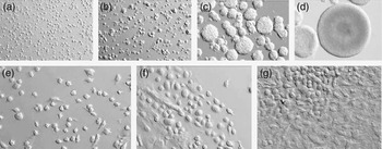
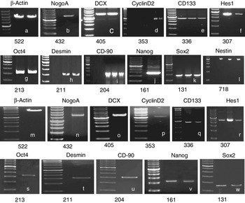
After 2 days of culture, NS cells proliferated as neurospheres (Figure 1a–d). The neurospheres were positive for the undifferentiated neural cell marker, Nestin and NS cells widely expressed NogoA, DCX, CyclinD2, CD133, Hes1, Oct4, Desmin, CD-90, Nanog and Sox2 (Figure 2a–l).
Porcine stem cells (a–d, NS cells; e–g, AFS cells), ×10. (a) Cultured for 0 h; (b) Cultured for 1 h; (c) Cultured for 3 days; (d) Cultured for 16 days; (e) Cultured for 1 h; (f) Cultured for 2 days; (g) Cultured for 3 days. NS cells, neural stem cells; AFS cells, amniotic fluid-derived stem cells.

NS (a–l) and AFS (m–w) cells markers by RT-PCR. Positive control was β-actin. Products are shown with a 50-bp or 100-bp ladder at the left of the gel and product sizes are under the margin. The 50 bp size marker is 50, 100, 150, 200, 250, 300, 350 (the brightest), 400, 450, 500, 550 and 600 bp (a–k, m–w); 100 bp size marker is 100, 200, 300, 400, 500, 600, 700, 800, 900 bp (l). NS cells, neural stem cells; AFS cells, amniotic fluid-derived stem cells; RT-PCR, reverse transcription-PCR.

We have established AFS cell lines with a typical doubling time of about 48 h and no need for feeder layers. Subconfluent cells show no evidence of spontaneous differentiation. After 2 days of culture, AFS cells proliferated as monolayer (Figure 1e–g). AFS cells widely expressed NogoA, DCX, CyclinD2, CD133, Hes1, Nanog, Sox2, Oct4, Desmin and CD-90 (Figure 2m–w).
Multilineage differentiation was characteristic of NS and AFS cells
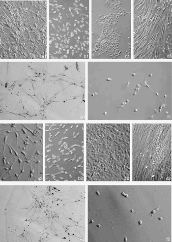
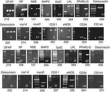
NS and AFS cells are broadly multipotent. Under specific inducing conditions they are able to give rise to lineages representative of the three embryonic germ layers. We found that NS and AFS cells were able to differentiate along neurogenic, adipogenic, osteogenic, myogenic and endothelial pathways (Figure 3). The induced differentiation to multiple fates could be documented by expression of mRNAs for lineage-specific genes. Both NS and AFS cells were differentiated into astrocyte (GFAP+), oligodendrocyte (GalC+), neuron (NF+, NSE+ and MAP2+), adipocyte (LPL+ and PPARγ-D+), osteoblast (Osteonectin+ and Osteocalcin+), myocyte (myf-6 +and myoD +) and endothelium (CD31+, CD34+, CD144+ and eNOS+) (Figure 4).
The differentiated cells of NS (a1–f1) and AFS (a2–f2) cells, ×10. (a1 and a2) Neurogenic cells; (b1 and b2) Adipogenic cells; (c1 and c2) Osteogenic cells; (d1 and d2) Myogenic cells; (e1 and e2) Endothelial cells; (f1 and f2) Control group cells. NS cells, neural stem cells; AFS cells, amniotic fluid-derived stem cells.

RT-PCR results of markers of the differentiated cells derived from NS (a1–o1) and AFS (a2–o2) cells. Products are shown with a 50-bp ladder at the left of the gel and product sizes are under the margin. GFAP, NF, NSE, MAP2 and GalC are neurogenic markers; LPL and PPARγ-D are adipogenic markers; Osteonectin and Osteocalcin are osteogenic markers; myf-6 and myoD are myogenic markers; CD31, eNOS, CD34 and CD144 are endothelial markers. The 50 bp size marker is 50, 100, 150, 200, 250, 300, 350 (the brightest), 400, 450, 500, 550, 600 bp. NS cells, neural stem cells; AFS cells, amniotic fluid-derived stem cells.

In vitro developmental ability of cloned embryos from different donor cells
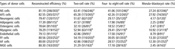
The results showed that there were no significant differences (P > 0.05) in reconstructed efficiency between the cloned embryos derived from different donor cells, but there were significant differences (P < 0.05) in in vitro developmental potential between the cloned embryos derived from stem cells (NS and AFS cells) and somatic cells (differentiated cells from NS and AFS cells, FF cells, AF cells and MGE cells) (Table 1). The in vitro developmental potential of the cloned embryos derived from stem cells were higher than that of the cloned embryos derived from somatic cells (Table 1).
In vitro development of cloned embryos from different donor cells

NS cells = neural stem cells; AFS cells = amniotic fluid-derived stem cells; FF cells = fetal fibroblast cells; AF cells = adult fibroblast cells; MGE cells = mammary gland epithelial cells.
Reconstructed efficiency is the number of cloned embryos divided by the number of enucleated oocytes. Within the same column, values with same superscripts (a, b, c and d) were not significantly different (P > 0.05), values with different superscripts (a, b, c and d) were significantly different (P < 0.05).
In vivo developmental ability of cloned embryos from different donor cells
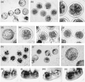
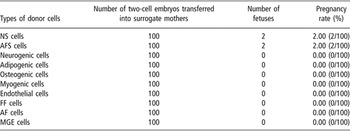
Four cloned fetuses (28 and 32 days) derived from NS (Figure 5k and l) and AFS (Figure 5m and n) cells were obtained. The pregnancy rate was higher in embryos cloned from NS and AFS cells nuclei than that of the embryos cloned from somatic cells (differentiated cells from NS and AFS cells, FF cells, AF cells and MGE cells) (Table 2), which demonstrated that in vivo developmental ability of embryos cloned from NS and AFS cells was higher than that of the embryos cloned from somatic cells.
Cloned embryos (×10) and fetuses derived from different donor cells. (a) Embryos from NS cells; (b) Embryos from AFS cells; (c) Embryos from neurogenic cells; (d) Embryos from adipogenic cells; (e) Embryos from osteogenic cells; (f) Embryos from myogenic cells; (g) Embryos from endothelial cells; (h) Embryos from FF cells; (i) Embryos from AF cells; (j) Embryos from MGE cells. (k and l) Fetuses from NS cells (28 days); (m and n) Fetuses from AFS cells (32 days). NS cells, neural stem cells; AFS cells, amniotic fluid-derived stem cells; FF cells, fetal fibroblast cells; AF cells, adult fibroblast cells; MGE cells, mammary gland epithelial cells.

In vivo development of cloned embryos from different donor cells

NS cells = neural stem cells; AFS cells = amniotic fluid-derived stem cells; FF cells = fetal fibroblast cells; AF cells = adult fibroblast cells; MGE cells = mammary gland epithelial cells.
Discussion
Production of cloned pigs by somatic cell nuclear transfer (SCNT) has unlimited value for developing critical biotechnology such as xenotransplantation (Prather et al., Reference Prather, Tao and Machaty1999; Polejaeva et al., Reference Polejaeva, Chen, Vaught, Page, Mullins, Ball, Dai, Boone, Walker, Ayares, Colman and Campbell2000). Various efforts have been made to establish this technology (Wolf et al., Reference Wolf, Zakhartchenko and Brem1998), and live piglets have been delivered after transfer of SCNT embryos (Betthauser et al., Reference Betthauser, Forsberg, Augenstein, Childs, Eilertsen, Enos, Forsythe, Golueke, Jurgella, Koppang, Lesmeister, Mallon, Mell, Misica, Pace, Pfister-Genskow, Strelchenko, Voelker, Watt, Thompson and Bishop2000; Onishi et al., Reference Onishi, Iwamoto, Akita, Mikawa, Takeda, Awata, Hanada and Perry2000; Polejaeva et al., Reference Polejaeva, Chen, Vaught, Page, Mullins, Ball, Dai, Boone, Walker, Ayares, Colman and Campbell2000). However, the viability of porcine SCNT embryos is poor, with an extremely low rate of cloned piglet production. Studies have demonstrated that many factors are involved in the development of porcine SCNT embryos (Polejaeva, Reference Polejaeva2001). The factors include donor cell types, recipient oocytes, fusion or activation methods and in vitro culture system. The success rate for cloning mice from ES cells is relatively high compared with that for differentiated somatic cells (Wakayama et al., Reference Wakayama, Rodriguez, Perry, Yanagimachi and Mombaerts1999; Rideout et al., Reference Rideout, Wakayama, Wutz, Eggan, Jackson-Grusby, Dausman, Yanagimachi and Jaenisch2000). This suggests that a cell in an undifferentiated state may be suitable as a donor cell for animal cloning. Although ES cells may be effective in NT in mice, this process is limited in other species, where definitive ES cells have not been established. Investigation of other undifferentiated cell types, such as somatic stem cells, in NT is important for animal cloning techniques.
NS cell is one type of well-defined somatic stem cell that can proliferate and differentiate in vitro. In this study, we isolated NS cells from porcine fetuses and the cells grown as spheres. The developmental potential differed between cloned embryos derived from neurospheres and from somatic cells, suggesting that the donor cells of the neurospheres, we used were undifferentiated. We have demonstrated that stem cells can be obtained from porcine amniotic fluid. The AFS cells grow easily in culture. They are capable of extensive self-renewal, a defining property of stem cells. The surface marker profile of AFS cells and their expression of the transcription factor Oct4 suggests that they represent an intermediate stage between pluripotent ES cells (Evans and Kaufman, Reference Evans and Kaufman1981; Martin, Reference Martin1981; Thomson et al., Reference Thomson, Itskovitz-Eldor, Shapiro, Waknitz, Swiergiel, Marshall and Jones1998) and lineage-restricted adult stem cells. Potential sources of AFS cells in the developing fetus are diverse (Gosden, Reference Gosden1983). AFS cells can serve as precursors to a broad spectrum of differentiated cell types. We have assessed their multipotent character.
Previous studies have reported nestin expression in proliferative precursor populations cultured from the porcine brain (Armstrong et al., Reference Armstrong, Hurelbrink, Tyers, Ratcliffe, Richards, Dunnett, Rosser and Barker2002; Uchida et al., Reference Uchida, Okano, Hayashi, Mine, Tanioka, Nomura and Kawase2003). In this study, NS cells were able to demonstrate nestin expression by RT-PCR. In addition to nestin, a number of other markers have been associated with NS and progenitor cells. This study shows expression of NogoA, DCX, Cyclin D2, CD133, Hes1, Oct4, Desmin, CD-90, Nanog and Sox2 (Figure 2a–l), some of which have been reported as expressed by CNS precursor cells, or subsets thereof (Cai et al., Reference Cai, Wu, Mirua, Pierce, Lucero, Albertine, Spangrude and Rao2002; Schwartz et al., Reference Schwartz, Bryant, Fuja, Su, O’Dowd and Klassen2003 andReference Schwartz, Nethercott, Kirov, Ziaeian, Young and Klassen2005). The expression of these markers (Figure 2a–l) by porcine NS cells argues for an important role for these genes in the neural development of large mammals. The study also shows expression of NogoA, DCX, Cyclin D2, CD133, Hes1, Oct4, Desmin, CD-90, Nanog and Sox2 (Figure 2m–w) in AFS cells. These results indicate that these cells were in an undifferentiated state.
Siddiqui and Atala (Reference Siddiqui and Atala2004) found that AFS cells were able to differentiate along adipogenic, osteogenic, myogenic, endothelial, neurogenic and hepatic pathways. This study shows that both NS and AFS cells are broadly multipotent, which were induced to differentiate into cell types representing each embryonic germ layer, including cells of neuronal, adipogenic, osteogenic, myogenic and endothelial lineages (Figure 3). The induced differentiation to multiple fates could be documented by expression of mRNAs for lineage-specific genes. Both NS and AFS cells could differentiate into astrocyte (GFAP+), oligodendrocyte (GalC+), neuron (NF+, NSE+ and MAP2+), adipocyte (LPL+ and PPARγ-D+), osteoblast (Osteonectin+ and Osteocalcin+), myocyte (myf-6 +and myoD +) and endothelium (CD31+, CD34+, CD144+ and eNOS+) (Figure 4). In addition to showing that induced stem cells express lineage-specific markers, it is important to confirm that they can give rise to cells with sufficient specialized function to have potential therapeutic utility in later research. Thus, NS and AFS cells may be useful in porcine cell transplantation studies potentially leading to the application of this strategy in the setting of nervous system and other systems disease and injury.
Our results showed that there were no significant differences (P > 0.05) in reconstructed efficiency between the cloned embryos derived from different donor cells, but there were significant differences (P < 0.05) in developmental potential between the cloned embryos derived from stem cells (NS and AFS cells) and somatic cells (differentiated cells from NS and AFS cells, FF cells, AF cells and MGE cells), which acted as donor nuclei. The developmental potential of the cloned embryos derived from stem cells (NS and AFS cells) were higher than that of the cloned embryos derived from somatic cells (Table 1). We found that porcine somatic cells used for NT could produce cloned embryos, but could not produce cloned fetuses. However, we found that using NS and AFS cells as donor cells could produce cloned fetuses (Figure 5).
NS (Mizutani et al., Reference Mizutani, Ohta, Kishigami, Van Thuan, Hikichi, Wakayama, Kosaka, Sato and Wakayama2006) and AFS cells may provide an alternative source of cells, without the need for ES cells, to generate cloned animals in porcine and other species. However, none of the studies on swine somatic cell cloning have compared the developmental ability between cloned embryos derived from NS, AFS cells and other donor cells such as differentiated cells from NS and AFS cells, FF cells, AF cells and MGE cells. This is the first report on production of cloned embryos using porcine NS and AFS cells. We conclude that porcine NS and AFS cells are broadly multipotent, which were induced to differentiate into cell types representing each embryonic germ layer, including cells of neuronal, adipogenic, osteogenic, myogenic and endothelial lineages. Thus, NS and AFS cells may be useful in porcine cell transplantation studies potentially leading to the application of this strategy in the setting of nervous system and other systems disease and injury. The developmental potential of the cloned embryos derived from stem cells (NS and AFS cells) were higher (P < 0.05) than that of the cloned embryos derived from somatic cells (differentiated cells from NS and AFS cells, FF cells, AF cells and MGE cells), which suggests that the undifferentiated state of the donor cell increases cloning efficiency.
Acknowledgements
The authors thank everyone for assistance with this project. This study was supported by a grant from Northwest A&F University Research Foundation (07ZR001).