INTRODUCTION
Reactive oxygen species (ROS), including superoxide anion radical (O2 •–), hydrogen peroxide (H2O2), hydroxyl radical (OH•), singlet oxygen (1O2) and lipid hydroperoxides are produced by incomplete reduction of oxygen during aerobic respiration and cellular metabolism. ROS play pivotal roles not only in control of cellular redox homeostasis but also in induction of cellular proliferation or apoptosis by activating signalling molecules (Hancok et al. Reference Hancok, Desikan and Neill2001). However, the unbalanced generation of ROS may cause chemical damage to the cells, which might induce breakage of DNA strands, protein oxidation, polysaccharide depolymerization, membrane-lipid peroxidation and impairment of signal transduction from membrane receptors in various physiological processes (Dröge, Reference Dröge2002). The aerobic organisms have evolved a series of multi-layered enzymatic and non-enzymatic defence systems, which can remove the aggressive ROS and/or repair ROS-mediated cell damage (Sies, Reference Sies1993). Distinct antioxidant enzymatic activities such as catalase, glutathione peroxidase (GPx) and peroxiredoxin (PRx) have been well characterized from numerous taxa. Trematode parasites are known to lack catalase, suggesting that GPx proteins play important antioxidant roles alone or together with other proteins (Mkoji et al. Reference Mkoji, Smith and Prichard1988).
GPx encompasses six distinct families of multiple isoenzymes, including classical/cytosolic/cellular GPx (GPx1/c-GPx), gastrointestinal GPx (GPx2/GPx-GI), plasma GPx (GPx3/p-GPx), phospholipid hydroperoxide GPx (GPx4/PHGPx; E.C. 1·11·1·12), epididymis-specific GPx (GPx5/e-GPx) and odourant metabolizing GPx (GPx6/o-GPx), which catalyse the reduction of H2O2, organic hydroperoxides and lipid hydroperoxides using glutathione as a reducing agent. These GPx families share similar structural and enzymatic properties with one another (Arthur, Reference Arthur2000). PHGPx proteins have many distinct features compared to the other GPx families, which perform functions in a monomeric form because they lack the subunit interaction domains (Epp et al. Reference Epp, Ladenstein and Wendel1983; Brigelius-Flohé et al. Reference Brigelius-Flohé, Aumann, Blöcker, Gross, Kiess, Klöppel, Maiorino, Roveri, Schuckelt, Usani, Wingender and Flohé1994; Arthur, Reference Arthur2000). These PHGPx molecules directly reduce hydroperoxidized phospholipids integrated into membranes (Ursini and Bindoli, Reference Ursini and Bindoli1987). Members of the other GPx families, however, interfere with lipid peroxidation only via a concerted operation with phospholipase, which implies that PHGPxs are deeply associated with the repair of disrupted biomembranes (Imai and Nakagawa, Reference Imai and Nakagawa2003). GPxs might also constitute the front line of enzymatic defence to ensure their survival against host immune cell-derived ROS in parasitic helminths, which cause chronic infections (Zelck and von Janowsky, Reference Zelck and von Janowsky2004). The parasitic helminth GPx families show a certain degree of biased distribution across taxa. GPx proteins homologous to mammalian GPx3 have been characterized in the filarial nematodes, including Brugia pahangi, Dirofilaria immitis and Wuchereria bancrofti (Henkle-Dührsen and Kampkötter, Reference Henkle-Dührsen and Kampkötter2001); whereas information on the PHGPx (GPx4) members is obtainable from trematodes such as Schistosoma mansoni (Mei and LoVerde, Reference Mei and LoVerde1995; Mei et al. Reference Mei, Thakur, Schwarts and LoVerde1996), Clonorchis sinensis (Cai et al. Reference Cai, Bae, Kim, Sohn, Lee, Jiang, Kim and Kong2008) and Paragonimus westermani (Kim et al. Reference Kim, Cai, Bae, Lee, Lee and Kong2009). Observations regarding the tissue distribution of the S. mansoni, C. sinensis and P. westermani PHGPxs were shown to be largely localized in vitellocytes within vitelline follicles and eggs (Roche et al. Reference Roche, Liu, LePresle, Capron and Pierce1996; Cai et al. Reference Cai, Bae, Kim, Sohn, Lee, Jiang, Kim and Kong2008; Kim et al. Reference Kim, Cai, Bae, Lee, Lee and Kong2009).
RNA interference (RNAi), which results in post-transcriptional gene silencing in a wide variety of organisms, was originally identified in Caenorhabditis elegans (Tabara et al. Reference Tabara, Grishok and Mello1998), and has since been used in human parasitic worms, including schistosome (Mourão et al. Reference Mourão, Dinguirard, Franco and Yoshino2009; Cheng et al. Reference Cheng, Fu, Lin, Shi, Zhou, Jin and Cai2009; Tchoubrieva et al. Reference Tchoubrieva, Ong, Pike, Brindley and Kalinna2010). In eukaryotic cells, double-stranded RNA (dsRNA) can be recognized by the RNA-induced silencing complex which effects degradation of the target mRNA, and abolishes expression of the corresponding protein (Zamore et al. Reference Zamore, Tuschl, Sharp and Bartel2000). dsRNAs, which can be synthesized in large quantities and transfected into target cells in the presence of lipofectamine reagents, are the most commonly used reagents for RNAi in cultured parasites (Morales et al. Reference Morales, Rinaldi, Gobert, Kines, Tort and Brindley2008).
Schistosoma japonicum is a major schistosome species in Asia, infecting not only humans but also wild or domestic animals. It is estimated that nearly one million people in China are presently afflicted with schistosomiasis, and 30 million are at risk of infection with S. japonicum (McManus et al. Reference McManus, Hu, Brindley, Feng and Han2004). Adult worms of S. japonicum settle in the mesenteric veins via the hepatic portal system. Most eggs lodge in the fine venules of intestine wall; others are carried retrograde by the portal venous system to the liver, in which they become trapped in the portal triad. The most prominent pathologic consequences of schistosomiasis are caused by inflammatory responses around the eggs which become trapped in the liver and other tissues of the host (Wyler, Reference Wyler1992). The parasite might be continuously exposed to oxidative stresses, from both ROS generated by endogenous metabolism and ROS generated by host inflammatory and immune cells (Selkirk et al. Reference Selkirk, Smith, Thomas and Gounaris1998; Zelck and von Janowsky, Reference Zelck and von Janowsky2004). In order to overcome these stressful conditions and to maintain its lifespan, the fluke is equipped with the antioxidant enzymes, such as peroxiredoxin (Kumagai et al. Reference Kumagai, Osada, Ohta and Kanazawa2009).
In this study, we isolated and characterized a novel PHGPx gene (SjGPx) from the blood fluke S. japonicum. Molecular characters of the gene were described, including selenocysteine (Sec) insertion sequence (SECIS) element, amino acid (aa) primary structure and phylogenetic position among various members of GPx families. The native S. japonicum glutathione peroxidase (SjGPx) was analysed by semi-quantitative reverse transcription PCR, Western blotting and enzyme relative activity assay. Finally, we employed a lipofectamine transfection procedure to introduce SjGPx dsRNA into cultured schistosome, and investigated the inhibitory effect of dsRNA-mediated gene knockdown of SjGPx on intrauterine egg formation in S. japonicum.
MATERIALS AND METHODS
Parasite
Positive Oncomelania hupensis snails were collected from the epidemic area of schistosomiasis in Hunan Province, People's Republic of China. Schistosoma japonicum (Chinese mainland strain) was maintained in O. hupensis. The 10-week-old specific pathogen free (SPF) female BALB/c mice, which were obtained from the Department of Animal, Hubei Academy of Medical Science, China, were infected percutaneously through shaven abdomen with 20 cercariae of S. japonicum by an adaptation of the ring method. The mice were sacrificed on the 42th day after infection, male and female adult worms were recovered by perfusion of the hepatic portal veins, and meantime, the eggs of S. japonicum were collected from the livers of infected mice (He et al. Reference He, Cai, Ni, Li, Zong and He2012). Then, all samples were washed repeatedly to remove host cells and debris with physiological saline. Eventually, some samples were either frozen and kept in liquid nitrogen or immediately used for nucleic acid extraction. Some adult worms collected for RNAi in vitro experiments were washed with sterile 0·9% NaCl solution containing 1000 U mL−1 penicillin and 1 mg mL−1 streptomycin and then cultured in the RPMI 1640 medium. The use of animals was approved by the Animal Ethics Committee of Wuhan University.
Isolation of S. japonicum gene encoding GPx homologues
The non-redundant GenBank database at National Center for Biotechnology Information (NCBI, http://www.ncbi.nlm.njh.gov) was screened with the nucleotide sequence of S. mansoni GPx (accession no. AY729668) used as query in the BLAST searches. A clone from S. japonicum database (accession no. CV697435) encoding GPx protein was selected based on BLAST algorithms. A cDNA library of S. japonicum was constructed using SMART® cDNA Library Construction Kit (Clontech, CA, USA) under the manufacturer's instruction. The S. japonicum cDNA library was screened by standard plaque lift hybridization using the DNA fragment as a probe. The insert cDNA from positive clone was amplified by PCR using universal T3 and T7 promoter primers, cloned into pMD18-T vector (Takara, Shiga, Japan) and subjected to nucleotide sequencing from both strands to obtain a full-length cDNA. The clone CV697435 was designated SjGPx. The coding profile and deduced aa sequence of the SjGPx gene was determined using the Open Reading Frame (ORF) Finder program at NCBI (http://www.ncbi.nlm.nih.gov/gorf/gorf.html). The SECIS was scanned using the SECISearch program (ver2·19; http://genome.unl.edu/SECISearch.html). The Compute pI/Mw program (http://www.expasy.ch/tools/pi_tool.html) was used for the calculation of the theoretical molecular weight (Mr) and isoelectric point (pI) value. The putative N-terminal hydrophobic signal peptide was predicted using the SignalP program (http://www.cbs.dtu.dk/services/SignalP).
The aa sequence alignment and phylogenetic analysis
The translated aa sequence of SjGPx was used as query in a series of BLAST searches, to retrieve the closely matched sequences from a variety of GenBank genomic databases. A total of 42 sequences were retrieved, according to their homology values and the taxonomical distributions of their donors. The aa sequences were aligned using ClustalX and optimized with GeneDoc (http://www.psc.edu./biomed/genedoc) programs, respectively. A phylogenetic analysis was conducted by the neighbour-joining (NJ) or maximum parsimony algorithm integrated into the PHYLIP package (ver3·6b). Alignment gaps were removed as missing data and the trees were displayed with TreeView. The statistical significance of each branching point was evaluated with 100 random samplings of the initial input using SEQBOOT software.
Semi-quantitative reverse transcription PCR (RT–PCR)
Forty-second-day live adult worms were chilled on ice for several minutes to facilitate separation of mated pairs. Total RNAs were extracted from eggs, fresh cercariae, male and female adults using Trizol reagents (Gibco, Carlsbad, CA). The RNA samples were treated with RNase-free DNase (GIBCO BRL, Rockvile, MD, USA). Poly(A)+ RNAs were prepared from the total RNAs by oligo(dT)-affinity chromatography (Qiagen, Valencia, CA). The first strand cDNAs were synthesized from 1 µg of poly(A)+ RNA and oligo-dT primers using a RNA PCR Kit (AMV) Ver. 3·0 (TaKaRa, Shiga, Japan) under the manufacturer's instruction. The semi-quantitative RT-PCR primers for SjGPx were designed based on the partial sequence. The primer sequences were SjGPx-F, 5′-GATCACTGGAAGTTCCGCAATG-3′ and SjGPx-R, 5′-CTTCTGTTTCAATAGCTCCATG-3′. The gene transcript was amplified by PCR with the specific primers using the cDNA as a template. The PCR was conducted with a thermal cycling profile of 94 °C for 4 min, 25 cycles of 50 s at 94 °C, 40 s at 59 °C, 1 min at 72 °C and a final extension of 10 min at 72 °C. The reaction product was resolved by 2% agarose gel electrophoresis and visualized by ethidium bromide staining. Reaction with a primer pair for the S. japonicum alpha-tubulin gene (GenBank accession no. AY815746, SjTubulin-F, 5′-CTACTGTAGTGGATGAAGTGCGAAC-3′ and SjTubulin-R, 5′-CAGCTGAAATTACTGGTGCATAAG-3′) was utilized as an internal quantity control, and the absence of any contaminating chromosomal DNA was verified via the preparation of reactions without reverse transcriptase during the first round of cDNA synthesis.
Cloning and expression of recombinant SjGPx protein
The gene segments spanning the ORF region of SjGPx was amplified by PCR using the first strand cDNA as templates, as described above, and then cloned into pGEM T-Easy vector (Promega). The unusual Sec codon (TGA) of SjGPx was converted into a sense Cys codon (TGC), using a complementary primer pair 5′-CTTGCAAGTGCGGCGCAACGGACAAAAATTATC-3′ and 5′-GATAATTTTTGTCCGTTGCGCCGCACTTGCAAG-3′, and the QuikChange Site-Directed Mutagenesis Kit (Stratagene, La Jolla, CA, USA). The mutated plasmid was transformed into competent Escherichia coli DH5α cells and the nucleotide sequence was verified by sequencing. Nucleotides corresponding to the mature domain of SjGPx were amplified from the plasmid construct using primers containing restriction enzyme sites for EcoRI and Xho I (5′-CCGAATTCCGCCAACTTCAGAAGATGCAC-3′ and 5′-GGCTCGAGTCACTTCTGTTTCAATAGCTCC-3′). After purification with a QIAquick PCR Purification Kit (Qiagen, Valencia, CA, USA) and digestion with the corresponding enzymes, the DNA was cloned into the pET-28a-c (+) vector (Novagen, Madison, WI, USA). The plasmid was then transformed into E. coli DH5α cells and the accuracy of the nucleotide sequence was verified by sequencing. The plasmid DNA exhibiting the correct codons was introduced into E. coli BL21 (DE3) cells. Expression of the recombinant protein was induced by treating the transformants with 0·5 mm isopropyl-β-D-thiogalactopyranoside (IPTG). The bacteria cells were sonicated and the recombinant proteins were purified by Ni-NTA agarose chromatography (Amersham Biosciences). The purified recombinant SjGPx (rSjGPx) protein was analysed by SDS-12% PAGE and used to generate specific antibody.
Generation of polyclonal antibody and Western blotting
Six-week-old, SPF female BALB/c mice were immunized subcutaneously 3 times (2-week interval) with the purified rSjGPx protein (30 µg each) mixed with Freund's adjuvant (Sigma). The mice finally received intravenously boosters with the proteins (10 µg). The mice were sacrificed 7 days after the final booster and blood was collected by heart puncture. The blood was centrifuged for 10 min at 3000 g at 4 °C and the antisera were stored at −70 °C until use.
The egg, cercariae and male and female adult worms of the parasite were ground with a Teflon pestle-homogenizer in phosphate-buffered saline (PBS, 140 mm NaCl, 2·7 mm KCl, 10 mm Na2HPO4 and 1·8 mm KH2PO4; pH 7·2) containing 2% SDS and a protease inhibitor cocktail (Roche, Mannheim, Germany). After centrifugation at 15 000 rpm for 30 min at 4 °C, the supernatants were recovered. SDS–PAGE was conducted using 12% gels with the native proteins and recombinant proteins under reducing condition. The gels were stained with Coomassie Brilliant Blue or transferred onto nitrocellulose membrane (Schleicher & Schuell BioScience, Dassel, Germany). The membrane was blocked for 1 h in Tris-buffered saline containing 0·05% (v/v) Tween-20 (TBS/T) and 5% (w/v) skim milk, followed by overnight incubation with the specific antibody diluted to 1:1000 in TBS/T containing 5% skim milk at 4 °C. The membrane was subsequently incubated for additional 1 h at room temperature with horseradish peroxidase (HRP)-conjugated goat anti-mouse IgG antibody (1:2000 dilutions; Cappel, West Chester, PA, USA). Signal was detected with an ECL system (Amersham Biosciences) under the manufacturer's instructions. An anti-SjTubulin antibody, which was produced similarly to the generation of anti-SjGPx antibody using its specific sequence, was used as an internal standard.
Enzyme activities of the native GPx of S. japonicum adult worm
Live adult worms were washed over than 10 times with cold physiological saline at 4 °C, after which homogenized with a Teflon-pestle homogenizer in PBS containing 5% Triton X-100. The homogenates were centrifuged at 15 000 rpm for 30 min at 4 °C and the supernatants containing GPx activities were stored. The total SjGPx activity was estimated using a Glutathione Peroxidase Assay Kit (Northwest Life Science Specialities, LLC, Canada) according to the manufacturer's instructions. The measurements were conducted with triplicate reactions and expressed as Mean ± s. d. of enzymatic unit mL−1 mg protein−1.
Synthesis of dsRNA of SjGPx
dsRNA was synthesized from cDNA encoding the full length of SjGPx using gene-targeted primers containing T7 promoter sequences 5′-TAATACGACTCACTATAGGGATGATCACTGGAAGTTCCGCAA-3′ and 5′-TAATACGACTCACTATAGGGTCACTTCTGTTTCTTTAGCTC-3′ spanning coding DNA positions of 33–563. One negative control (NC) dsRNA against Firefly Luciferaseds was also synthesized from plasmid pGL3-Basic (Promega, Madison, WI). RNAs were synthesized and purified using the Megascript RNAi kit (Ambion, Austin, TX) according to the manufacturer's instructions. dsRNAs were precipitated with 5 m ammonium acetate and 95% ethanol, after which the RNA pellets were dissolved in water. Integrity of the dsRNAs was verified by non-denaturing 1% agarose gel electrophoresis, and concentration and purity determined with a spectrophotometer (ND-1000, NanoDrop Technologies, Wilmington, DE).
Treatment of Schistosomes with dsRNA in vitro culture
The Lipofectamine™ 2000 transfection reagent (Invitrogen, USA) was used in the dsRNA selection experiment to improve transfection efficiency. 800 µL of OPTI-MEM (Invitrogen, USA) containing 8 µL of Lipofectamine™ 2000 and dsRNA was added into 3·2 mL of antibiotic-free RPMI 1640 medium, in which 50 mated worms were cultured. Then the worms were cultured for 6 h at 37 °C before media were replaced with fresh RPMI 1640 medium containing 10% (v/v) fetal bovine serum and antibiotics (He et al. Reference He, Cai, Ni, Li, Zong and He2012).
To examine the optimal concentration of dsRNA in mediating SjGPx knockdown, 10 and 50 nm of the dsRNAs were added into the culture. The RNA against Firefly Luciferaseds was as NC group. After 5 days of cultivation, the parasites and eggs from culture medium were collected. Parasites were used for determination of SjGPx transcript level using semi-quantitative RT–PCR. The protein levels of SjGPx were determined by Western blotting and GPx activity assays. The morphological characteristics of intrauterine eggs and collected eggs from the culture medium were observed using a light microscope (Olympus, CKX41SF, Japan) from the 1st to 5th day.
RESULTS
Isolation of a selenium-dependent GPx gene from S. japonicum
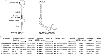
A clone (accession no. CV697435) from S. japonicum database was selected, which exhibited a significant degree of sequence identity to PHGPx isolated from various taxa including S. mansoni (AAU34080 and AAA29885), C. sinensis (ABK58679-ABK58682), P. westermani (ABE68811 and ABE68812), Homo sapiens (CAA50793), Mus musculus (BAA22780) and Drosophila melanogaster (AAR96123) (E-values < 7 × 10⁻³²) during the BLASTX analysis. The complete cDNA sequence was determined by consecutive cDNA library screening using the clone as probe. The cDNA, designated SjGPx, was comprised of 724 bp, and contained 531 bp ORFs, from an ATG at nucleotide position 33–35 to a TGA at positions 561–563. In addition to the conventional stop codon, the SjGPx gene contained a second in-frame stop codon within its ORF at nucleotide position 180–182, which might decode the 21st aa, Sec (Stadtman, Reference Stadtman1996). The SECIS predicted within the 3′-untranslated regions (UTR) also suggested that the opal TGA codon encodes Sec (Fig. 1A). The SECIS conserved secondary structures characteristic to those of selenium-dependent homologues: two helixes separated by an internal loop, a quartet of non-Watson–Crick base pairs located at the base of helix II and an apical loop (Kryukov et al. Reference Kryukov, Castellano, Novoselov, Lobanov, Zehtab, Guigó and Gladyshev2003). Nucleotide signatures in the quarter were found to be ATGA_AA_GA in other trematode genes. SjGPx harboured SECIS with a different sequence conservation GTGA_AA_GA, in that the adenosine preceding the quarter is replaced with guanosine (Fig. 1B). The full-length cDNA sequence of SjGPx was registered in GenBank under accession no. KJ941008.
Analysis of SECIS elements in SjGPx of S. japonicum. (A) SECIS elements in SjGPx. The 3′-UTR of SjGPx mRNA was predicted to have a secondary structure characteristic to SECIS, including an apical loop, an internal loop, a quartet (SECIS core) and two helices (SECISearch program, ver2·19). The common structure of SECIS, as predicted using the bovine GPx gene (XP_612375), is shown for comparison (leftmost panel). The nucleotide positions are numbered at both start and end points of the elements; (B) comparison of the primary SECIS structures. Nucleotide sequences constituting the SECIS motif were retrieved from the PHGPx genes of C. sinensis, P. westermani, S. mansoni, H. sapiens and M. musculus, and aligned in accordance with the predicted secondary elements. Nucleotides conserved within the invariant quartet and apical loop are highlighted in boldface.

Primary structure of SjGPx and its phylogenetic position within the GPx family
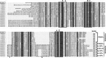
The decoded polypeptide of SjGPx was found to be composed of 176 amino acids with predicted Mw/pI of 19·7 kDa/9·08, and shared a significant degree of sequence identity with other trematode GPx (39–75%, Fig. 2). SjGPx contained three conserved functional domains found in the GPx family and the aa residues constituting the catalytic triad (Sec, Gln and Trp) were positioned separately within each of the domains (Epp et al. Reference Epp, Ladenstein and Wendel1983). The SjGPx protein did not harbour the subunit interaction domains, which are required for the formation of an active tetramer in GPx isozymes other than PHGPxs (Fig. 2, Brigelius-Flohé et al. Reference Brigelius-Flohé, Aumann, Blöcker, Gross, Kiess, Klöppel, Maiorino, Roveri, Schuckelt, Usani, Wingender and Flohé1994). Amino acid sequence of SjGPx was registered in GenBank under accession no. AJD79086.
Primary structure of the PHGPx protein of S. japonicum. The deduced aa sequence of SjGPx was aligned with those of other trematode PHGPxs and vertebrate 6 GPx members retrieved from the GenBank database, using the ClustalX program, then optimized with GeneDoc. Identical aa in the alignment are highlighted in black, while similar residues are shown in grey. Boxes A, B and C represent three highly conserved domains in GPx families. Amino acid residues forming the catalytic triad are indicated by solid arrowheads. The Sec residues in box A are denoted by ‘U’ or ‘X’. Dotted boxes I and II mark subunit-binding domains found in the multiple GPx members, rather than in PHGPxs. The abbreviated names of six GPx families are provided at the head of the alignment. The identity value of each enzyme against SjGPx, which was calculated from the alignment after removing positions with a gap as missing data, is provided at the end of the alignment. The proteins used in this analysis were selected from various trematode organisms as follows: S. mansoni, SmGPx1 (AAA29885) and SmGPx2 (AAU34080); C. sinensis, CsGPx1–4 (ABK58679–ABK58682); P. westermani, PwGPx1 (ABE68811) and PwGPx2 (ABE68812). The vertebrate 6 GPx families proteins were selected from H. sapiens (Hs) and M. musculus (Mm): HmGPx4 and MmGPx4 (PHGPx, CAA50793 and BAA22780); HmGPx1 and MmGPx1 (c-GPx, CAA68491 and AAH86649); HmGPx2 and MmGPx2 (GPx-GI, CAA48394 and AAH54848); HmGPx3 and MmGPx3 (p-GPx, AAP50261 and AAH37027); HmGPx5 and MmGPx5 (e-GPx, NP_001500 and NP_034473); HmGPx6 and MmGPx6 (o-GPx, NP_874360 and AAH13526).

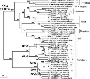
The aa sequence of SjGPx was used in homology searches with BLAST algorithms from the GenBank database, and several hundred PHGPx-like proteins were retrieved. These sequences revealed various degrees of sequence identity to SjGPx ranging 35–58% (E-values < 1 × 10⁻²⁰). Information regarding the other GPx families was obtained by a series of subsequent BLAST searches using the aa sequence of human GPx3 (AAP50261). A total of 42 entries were finally selected, by considering both redundancy and taxonomical distributions and used in a phylogenetic analysis. As shown in Fig. 3, six classes of the GPx family were well separated in the phylogenetic tree constructed by the NJ method. A similar clustering pattern was also observed in a tree based on the maximum parsimony algorithm, of which bootstrap values were incorporated into the NJ tree. The members of PHGPx had a highly diverse donor source and the GPxs identified from free-living nematodes, insects and plants, as well as trematodes and vertebrates, were categorized into this group, including SjGPx protein. The Sec codon and the associated SECIS motif were recognized exclusively within the mRNA sequences coding for the trematode and vertebrate GPx genes (indicated by † and ‡, respectively, in Fig. 3). Among the arthropod proteins examined, only an arachnidal tick PHGPx (Boophilus microplus, ABA25916) was expected to be Sec-dependent.
Phylogenetic relationships of SjGPx among various members of GPx families. A phylogenetic analysis was conducted based on a multiple alignment of aa sequences retrieved from the GenBank database. The tree was constructed by a maximum parsimony algorithm using PHYLIP after excluding positions with gaps and was unrooted. Numbers at branching nodes indicate their percentages of appearance in 100 bootstrap replicates and values of >50% are marked. The branches separating each of the GPx families are presented with thick bars. The presences of Sec codon (†) and SECIS (‡) within each of the corresponding mRNA sequences are indicated at the end of each sequence names. The identity of each analysed sequence is distinguished by a GenBank accession number followed by the species name, from which it has been isolated.

Expression patterns of SjGPx and the native GPx activity in S. japonicum
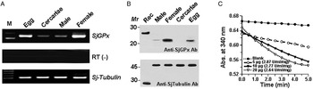
Expression level of the SjGPx mRNA was examined by semi-quantitative RT–PCR using RNAs isolated from S. japonicum egg, cercariae, male and female adult worms. As shown in Fig. 4A, the amplification of the target gene clearly revealed that the SjGPx gene was strictly transcribed in the egg and mature female worm. The gene of cercariae and adult male worm showed a very low transcription levels. The Sj-Tubulin, used as an internal control, exhibited a constant expression level during the stages (Fig. 4A).
Identification of the native SjGPx in S. japonicum. (A) Transcription profile of SjGPx gene in S. japonicum parasite. The transcription level of SjGPx was determined with the gene-specific primers via a semi-quantitative RT–PCR. Total RNAs were extracted from egg, cercariae, male and female adult stages of the parasite, and used as templates to examine mRNA transcripts of SjGPx gene. The amplified products were separated on 2% agarose gels and stained with ethidium bromide. The Sj-Tubulin (GenBank accession no. AY815746) gene was also amplified using the specific primers as an internal control. The absence of any contaminated genomic DNA was confirmed by preparing reactions without reverse transcriptase during the first reverse transcription step (RT−). The position of 100-bp DNA size standards (M) was shown on the left; (B) Western blotting of the native SjGPx protein. A total of 30 µg of proteins extracted from male adult, female adult, cercariae and egg were separated on a SDS–12% PAGE gels under reducing conditions and transferred onto nitrocellulose membranes. The membranes were immuno-reacted against the recombinant SjGPx-specific mouse antisera. The rSjGPx (50 ng) was employed as a positive control. Molecular weight marker (Mr) in kDa was shown on the left. An antibody specific to the SjTubulin was used as an internal standard. Rec, recombinant SjGPx/SjTubulin protein; (C) detection of GPx activities in S. japonicum. The specific activity was assayed using a specific Glutathione Peroxidase Assay Kit as described in the section ‘Materials and methods’, and the value of GPx activity was calculated using an equation as described in the section ‘Results’. The measurements were conducted with triplicate reactions employing the worm extracts (5, 10 and 20 µg of each), and the result from one reaction is presented as a representative.

To observe their expression proteins of SjGPx, we conducted Western blotting, employing the egg, cercariae, male and female adult worm extracts, using anti-SjGPx specific mouse sera (Fig. 4B). The cercariae proteins were not found to react with the SjGPx antibody. However, the blot containing the female and egg whole proteins, which had been extracted in the presence of 2% SDS, exhibited strong positive reactions. The results were well matched to the mRNA expression levels. As shown in Fig. 4B, a strong band (migrated approximately 20 kDa), which might be well matched with the size of the SjGPx protein (the predicted theoretical Mr 19·7 kDa by expasy program), was determined against SjGPx antisera. The S. japonicum alpha-tubulin protein (SjTubulin), used as an internal control, exhibited a constant expression level during the stages. As controls, the antibodies were strongly reactive with the recombinant SjGPx/SjTubulin proteins.
The relative enzymatic activity of GPx proteins in S. japonicum whole adult worm extracts was 2·76 ± 0·12 U mL−1 mg−1 (3 replicates) using a specific Glutathione Peroxidase Assay Kit (Fig. 4C). The specific enzymatic activity was calculated using the equation,
 $$[{\rm GPx}]\, = \,\{ {\rm 2}({\rm mRate}_{\rm s} - {\rm mRate}_{\rm b} )V_{{\rm Rxm}} /{\rm 2} \cdot {\rm 74}V_{\rm s} \}\, {\rm df},$$
in which mRates = 1000 ΔOD340 min−1 of sample, mRateb = 1000 ΔOD340 min−1 of blank, V Rxm = volume of reaction mixture, V s = volume of sample, and df = sample dilution factor.
RNAi-mediated knockdown of SjGPx and morphological observation of intrauterine eggs
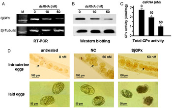
Effects of RNAi-mediated knockdown on SjGPx were examined using semi-quantitative RT–PCR, Western blotting and GPx activity assays. As shown in Fig. 5, the effect of dsRNA was dose-dependent. SjGPx dsRNA at concentration of 50 nm was significantly reduced the transcript level of the target gene (Fig. 5A). The reduced level of SjGPx protein caused by dsRNA in schistosomes paralleled reduction in mRNA level (Fig. 5B). The whole GPx enzyme activities were reduced approximately 29·35 and 63·04% after adding 10 and 50 nm dsRNA, respectively, as shown in Fig. 5C.
(colour online). Inhibitory effects of dsRNA-mediated gene knockdown of SjGPx. 0, 10 and 50 nm of the dsRNAs were added into the schistosomes culture, After 5 days of RNAi, mated worms were collected for determination of (A) SjGPx transcript level using semi-quantitative RT–PCR with the gene-specific primers; (B) SjGPx protein level using Western blotting with the SjGPx-specific mouse antisera; (C) the GPx activity using a Glutathione Peroxidase Assay Kit, the assays were conducted in triplicate employing the worm extracts (10 µg of each) and expressed as mean ± s.d. *P < 0·01 by Student's t test; and (D) RNAi effects of 50 nm SjGPx dsRNA on intrauterine eggs formation in S. japonicum female adult worms cultured in vitro for 5 days. Intrauterine eggs within female adult worms and laid eggs from the culture medium in the untreated group, NC group (the RNA against Firefly Luciferaseds from pGL3-Basic plasmid) and SjGPx group were observed using a light microscope under the common light (scale bars were showed on the left side of each panel).

From the 1st to 5th day after SjGPx gene knockdown, the morphology of intrauterine eggs was continuously observed under a light microscope. On the 1st to 2rd day, the intrauterine eggs maintained a normal shape. On the 3rd day post-treatment, intrauterine eggs resembling spindles began to appear in the SjGPx gene knockdown group (data not shown), and on the 5th day, the eggs from intrauterine of females and eggs from culture medium were more severely deformed. Many irregular eggs, including spindle-like eggs and eggs with invaginated eggshells adhering to each other were observed in the female uteri (Fig. 5D). The majority of eggs from the untreated group and NC group were morphologically normal.
DISCUSSION
Among six distinct GPx families, PHGPx (GPx4) is a major antioxidant enzyme, which plays unique roles in the repairing of disrupted biomembranes by directly reducing hydroperoxidized phospholipids integrated into membranes (Ursini and Bindoli, Reference Ursini and Bindoli1987; Imai and Nakagawa, Reference Imai and Nakagawa2003). PHGPx proteins have been isolated from most eukaryotes, including vertebrates, insects, nematodes, trematodes and plants, whereas the other GPx isoenzymes show a more restricted taxonomical distribution. In addition to vertebrates, a number of filarial nematodes are known to express GPx3-like enzymes (Henkle-Dührsen and Kampkötter, Reference Henkle-Dührsen and Kampkötter2001). Among the reported parasitic trematodes, two GPx proteins in S. mansoni (Williams et al. Reference Williams, Pierce, Cookson and Capron1992; Mei and LoVerde, Reference Mei and LoVerde1995), four GPx proteins in C. sinensis (Cai et al. Reference Cai, Bae, Kim, Sohn, Lee, Jiang, Kim and Kong2008) and two GPx proteins in P. westermani (Kim et al. Reference Kim, Cai, Bae, Lee, Lee and Kong2009), showing strong phylogenetic affinities towards the vertebrate PHGPx, have been described. In the present study, we isolated and characterized a novel PHGPx gene from a blood fluke S. japonicum. The deduced aa sequence of the gene also revealed the primary structure characteristic to the PHGPx (GPx4) family, including aa conservation, absence of the subunit interaction domains and well-preserved catalytic and glutathione-binding domains (Fig. 2). In a phylogenetic analysis, SjGPx protein also showed tight relationships with the other PHGPx-related members (Fig. 3).
Many GPx proteins have been known to contain an opal UGA codon, a stop signal in the standard genetic code, encodes Sec at their catalytic site. The alternative decoding of UGA generally depends on a cis-factor, called SECIS, which is located in the 3′-UTR of these selenoprotein genes (Stadtman, Reference Stadtman1996). A biased distribution of Sec-dependent PHGPxs was detected along with the taxonomical positions of their donor species (Fig. 3). The Sec codon and concurrent SECIS motif were exclusively identified within the mRNA sequences of trematode and vertebrate GPx genes, except for one sequence identified from an arachnid (ABA25916). Among the trematode GPx enzymes obtainable, only P. westermani GPx1 (ABE68811) was found to lose Sec-related sequence factors and was determined to be a selenium-independent PHGPx (Fig. 2). Our results suggested that SjGPx gene is selenium-dependent PHGPx, which contain Sec codon and SECIS motif within its mRNA sequence (Fig. 1).
Helminth parasites may be continuously exposed to dual oxidative stresses, from both ROS generated by endogenous intracellular metabolism and ROS generated by host inflammatory and immune cells (Selkirk et al. Reference Selkirk, Smith, Thomas and Gounaris1998; Zelck and von Janowsky, Reference Zelck and von Janowsky2004). During their lifespan in host tissues, removal of ROS and protection of macromolecules from the oxidative attacks would be inevitable. The antioxidant enzymes might also constitute a major defensive system in these causative parasites (Callahan et al. Reference Callahan, Crouch and James1988). The adults of S. japonicum settle in the mesenteric veins via the hepatic portal system, and their eggs lodge in the fine venules of intestine wall and are carried retrograde by the portal venous system to the liver, where they become trapped in the portal triad. The parasite might be continuously exposed to oxidative stresses. In order to overcome these stressful conditions and to maintain its life-span, the parasite is equipped with the antioxidant enzymes. Furthermore, phenol oxidase, which is secreted from the vitellocytes, acts on eggshell precursors, thus converting tyrosine into quinone in the presence of oxygen. During this tanning process, incomplete reduction of the oxygen may result in a local increase of harmful ROS (Cordingley, Reference Cordingley1987; Cai et al. Reference Cai, Bae, Zhang, He, Jiang and He2009; Bae et al. Reference Bae, Kim, Ahn, Kim and Kong2015). Therefore, effective antioxidant enzymes, such as GPx, might be required for the protection of macromolecules and/or the fertilized ovum. Among the reported trematode GPx enzymes, we noticed S. mansoni GPx proteins were shown to be localized on vitelline follicles and eggs (Roche et al. Reference Roche, Liu, LePresle, Capron and Pierce1996). The PHGPx-like proteins from C. sinensis and P. westermani parasites were also found localized in vitellocytes within vitelline follicles and premature eggs (Cai et al. Reference Cai, Bae, Kim, Sohn, Lee, Jiang, Kim and Kong2008; Kim et al. Reference Kim, Cai, Bae, Lee, Lee and Kong2009). In this study, our RT–PCR and Western blots results showed that the S. japonicum GPx was found to be confined express in the adult female worms and eggs (Fig. 4). These results indicated that trematodes GPx proteins might play key roles during the parasite egg production/eggshell formation.
Increasing evidence indicates that loss-of-function genetic manipulation through RNAi may be generally applicable for investigation of schistosome genes (Krautz-Peterson et al. Reference Krautz-Peterson, Radwanska, Ndegwa, Shoemaker and Skelly2007; Morales et al. Reference Morales, Rinaldi, Gobert, Kines, Tort and Brindley2008). After processing of dsRNA by dicer, the resulting short interfering RNAs join with the effector nuclease complex which recognizes and attacks the homologous target mRNAs, leading to gene silencing (Tavernarakis et al. Reference Tavernarakis, Wang, Dorovkov, Ryazanov and Driscoll2000). In schistosomes this can result in long-term knockdown and silencing of the target gene (Morales et al. Reference Morales, Rinaldi, Gobert, Kines, Tort and Brindley2008). To investigate the involvement of the GPx gene of schistosome egg formation, we performed a RNAi experiment, delivering SjGPx dsRNA into schistosomes by lipofectamine transfection assay. The results of RNAi-mediated knockdown of SjGPx in transcript showed that the GPx enzymatic activities were specifically suppressed, and the undeveloped intrauterine eggs of females were more severely deformed. Many irregular eggs were observed in the female uteri and culture medium (Fig. 5D). Therefore, effective antioxidant enzymes, such as GPx, may be required for the protection of macromolecules and/or the fertilized ovum during the trematode egg production/maturation. Gene knockdown of SjGPx mRNA might be represented a novel and potentially effective strategy for combating schistosomiasis.
FINANCIAL SUPPORT
This study was supported by a National Science Foundation of China (grant no. 30972568 to L.H. and G.B.C.) and also by a research grant from Health Department of Hubei Province, P. R. China (grant no. XF2010-17 to G.B.C.).