Introduction
Epidemiological and experimental studies indicate a relationship between the periconceptional, fetal and early infant phases of life and the subsequent development of adult diseases.Reference Barker, Bagby and Hanson 1 For instance, low birth weight and disproportion have been linked to increased risks of hypertension,Reference Lamireau, Nuyt and Hou 2 renal diseaseReference Tulassay and Vásárhelyi 3 and diabetes.Reference Curhan, Willett and Rimm 4
Previous studies have reported that maternal undernutrition or restriction of dietary protein during pregnancy leads to elevated blood pressure and vascular and renal dysfunction in the offspring.Reference Langley and Jackson 5 – Reference Cambonie, Comte and Yzydorczyk 7 Although an unfavorable prenatal environment is believed to trigger the risk of chronic diseases such as metabolic syndrome in adulthood, it is unclear if maternal undernutrition followed by an unfavorable postnatal and/or weanling environment is associated with a greater risk of such diseases. Studies have shown that programmed hypertension is associated with vascular and renal dysfunction,Reference Kawamura, Itoh and Yura 8 , Reference Rocha, Gomes and Forti 9 which are characterized by impaired nitric oxide (NO)-mediated vasodilatation and vasoconstrictors such as endothelin-1 (ET-1). NO is an important molecule that functions as an endogenous vasodilator, and plays a role in the pathogenesis and maintenance of hypertension. However, excessive production of reactive oxygen species (ROS), especially superoxide (O2 −), associated with hypertension can enhance NO inactivation and reduce bioactive NO.Reference McIntyre, Bohr and Dominiczak 10 , Reference Taniyama and Griendling 11 In addition, the mechanisms involved in the programming of hypertension have remained unclear, although a significant correlation between maternal undernutrition and vascular and renal dysfunction has been reported.Reference Barker, Bagby and Hanson 1 , Reference Lamireau, Nuyt and Hou 2 , Reference Alexander 6
Fructose is a monosaccharide commonly found in the Western diet. There is epidemiological and experimental evidence that suggests a progressive association between fructose consumption and the development of metabolic syndrome.Reference Thorburn, Storlien and Jenkins 12 – Reference Brown, Dulloo and Yepuri 14 A high-fructose diet or adding fructose to drinking water in animal models has been known to lead to systemic hypertension.Reference Hwang, Ho and Hoffman 15 , Reference Takagawa, Berger and Hori 16 In addition, the administration of fructose resulted in the development of renal hypertrophy, glomerular hypertension and cortical vasoconstriction.Reference Sanchez-Lozada, Tapia and Jimenez 13 , Reference Johnson, Segal and Sautin 17 However, little is known about the long-term effects of a postnatal fructose diet on programmed hypertension.
This study was designed to examine whether a postnatal fructose diet would enhance the programmed hypertension and vascular and renal dysfunction in offspring from dams exposed to protein restriction during the gestation and suckling periods. In this study, we showed that maternal protein restriction during the gestation and suckling periods elevated the blood pressure and downregulated NO production and endothelial nitric oxide synthase (eNOS) phosphorylation in the offspring. The postnatal fructose diet made no significant difference to these alterations.
Methods
Animals
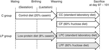
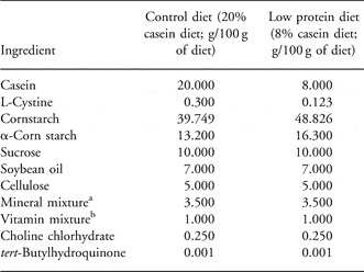
All procedures were performed according to the Guidelines for Animal Experimentation, Aomori University of Health and Welfare. Virgin female Wistar rats aged 10–11 weeks, weighing 226–256 g, were obtained from Charles River Laboratories Japan, Inc. (Yokohama, Japan). They were maintained at a temperature of 23 ± 1°C under a 12 h light–dark cycle and had access to food and tap water ad libitum. A vaginal impedance reader (Model MK-10C; Muromachi Kikai Co. Ltd, Osaka, Japan) was used to determine whether the female rats were in the appropriate stage of the estrous cycle for mating as described previously.Reference Wlodek, Mibus and Tan 18 This was routinely performed in the afternoon, and a reading of >3 kΩ indicated that the females were in proestrus and presumably in estrus. One appropriate female was mated with one male overnight. The next morning, the presence of a vaginal plug indicated successful mating and was taken as day 0 of gestation. Pregnant rats were randomly allocated to feed ad libitum on a diet containing either a 20% (control group: C, n = 6) or 8% (low-protein group: LP, n = 7) casein diet during gestation and the suckling period (Fig. 1). The diets were isocaloric, as described in Table 1. Following delivery, the pups were weighed at 4-day post partum and each lactating dam was kept with eight pups to ensure adequate nutrition until weaning. At the end of lactation (days 21–26), the male pups were separated from the control or restricted dams and received a standard commercial laboratory diet (MF diet; Oriental Yeast, Tokyo, Japan) or a 60% fructose diet (F2HFrD high-fructose diet; Oriental Yeast) as follows: control-on-control (CC, n = 7), 60% fructose diet-on-control (CF, n = 7), control-on-low-protein diet (LPC, n = 6) and 60% fructose diet-on-low-protein diet (LPF, n = 8). The pups were weighed every week, and the systolic blood pressure (SBP) was measured once every 2 weeks. At postnatal days 94–98, 24 h urine samples were collected using metabolic cages. Before sacrificing at postnatal days 97–101, the animals were fasted overnight and then weighed, and blood samples were collected under ether anesthesia. The kidney, heart, liver and aorta were immediately removed and weighed. The aorta tissues were sectioned and kept in saline at 4°C for measurement of O2 − production or stored at −80°C for evaluation of mRNA and protein expression.
Experimental design. Pregnant Wistar rats were fed control (20% casein) and low-protein (8% casein) diets during the gestation and suckling periods. Experimental details are described in Methods. CC, control-on-control group; CF, 60% fructose diet-on-control; LPC, control-on-low-protein diet; LPF, 60% fructose diet-on-low-protein diet.

Composition of the control and low protein diets

aAIN-93G mineral mixture (Oriental Yeast, Tokyo, Japan).
bAIN-93G vitamin mixture (Oriental Yeast).
Determination of blood pressure
The SBP was measured in unanesthetized, prewarmed, restrained rats by the tail-cuff plethysmography method (model MK-1100, Muromachi Kikai Co. Ltd, Tokyo, Japan). The final SBP value is presented as the mean of five determinations. Experimental measurements were performed between 9:00 and 13:00 hours.
Blood chemistry
Plasma samples were separated by centrifugation at 800 g for 10 min at 4°C, and tested for glucose (GLC), triglyceride (TG), total cholesterol and blood urea nitrogen by using an autoanalyzer for blood chemistry (Fuji Dry-Chem 3500V, Fujifilm, Tokyo, Japan). Insulin was measured using a Rat Insulin ELISA kit (TMB) (AKRIN-010T, Shibayagi, Gunma, Japan).
Real-time polymerase chain reaction (PCR)
Total RNA in the thoracic aorta and kidney was extracted using the SV Total RNA Isolation System (Promega Corp., Madison, WI, USA) according to the manufacturer's instructions, and cDNA was synthesized using the High Capacity cDNA Reverse Transcription Kit (Applied Biosystems, Foster City, CA, USA). Vascular and renal mRNA levels of eNOS, p47phox, Nox4, ET-1 and endothelin receptor type B (ETB) were analyzed using the inventoried primers TaqMan gene expression assays Rn02132634_s1, Rn00586945_m1, Rn00585380_m1, Rn00561129_m1 and Rn00569139_m1, respectively (Applied Biosystems). GAPDH mRNA was also analyzed using the TaqMan Rodent GAPDH Control Reagent Kit (Applied Biosystems) as an endogenous control. Real-time PCR was performed using the TaqMan Gene Expression Master Mix (Applied Biosystems) on an Applied Biosystems 7000 sequence detection system, according to the manufacturer's instructions. The gene expression levels were expressed relative to the intensity of GAPDH in each co-amplified sample.
Western blot analysis
Tissues of the abdominal aorta and kidney were homogenized in six times its volume of homogenizing buffer (50 mM N-2-hydroxyethylpiperazine-N-2-ethanesulfonic acid, 150 mM NaCl, 1 mM dithiothreitol and 0.5% (v/v) Tween-20; pH 7.4) containing protease inhibitor cocktail tablets (Roche Applied Science, Indianapolis, IN, USA). The homogenates were centrifuged at 5000 g for 45 min at 4°C. Supernatants were collected, and protein concentration was determined using the BCA™ Protein Assay Kit (Pierce, Rockford, IL, USA). For Western blot analysis, the proteins were electrophoresed on 10% SDS-polyacrylamide gel and then electrotransferred onto polyvinylidene difluoride membranes (GE Healthcare UK Ltd, Buckinghamshire, UK). The membranes were blocked by incubation in PBS containing 0.1% v/v Tween 20 and 5% nonfat dry milk for 8 h, followed by overnight incubation at 4°C with mouse eNOS monoclonal antibody (1:500, BD Biosciences, San Jose, CA, US) and rabbit phospho-eNOS-Ser1177 (1:1000, Cell Signaling Technology, Danvers, MA, USA). The specifically bound primary antibodies were detected with horseradish peroxidase-conjugated rabbit anti-mouse IgG, goat anti-rabbit IgG or swine anti-rabbit IgG antibodies (1:2000; DAKO, Glostrup, DK), and enhanced with ECL Western Blotting Detection Reagents (GE Healthcare UK Ltd) on Hyperfilm (GE Healthcare UK Ltd). Quantitative analysis of the specific band density was performed using ATTO densitometry software (ATTO Corporation, Tokyo, Japan). Protein levels were normalized to the β-actin expression from the same sample.
Determining O2 − production and urinary 24 h nitrate/nitrite (NO x ) excretion
O2 − production in the thoracic aorta was determined using a lucigenin (bis-N-methyacidinium nitrate)-enhanced chemiluminescence technique.Reference Kerr, Brosnan and McIntyre 19 A ring segment of thoracic aorta (3–4 mm) was incubated in 1 ml lucigenin (5 μmol/l)-phosphate buffer (pH 7.8) at room temperature to determine the response to the addition of either 100 μmol/l NADH (nicotineamide adenine dinucleotide) or 100 μmol/l NADPH (nicotineamide adenine dinucleotide phosphate) for 3 min; then, chemiluminescence was measured for 30 s using a sensitive luminometer (TD-20/20; Turner Designs, Sunnyvale, CA, USA). Results were reported as relative light units per minute and normalized to the dry weight of the aorta.
The NO x content in the 24 h urine samples was measured by the Griess method with the NO2/NO3 Assay Kit-C II (Dojindo Laboratories, Kumamoto, Japan). Samples were read at 540 nm with a Microplate Reader (Model 680; Bio-Rad Laboratories Inc., Hercules, CA, USA).
Statistical analysis
Statistical analyses were performed by one-way analysis of variance (ANOVA), followed by the Tukey test. Each value was expressed as mean ± s.e.m.
Results
Body weights
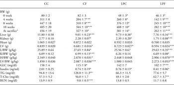
During pregnancy, there was no significant difference in the body weights between the control (C) and low-protein (LP) groups (C, 389 ± 3 g, n = 6 v. LP, 391 ± 5 g, n = 7 at day 20 of gestation). Maternal undernutrition resulted in fetal growth retardation reflected by the significantly decreased neonatal body weight at 4 d (C, 7.17 ± 0.70 g, n = 6 v. LP, 5.20 ± 0.55 g, n = 7; P < 0.05). Litter size was similar between groups (C, 13.0 ± 1.0 v. LP, 14.0 ± 0.4). At weaning, body weights of offspring from dams exposed to the low-protein diet were lower than that of those exposed to control diet-fed dams (Table 2). The body weights of the CF, LPC and LPF groups remained significantly lighter than those of the CC group throughout the 60% fructose diet treatment.
Morphological characteristics and plasma parameters of offspring

CC, control-on-control group; CF, 60% fructose diet-on-control; LPC, control-on-low-protein diet; LPF, 60% fructose diet-on-low-protein diet; BW, body weight; L, liver; K, kidney; H, heart; LV, left ventricular; GLC, glucose; TG, triglyceride; T-Cho, total cholesterol; BUN, blood urea nitrogen.
aAt sacrifice after fasting overnight. Values are expressed as means ± s.e.m. (n = 5–8)
*P < 0.05 compared with CC, **P < 0.05 compared with LPC.
The food intake per day at weeks 4–5 was 7.4 ± 0.3 g diet/100 g of body weight, 8.1 ± 0.3 g/100 g, 7.5 ± 0.1 g/100 g and 9.7 ± 0.8 g/100 g in the CC, CF, LPC and LPF groups, respectively; at weeks 9–10, it was 5.2 ± 0.2 g/100 g, 6.0 ± 0.1 g/100 g, 5.2 ± 0.1 g/100 g and 6.7 ± 0.3 g/100 g in the CC, CF, LPC and LPF groups, respectively (n = 6–8). The levels of food intake in the LPF group at weeks 4–5 and 9–10 were significantly higher than those in the CC and LPC groups (P < 0.05).
Tissue weights and plasma parameters of offspring
The relative weights of the liver, heart and left ventricle in the LPF group were significantly higher than those in the CC and LPC groups (Table 2). Although there was no significant difference between the CC and LPC groups in the relative weights of the kidney, the 60% fructose diet groups (CF and LPF) showed a significant difference compared with CC.
There was no significant difference between CC and LPC in the plasma GLC levels; however, the levels of CF and LPF were significantly lower than those of CC (Table 2). The levels of plasma insulin in CF, LPC and LPF were decreased significantly compared with those in CC. The TG levels in CF were significantly lower than those in CC, and the TG levels in LPC and LPF tended to be lower than those in CC.
Effect of fructose diet on blood pressure
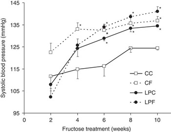
As expected, SBP increased in the fructose-fed offspring (CF and LPF), compared with CC offspring (Fig. 2), indicating that the 60% fructose diet induced elevated blood pressure. However, from 6 weeks, the SBP of LPC offspring was also higher than that of CC offspring throughout the treatment period. These data indicated that SBP in adult offspring from dams exposed to low protein increased independent of the postnatal 60% fructose diet.
Systolic blood pressure of adult male offspring. Values are expressed as mean ± s.e.m. (n = 6–8). *P < 0.05 compared with CC. CC, control-on-control group; CF, 60% fructose diet-on-control; LPC, control-on-low-protein diet; LPF, 60% fructose diet-on-low-protein diet.

Urinary NO x excretion
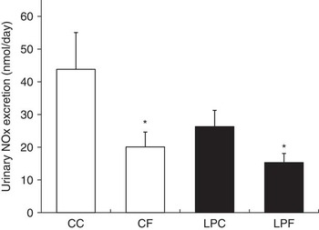
Since NO metabolites reflect the amount of systemic NO, the content of 24 h urinary NO x was measured (Fig. 3). The levels of 24 h urinary NO x were significantly lower in CF and LPF than in CC offspring, and the NO x levels in LPC tended to be lower than those in CC.
Urinary NO x excretion. Values are expressed as mean ± s.e.m. (n = 5–8). *P < 0.05 compared with control-on-control (CC).

Expression of mRNA and protein of eNOS in the aorta and kidney
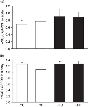
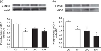
In the aorta, there was no significant difference in the levels of eNOS mRNA expression among all of the groups (Fig. 4a). However, a significant decrease in phosphorylated eNOS protein abundance was found in the CF, LPC and LPF offspring compared with CC offspring (Fig. 5a). Likewise, in the kidney, although there was no significant difference in the expression levels of eNOS mRNA among all of the groups (Fig. 4b), phosphorylated eNOS protein abundance in CF, LPC and LPF offspring decreased significantly compared with that in CC offspring (Fig. 5b).
Expression of endothelial nitric oxide synthase (eNOS) mRNA in the aorta (a) and kidney (b). Values are indicated as arbitrary ratios of the amount of expressed mRNA of each factor to that of GADPH. Values are expressed as mean ± s.e.m. (n = 6–8).

Protein abundance of total and phosphorylated endothelial nitric oxide synthase (eNOS) in the aorta (a) and kidney (b). Values are expressed as mean ± s.e.m. (n = 5–7). *P < 0.05 compared with control-on-control (CC).

Expression of p47phox and Nox4 mRNA in the aorta
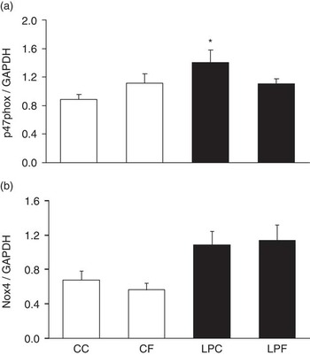
The levels of p47phox mRNA in LPC offspring increased significantly compared with those in CC (Fig. 6). The levels of p47phox mRNA in LPF offspring were similar to those in CF. The data indicated that vascular p47phox mRNA expression in maternal protein-restricted offspring may be upregulated independent of the fructose diet. Although no significant difference was found, the levels of Nox4 mRNA in LPC and LPF tended to be higher than those in CC.
The mRNA expression of p47phox (a) and Nox4 (b) of NAD(P)H oxidase subunits in the aorta. Values are indicated as arbitrary ratios of the amount of expressed mRNA of each factor to that of GAPDH. Values are expressed as mean ± s.e.m. (n = 5–7). *P < 0.05 compared with control-on-control (CC).

Superoxide production in the aorta
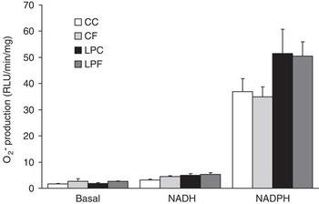
Both NADH- and NADPH-stimulated O2 − production were enhanced in the aorta in all of the groups, compared with the O2 − production in the absence of NADH or NADPH (Fig. 7). Although a statistically significant difference was not reached, a tendency toward increased NADPH-stimulated O2 − levels in LPC offspring was observed compared with CC and CF offspring.
Superoxide (O2 −) production in the aorta. NAD(P)H oxidase-derived O2 − production was determined in the aorta rings by lucigenin chemiluminescence under basal (0 μmol/l), NADH (100 μmol/l) and NADPH (100 μmol/l)-stimulated conditions. Values are expressed as mean ± s.e.m. (n = 6–8).

Expression of ET-1 and ETB mRNA in the aorta and kidney
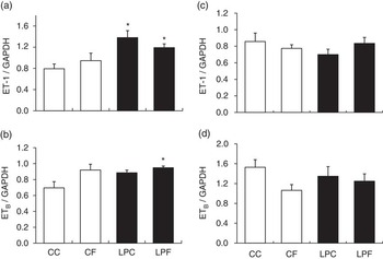
In the aorta, a significant increase in the expression of ET-1 mRNA was found in LPC and LPF offspring compared with CC offspring (Fig. 8). The expressions of ETB mRNA in LPF were significantly higher than that in the CC. In addition, a tendency toward higher ETB mRNA expression levels in CF and LPC was observed. In the kidney, there was no significant difference in ET-1 and ETB mRNA expression between the groups.
Expression of endothelin-1 (ET-1) and endothelin receptor type B (ETB) mRNA in the aorta (a and b) and kidney (c and d). Values are indicated as arbitrary ratios of the amount of expressed mRNA of each factor to that of GAPDH. Values are expressed as mean ± s.e.m. (n = 4–8). *P < 0.05 compared with control-on-control (CC).

Discussion
Maternal undernutrition or dietary protein restriction during pregnancy and lactation is believed to be associated with long-term metabolic consequences such as hypertension, cardiovascular diseases and diabetes. However, there is limited information about whether these adult diseases in the resulting growth-restricted offspring would be exacerbated by an unfavorable postnatal environment. The key findings of this study are as follows: (i) SBP increased not only in the 60% fructose-fed offspring (CF and LPF) but also in the maternal protein-restricted offspring (LPC) compared with CC offspring; (ii) eNOS phosphorylation in the aorta and kidney of CF, LPC and LPF offspring decreased significantly; and (iii) in the aorta, mRNA levels of p47phox, NAD(P)H oxidase subunits in LPC, and ET-1 in LPC and LPF offspring increased significantly.
Previous reports have described that maternal undernutrition or dietary protein restriction during pregnancy led to hypertension.Reference Langley and Jackson 5 – Reference Cambonie, Comte and Yzydorczyk 7 We also showed elevated SBP in LPC offspring from 6 weeks. In addition, the SBP of fructose-fed offspring (CF and LPF) was also higher than that of CC offspring throughout the treatment period, supporting the previous report that blood pressure was significantly higher in high fructose-fed rats.Reference Takagawa, Berger and Hori 16 , Reference Miatello, Risler and Castro 20 However, no significant difference in SBP between LPC and LPF was observed in this study, suggesting that a fructose diet may not enhance the elevation of blood pressure in maternal protein-restricted offspring.
In this study, we found a decrease in the urinary NO x contents of CF and LPF offspring (Fig. 3). Previous studies have reported that NO production in fructose-fed rats decreased both at the vascular and renal levels.Reference Johnson, Segal and Sautin 17 , Reference Miatello, Risler and Castro 20 , Reference Zhao, Xu and Cui 21 Considering that NO is an important molecule that functions as an endogenous vasodilator and is associated with endothelial function,Reference McIntyre, Bohr and Dominiczak 10 , Reference Taniyama and Griendling 11 our results suggest the impairment of NO production in the elevation of blood pressure.
As several studies on maternal undernutrition have shown that endothelium-dependent and -independent vasodilatation were impaired in later stages of childhood and in adulthood,Reference Rodford, Torrens and Siow 22 , Reference Torrens, Hanson and Gluckman 23 we examined whether the expression levels of vascular and renal eNOS were altered. Although the levels of eNOS mRNA expression among all of the groups were not significantly altered in the aorta and kidney, the ratio of phosphorylated eNOS at Ser1177 /total eNOS in the aorta and kidney was reduced in high fructose-fed offspring (CF and LPF) as well as in maternal protein-restricted offspring (LPC), compared with CC. The endothelial dysfunction is associated with a decrease in activity and expression of eNOS in the aorta and kidney from male offspring of nutritionally restricted damsReference Franco Mdo, Arruda and Dantas 24 and fructose-treated rats.Reference Zhao, Xu and Cui 21 , Reference Nishimoto, Tomida and Matsui 25 Therefore, we assumed that the inactivation of eNOS in the aorta and kidney may be, at least in part, likely to lead to elevated blood pressure in CF, LPC and LPF offspring.
In this study, a postnatal high-fructose diet affected the blood pressure in offspring (CF) from control dams, whereas the fructose diet showed no alternation in the blood pressure between LPC and LPF offspring from dams exposed to a low-protein diet during the gestation and suckling periods. The reasons for this phenomenon in maternal protein-restricted offspring remain as speculation at present. One possibility is that the lower levels of plasma insulin in LPC and LPF offspring may be associated with the impairment of pancreatic islet function. Several studies have reported the increase of islet cells vulnerability of adult rat offspring from protein-restricted dam.Reference Merezak, Reusens and Renard 26 , Reference Goosse, Bouckenooghe and Balteau 27 Theys et al.Reference Theys, Clippe and Bouckenooghe 28 showed lower plasma insulin level and islet dysfunction in the offspring exposed to maternal protein restriction during the pregnancy and suckling periods. Since insulin signaling is known to activate the PI3K/Akt/eNOS signal transduction pathway,Reference Fleming and Busse 29 – Reference Motley, Eguchi and Patterson 31 we hypothesized that the lower levels of plasma insulin from the islet in LPC and LPF offspring may affect inactivation of eNOS phosphorylation in the aorta and kidney, leading to elevated blood pressure. Another possibility is that maternal protein restriction may upregulate the expression of NAD(P)H oxidase subunits and ET-1 in the aorta of the offspring. In this study, we showed that p47phox mRNA expression in the aorta was upregulated, and that Nox4 mRNA expression tended to be higher in parallel with an increase in O2 − production in LPC and LPF offspring from dams exposed to a low-protein diet. Our results were in agreement with those of several studies, stating that offspring after exposure to intrauterine malnutrition present an increased production of O2 −on the endothelium-dependent vasorelaxation in the mesenteric microvessels.Reference Franco Mdo, Arruda and Dantas 24 , Reference Franco, Akamine and Reboucas 32 , Reference Roghair, Segar and Volk 33 NAD(P)H oxidase is known to be one of the major enzymes actively involved in O2 − production. The excessive production of O2 − can enhance NO inactivation, which as a result may reduce bioactive NO.Reference McIntyre, Bohr and Dominiczak 10 , Reference Taniyama and Griendling 11 Therefore, this upregulated expression of NAD(P)H oxidase subunits in the aorta may be causally associated with the elevation of blood pressure. Moreover, we found that the vascular levels of mRNA expression of ET-1 in LPC and LPF, and of ETB in LPF offspring were significantly higher than those in CC offspring. Vascular dysfunction associated with hypertension is characterized by an imbalance between NO and ET-1 production. From our findings, we assumed that maternal protein restriction may contribute to an imbalance between the vasodilator NO and the vasoconstrictor ET-1 and may be linked to elevated blood pressure independent of a postnatal fructose diet.
In conclusion, we showed herein that maternal protein restriction during the gestation and suckling periods elevated the blood pressure, downregulated NO production and eNOS phosphorylation, whereas these were unaffected by a postnatal fructose diet. Although further studies are necessary to elucidate the precise molecular signal(s), our results from this study suggest that either maternal protein-restricted diet or a postnatal fructose diet in isolation may enhance blood pressure elevation and vascular and renal dysfunction in adult offspring. Thus, these effects seem unlikely to be cumulative in adult offspring in our animal model.
Acknowledgements
This study was supported by a Grant-in-Aid (No. 20500710) for Scientific Research from the Ministry of Education, Culture, Sports, Science and Technology of Japan.
Statement of interest
None.