I. INTRODUCTION
Cubic silicon carbide (3C–SiC) is an attractive wide-band gap semiconductor material for high frequency, high temperature, and high power electronic devices due to its excellent physical and chemical properties.Reference Ferro, Monteil, Vincent, Thevenot, Tran, Cauwet and Bouix1, Reference Severino, D’Arrigo, Bongiorno, Scalese, La Via and Foti2 Heteroepitaxial growth of 3C–SiC films on Si substrates by chemical vapor deposition (CVD) is a promising method, which not only can supply large-size low-cost substrate for heteroepitaxial growth of III–V compound semiconductors but also can be integrated with the Si-based devices. Due to the large lattice mismatch (∼20%) and difference in thermal expansion coefficient (∼8%) between 3C–SiC and Si, a severe strain exists at the interface and in the epitaxial layer. The strain relaxation will result in crystalline defects, such as misfit dislocations, stacking faults, and twins, at the SiC/Si interface and in the SiC epilayers.Reference Severino, D’Arrigo, Bongiorno, Scalese, La Via and Foti2, Reference Portail, Zielinski, Chassagne, Roy and Nemoz3 These defects have severely restricted the applications of 3C–SiC in electronic devices compared with commercial 4H– and 6H–SiC materials.
To partially accommodate the large lattice mismatch, a carbonization process before epitaxial growth has been proved to be a helpful step to obtain high quality 3C–SiC film on Si substrate.Reference Nishino, Powell and Will4 Carbonization is a chemical reaction process between Si substrate and C-bearing molecules from the gaseous phase to form an ultrathin 3C–SiC buffer layer. Many people have studied on the buffer layer grown by different techniques with various C-bearing precursors, in terms of morphology, growth kinetics, chemical nature, crystalline structure, and several growth models have been proposed.Reference Ferro, Monteil, Vincent, Thevenot, Tran, Cauwet and Bouix1, Reference Mogab and Leamy5–Reference Cimalla, Pezoldt, Ecke, Rossler and Eichhorn14 For the buffer layer morphology, people mainly concentrated on the void, which is the most common interfacial defect associated with heteroepitaxial growth of 3C–SiC on Si. It has been shown that voids can locally favor the formation and propagation of the process-related defects in the 3C–SiC films and result in a rough SiC/Si interface.Reference Severino, D’Arrigo, Bongiorno, Scalese, La Via and Foti2, Reference Portail, Zielinski, Chassagne, Roy and Nemoz3 As a result, a problem is posed in fabricating devices, such as metal oxide semiconductor field-effect transistor and heterojunction bipolar transistor.Reference Gupta, Sengupta and Jacob15 Therefore, elucidating and suppressing the formation of voids are crucial for growth of high quality 3C–SiC film. The evolution of void density and surface roughness has been intensively investigated as a function of temperature, processing time, and C-bearing species flow rates.Reference Portail, Zielinski, Chassagne, Roy and Nemoz3, Reference Mogab and Leamy5, Reference Kordina, Bjorketun, Henry, Hallin, Glass, Hultman, Sundgren and Janzen13, Reference Yoshinobu, Mitsui, Tarui, Fuyuki and Matsunami16 However, the void size including width and depth has not been systematically and quantitatively studied, which is essential for understanding the void formation and evolution. Furthermore, few researchers have paid attention to the roles of temperature ramp-up rate and carrier gas (hydrogen) in this process. In this paper, we perform an in-depth study on the dependences of void density, void width, void depth, and surface roughness on the carbonization parameters using atomic force microscopy (AFM), and a buffer layer growth model is proposed based on the experimental results.
II. EXPERIMENTAL DETAILS
All experiments were carried out in a homemade horizontal cold-wall atmospheric pressure CVD system. The system includes an 86-mm-diameter quartz reactor and a SiC-coated graphite susceptor heated by a radio frequency induction power supply. The on-axis p-type Si (100) was used as substrate. Hydrogen (H2, >99.999%) and propane (C3H8, 1% in H2) were used as carrier gas and C-bearing precursor, respectively. Before carbonization, the substrate was in situ etched by a mixture gas of H2 (2.5 slm)/HCl (10 sccm) at 1020 °C for 10 min. Subsequently, the substrate was cooled down to room temperature. Then, the C3H8 was introduced into the chamber and mixed with H2. Once the mixed gas flow was stable in the reactor, the temperature was ramped up to the carbonization temperature and kept for several minutes to form SiC buffer layer. After the carbonization process, the heating system was switched off and cooled down to room temperature in H2. Typical process conditions were as follows: (i) carbonization temperature: 1100–1250 °C; (ii) processing time: 2.5–10 min (here, it means the duration from temperature ramping to power off); (iii) temperature ramp-up rate: 15–34 °C/s; (iv) C3H8 flow rate: 0.5–0.9 sccm (it means the real value); (v) H2 flow rate: 2.0–3.6 slm. For each sample, four process parameters were kept the same, while the fifth parameter was systematically varied. All the samples were characterized by AFM (Veeco Dimension Icon, Plainview, NY) in a tapping mode. The void width, depth, and surface roughness were obtained by the NanoScope Analysis software. To further evaluate the microstructure, one typical sample was characterized by transmission electron microscopy (TEM, Tecnai G2 F20, FEI Co., Hillsboro, OR).
III. RESULTS AND DISCUSSION
A. Effect of temperature
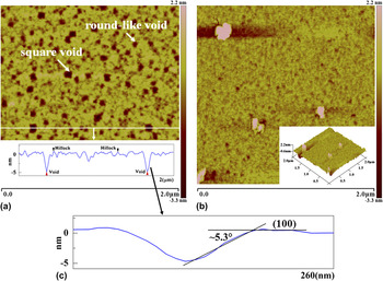
Four samples were carbonized at different temperatures of 1100, 1150, 1200, and 1250 °C, respectively, while the other parameters were kept the same (2.5 min, 22 °C/s, 0.9 sccm C3H8 in 2 slm H2). The typical morphologies are characterized by AFM and shown in Fig. 1. As presented in Fig. 1(a), a large number of square or round-like voids are observed when the sample is carbonized at 1200 °C. The sectional profile shows that the voids are in inverted unsealed pyramidal shape, and small hillocks can also be detected between these voids. It has been widely proved that such square voids are formed during the carbonization stage due to the Si atoms out-diffusion from the heated Si (100) substrate, and all the lateral faces of the voids should be oriented along the {111} planes due to the lowest surface energy of Si.Reference Severino, D’Arrigo, Bongiorno, Scalese, La Via and Foti2, Reference Gupta, Sengupta and Jacob15 However, such lateral faces are not {111} planes in this study, which can be proved by the fact that the angle between the surface plane of Si substrate and the lateral face of the voids is about 5.3°, as shown in Fig. 1(c), differing from the value of 54.5° reported by Gupta et al.Reference Gupta, Sengupta and Jacob15 This indicates that the voids are not in thermodynamic equilibrium state, and they will enlarge with increasing time. The round-like voids could be the boundaries when neighboring 3C–SiC nuclei coalesce or some voids, which are not completely sealed with a small aperture in the Si substrate, while the small hillocks are recognized as initial SiC crystallites. For the samples carbonized below 1200 °C, besides the voids, the central zone presents grayer and blurry features observed by naked eyes. The blurry area is characterized by AFM and shown in Fig. 1(b). It is noted that there are several particles with near-squared base embedded in the substrate, which are deleterious for the subsequent epitaxial growth of 3C–SiC film.
The AFM surface morphology (2 × 2 μm2) of buffer layers (a) carbonized at 1200 °C, the sectional profile is inserted in the bottom; (b) carbonized at 1150 °C, the 3D image is inserted in the lower right corner; (c) one of the magnified sectional profile of the square voids shown in (a).

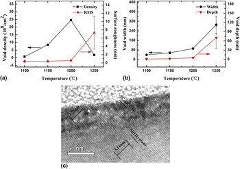
The dependences of void density, width, depth, and surface roughness on carbonization temperature are shown in Figs. 2(a) and 2(b). The void density increases rapidly as the temperature rises from 1100 to 1200 °C and then decreases abruptly when the temperature further increases to 1250 °C, while the void width, depth, and surface roughness monotonically increase with increasing temperature. The trends of void size and surface roughness with varying carbonization temperature are quite consistent with the former results. For the trend of void density, however, there is something different with the previous reports for the same temperature range.Reference Ferro, Monteil, Vincent, Thevenot, Tran, Cauwet and Bouix1, Reference Severino, D’Arrigo, Bongiorno, Scalese, La Via and Foti2, Reference Kordina, Bjorketun, Henry, Hallin, Glass, Hultman, Sundgren and Janzen13 It was reported that,Reference Bittencourt17 at the initial stage of carbonization, the adsorbed C atoms can break the superficial Si–Si bonds through inserting the C atoms and giving rise to shrinkages of Si atomic row spacing. These shrinkages lead to the formation of three-dimensional (3D) configurations, which act as the sites for initial SiC nuclei formation. Therefore, a higher density of adsorbed C-bearing species on the Si substrate can lead to a higher density of initial nuclei. According to Ref. Reference Gupta, Sengupta and Jacob15, the out-diffusion of Si atoms is effective when the temperature is above 1100 °C, while the decomposition efficiency of C3H8 is quite low in the temperature range of 1100–1200 °C. When the temperature is below 1200 °C, there are not enough C-bearing species to react with the superfluous and active Si atoms to form initial SiC nuclei, probably giving rise to the deposition of silicon particles [seen in Fig. 1(b)] and lower coverage of SiC on the Si substrate. When temperature is increased to 1200 °C, the higher decomposition rate of C3H8 leads to a higher initial nuclei density, and most parts of Si substrate are covered by SiC nuclei. As reported by Li and Steckl,Reference Li and Steckl6 once the initial SiC nuclei are formed, the supply of Si atoms comes from the uncovered areas of Si substrate leading to the formation of initial voids, which are localized at these uncovered areas and surrounded by SiC nuclei. These initial voids can enlarge both in horizontal and vertical directions as the growth of initial nuclei. For lower temperature (below 1200 °C), this enlargement process is much weaker due to the slower nuclei growth rate and out-diffusion rate of Si atoms. Therefore, the density and size are smaller for lower temperature. However, when temperature is further increased to 1250 °C, the growth of initial nuclei dominates and initial voids will be sealed by coalescence of neighboring 3C–SiC nuclei, leading to a lower void density. To evaluate the microstructure, the sample carbonized at 1200 °C was characterized by cross-sectional high-resolution TEM as shown in Fig. 2(c). The buffer layer thickness is about 4 nm and the buffer layer possesses epitaxial nature with the Si substrate. This epitaxial nature is beneficial for subsequent epitaxial growth of 3C–SiC film. Furthermore, it is obvious that every four Si lattice planes inserted additional SiC lattice plane at the SiC/Si interface as indicated by the triangle symbol in Fig. 2(c); this is consistent with the analysis in Ref. Reference Zielinski, Ndiaye, Chassagne, Juillaguet, Lewandowska, Portail, Leycuras and Camassel18.
The dependences of void density and surface roughness (a), void width and depth (b) on carbonization temperature. A typical high-resolution TEM image of buffer layer characterized along the [011] zone axis (c).

B. Effect of processing time
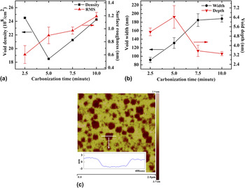
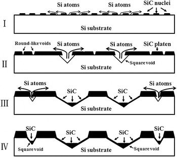
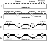
Figures 3(a) and 3(b) show the dependences of void density, width, depth, and surface roughness on processing time, which were carbonized for 2.5, 5.0, 7.5, and 10.0 min, respectively, keeping the other parameters constant (1200 °C, 22 °C/s, 0.9 sccm C3H8 in 2 slm H2). It shows that the void density initially decreases to a minimum value after 5 min carbonization and then gradually increases with increasing processing time, while the void depth follows the inverse trend compared with the void density. It is interesting to note that the void width and depth appear to be saturated after 7.5 min carbonization. The AFM image shows that the largest voids located at the bottoms are flat as shown in Fig. 3(c). This indicates that larger voids have been covered by SiC, while the smaller voids are not sealed after 7.5 min carbonization. Based on these observations, the growth model of buffer layer with processing time is shown in Fig. 4. It shows that at the initial stage of carbonization, a large number of small SiC nuclei are homogenously distributed on the substrate. As processing time increases, initial voids appear between the nuclei and extend both in horizontal and vertical directions to form square voids with inverted pyramidal shape and a squared base due to out-diffusion of Si atoms. At the same time, the initial nuclei grow laterally and vertically. The growth rate of nuclei near the voids is higher than that of nuclei far away from the square voids because of the shorter diffusion path for Si atoms. Gradually, the neighboring nuclei near the square voids coalesce with each other and form SiC platens. However, the nuclei far away from the square voids only partially connect with each other. The round-like voids form between neighboring nuclei boundaries. As the processing time is further increased, the square voids are sealed; meanwhile, the supplying of Si atoms are from the small round-like voids, as shown in Fig. 4(c). Finally, the small voids become larger and then the whole surface is covered by SiC thin films as shown in Fig. 4(d).
The dependences of void density and surface roughness (a), void width and depth (b) on processing time. The AFM image of the buffer layer carbonized for 7.5 min (c).

A proposed model for the growth of SiC buffer layer.

C. Effect of ramp-up rate
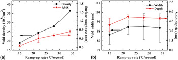
To investigate the effects of temperature ramp-up rate on the buffer layer evolution, the processing time was fixed at 2.5 min because the voids were not sealed under this condition. Four samples were carbonized with the temperature ramp-up rate varied from 15 to 34 °C/s, keeping the other parameters constant (1200 °C, 2.5 min, 0.9 sccm C3H8 in 2 slm H2). It is interesting that the ramp-up rate slightly affects the void width and depth but strongly affects the void density and surface roughness, as shown in Figs. 5(a) and 5(b). As reported by Li and Steckl,Reference Li and Steckl6 the void density was dependent on initial 3C–SiC nuclei density. For higher initial nuclei density, the coalescence of neighboring nuclei takes place within a short time to form a continuous film, and Si atoms out-diffusion could be prevented, resulting in lower density of voids. Thus, the increase in void density and surface roughness at higher ramp-up rate can be ascribed to the decrease in the initial nuclei density. The formation of initial nuclei is dependent on two factors. One factor is the number of active Si atoms out-diffused from Si substrate surface. The other factor is the number of C-bearing species adsorbed on the substrate. Above 780 °C, C3H8 starts to decompose, and then, the effective nucleation process takes place in the temperature range of 780–1200 °C.Reference Stinespring and Wormhoudt7 As reported in Ref. Reference Hens, Wagner, Hölzing, Hock and Wellmann19, the nucleation dominates in the temperature range below 1200 °C, while above this temperature, nuclei growth dominates. Therefore, the effective nucleation time is shorter for higher ramp-up rate, and there is no enough time for C-bearing species absorbed on the Si substrate to react with active Si atoms. Therefore, the density of initial nuclei is lower at higher ramp-up rate.
The dependences of void density and surface roughness (a), void width and depth (b) on temperature ramp-up rate.

D. Effect of C3H8 and H2
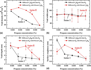
In previous studies, people mainly focused on the effects of C-bearing species flow rates on the buffer layer morphology in a constant H2 flow rate, and few researchers paid attention to the effect of H2 on the buffer layer morphology with a constant C-bearing species flow rate. To comparatively study the roles of C3H8 and H2 during the carbonization process, two series of experiments were carried out. The H2 flow rate was fixed at 2 slm, while the C3H8 flow rate was varied from 0.5 to 0.9 sccm (Series I). The C3H8 flow rate was fixed at 0.9 sccm, while the H2 flow rate was varied from 3.6 to 2.0 slm (Series II). The C3H8 concentration defined as the ratio of C3H8 to H2flow rates was in the range of 0.024–0.043%, and the other parameters remained the same for each experiment (1200 °C, 2.5 min, 22 °C/s).
Figures 6(a)–6(d) show the dependences of void density, width, depth, and surface roughness on C3H8 concentration, the square and circle symbols corresponding to the changes in C3H8 (Series I) and H2 (Series II), respectively. From Fig. 6(a), it can be seen that the void density decreases with increasing C3H8 concentration. This trend is consistent with previous reports.Reference Ferro, Monteil, Vincent, Thevenot, Tran, Cauwet and Bouix1, Reference Li and Steckl6 Increasing the C3H8 concentration enhances the initial SiC nuclei coverage of the Si substrate. As a result, the out-diffusion sites for Si atoms are reduced and then the density of voids is decreased. For the void width, it almost keeps constant with an average value of 92 nm as shown in Fig. 6(b). This indicates that the void width is not sensitive to the C3H8 and H2 flow rates with these conditions. As shown in Fig. 6(c), the void depth decreases as the C3H8 concentration increases, except for a singular point (0.034%) of C3H8 concentration for Series II. The surface roughness shows a similar trend as the void density and depth with increasing C3H8 concentration, as shown in Fig. 6(d). In this study, the surface roughness is defined as root-mean-square average of height deviations taken from the mean image data plane. Therefore, the surface roughness should be a comprehensive reflection of the void density, width, and depth.
The dependences of void density (a), void width (b), void depth (c), and surface roughness (d) on C3H8 concentration.

For each C3H8 concentration, it is evident that the values of void density, depth, and surface roughness of Series II are much larger than those of Series I. As mentioned in previous section, the void density is dependent on the initial SiC nuclei density. Such nuclei density is proportional to the hydrocarbon concentration, which has been reported by many researchers. Therefore, the void density should show a little difference for Series I and Series II. However, the result shown in Fig. 6(a) is contrary to the prediction. To explain this difference, we presume that the consumption of Si atoms out-diffused from Si substrate is a competition process among three approaches. The first approach is reaction with C-bearing species to form SiC. The second approach is direct diffusion to free gas stream. The last approach is reaction with H2 to form volatile gaseous species. According to the boundary layer theory, high velocity of gas stream helps to reduce the boundary layer thickness.Reference Chen20 For Series II, the total flow rate is higher than that of Series I. It means that the boundary layer thickness is thinner for Series II. Thus, the flux of C-bearing and H2 species arriving at the substrate is much larger than that of Series I, so does the diffusion of Si atoms to the free gas stream. Thus, for Series II, most of Si atoms take part in the reaction with H2 or diffuse to the free gas stream, giving rise to a lower initial SiC nuclei density, although the C3H8 concentration is kept the same. For the void depth, it is defined as the vertical height from sample surface to the bottom of voids including the buffer layer thickness. The decrease in void depth could be due to the decrease in layer thickness as the C3H8 concentration is increased. This is supported by the studies on growth kinetics of buffer layer. On the other hand, the void is in nonequilibrium state and its width is constant in the investigated range, making the Si atoms out-diffusion is prone to be along the vertical direction. This process will be accelerated if the consumption of Si atoms enhanced. Thus, the depth is deeper for Series II.
IV. CONCLUSIONS
The influence of carbonization parameters on the morphology of ultrathin SiC buffer layer grown on Si (100) substrate has been systematically investigated by AFM. The buffer layer morphology is mainly decorated with square or round-like voids. The quantitative analysis of the evolution of voids with processing time indicates that the voids are not in thermodynamic equilibrium state and can be sealed by SiC after 7.5 min carbonization. Based on these observations, a SiC buffer layer growth model is proposed. Furthermore, it is found that the void density is mainly dependent on the initial SiC nuclei density and related to the temperature, temperature ramp-up rate, and C3H8 concentration. A higher initial SiC nuclei density produces a lower void density. The void size is evolved with processing time, while it is related to temperature, C3H8 concentration, and H2 flow rate. The void becomes wider and deeper with increasing temperature, while C3H8 and H2 are not sensitive to the void width but slightly affect the depth. The buffer layer morphology is deteriorated with increasing H2 flow rate when fixing the C3H8 concentration.
ACKNOWLEDGMENTS
This work is supported by the Young Scientists Fund of the National Natural Science Foundation of China (Grant No. 51002176) and the Innovation Program of the Chinese Academy of Sciences (Grant No. KJCX2-EW-W10).