Introduction
The bone-anchored hearing aid (BAHA) represents an alternative to the conventional bone-conduction hearing aid (fitted to a headband or onto a specially strengthened spectacle). The BAHA system, in which the transducer is fitted onto a percutaneous coupling device attached to a titanium fixture placed in the temporal bone, has both cosmetic and acoustic advantages over the conventional bone conduction hearing aid, and has known quality of life benefits.Reference Hol, Spath, Krabbe, van der Pouw, Snik and Cremers1
In the early 1980s, Tjellström and Håkansson published the first clinical BAHA studies.Reference Tjellström and Håkansson2 Over time, the implantation procedure has been simplified. For adults, the standard two-stage surgical procedure for inserting the percutaneous implant into the temporal bone has been converted to a single-stage procedure in which the percutaneous coupling device (abutment) is premounted onto a self-tapping screw fixture. As a result of positive experience with the BAHA for conventional indications, and the low rate of predominantly minor complications,Reference Granström, Bergström, Odersjo and Tjellström3–Reference van der Pouw, Mylanus and Cremers5 the indications for BAHA continue to grow, extending to severe unilateral hearing loss and to cases of unilateral conductive or mixed hearing loss which traditionally have been left unaided.Reference Kunst, Leijendeckers, Mylanus, Hol, Snik and Cremers6, Reference Wazen, Spitzer, Ghossaini, Kacker and Zschommler7
Insertion of the percutaneous implant may sometimes give rise to adverse skin reactions. Several large studies reported an average occurrence of adverse skin reactions in adults of 15 per cent (10–20 per cent), most of which can be successfully treated in an office setting. Implant extrusion is the most severe complication, although rare (average occurrence 5 per cent (2.5–10 per cent)), and may be the result of trauma, inadequate osseo-integration or inflammatory processes.Reference Granström, Bergström, Odersjo and Tjellström3–Reference van der Pouw, Mylanus and Cremers5 An important predisposing factor for adverse skin reactions is the mobility of the skin surrounding the abutment. Therefore, a vital part of the surgical procedure is the reduction of subcutaneous tissue so that the skin is sufficiently thin, firmly attached to the underlying periosteum and without hair follicles.Reference Tjellström and Granström8 In some patients, however, independent of the technique used to reduce subcutaneous tissue,Reference Lekakis, Najuko and Gluckman9–Reference Snyder, Moore and Johnson11 the skin hypertrophies and pushes up against the implant abutment, in some cases even growing over it, jeopardising the accessibility of the abutment for the transducer coupling. If hypertrophic skin persists and does not respond to conservative treatment (e.g. application of antibiotic and corticosteroid ointment), revision surgery is needed. At the department of otorhinolaryngology of the Radboud University Medical Center, approximately 6 per cent of the patients have required revision surgery of the surrounding skin for this indication.
Revision surgery may not be applicable or desirable in some patients, and alternative methods of treatment are required. We present a patient with persistent hypertrophic skin surrounding the percutaneous implant, successfully treated with intralesional applied corticosteroids.
Case report
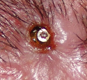
Eight months after his initial BAHA implant, a 62-year-old man underwent revision surgery to reduce and thin the surplus of skin and subcutaneous scar tissue overgrowing the abutment. The pre-operative condition is shown in Figure 1.
Pre-operative condition. The skin surrounding the abutment is hypertrophic and has partly covered the percutaneous implant.

After local anaesthesia, a straight incision of approximately 3 cm was made, with the percutaneous abutment at the centre of the incision. Both sides of the incision were thinned out, removing all subcutaneous tissue but leaving the periosteum.
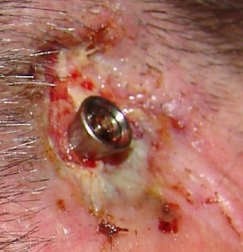
Post-operative care consisted of a bolster dressing under a healing cap for one week. Figure 2 shows the post-operative condition directly after removal of the healing cap and bolster dressing.
One week post-operative appearance, immediately after removing the healing cap and bolster dressing.

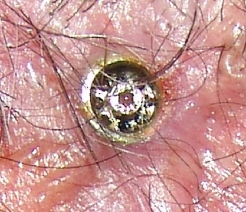
Despite a good initial result, with the patient being able to wear his BAHA without any problems or complaints, after six to eight weeks the skin and subcutaneous scar tissue overgrowth reoccurred (Figure 3). As an alternative to yet another surgical procedure, the hypertrophic skin was treated with intralesional injections of a total of 1 ml of triamcinolone acetonide 40 mg/ml, using a 30-gauge needle on a Luer-lock syringe, without any preceding local anaesthesia. The injection was well tolerated by the patient. After just a few days, he noticed that coupling the BAHA transducer on the abutment became easier.
Eight week post-operative appearance. The hypertrophic scar tissue has recurred.

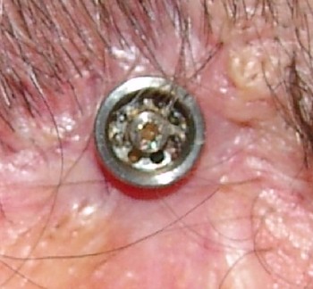
Three weeks after the triamcinolone treatment, a satisfying result was seen (Figure 4). No relapse was seen over the subsequent six months.
Appearance three weeks after intralesional treatment with 1 ml triamcinolone acetonide 40 mg/ml (three weeks after Figure 3).

Discussion
The use of triamcinolone acetonide injections for the treatment of hypertrophic scars and keloids has been shown to be effective in some patients, and has been in use for several decades.Reference Kiil12, Reference Muneuchi, Suzuki, Onodera, Ito, Hata and Igawa13 In the majority of patients, multiple intralesional injections are needed to obtain the desired clinical effect, i.e. flattening of the lesion and cessation of itching. In the long term, the recurrence rate may rise to 50 per cent.Reference Kiil12 The main effect of intralesional steroids on the connective tissue of keloids is a decrease in viscosity due to a loss of ground substance.Reference Krusche and Worret14 Few complications have been documented, and are limited to local skin changes or pain as a result of the injection. Rarely, Cushing's syndrome has been described, usually self-limiting. However, in children, features of Cushing's syndrome may be protracted. Therefore, the dosages of intralesional steroids normally recommended for adults are inappropriate for children.Reference Teelucksingh, Balkaran, Ganeshmoorthi and Arthur15
• Siting of a percutaneous bone-anchored hearing aid (BAHA) implant may give rise to adverse skin reactions
• This paper describes a patient with persistent hypertrophic skin surrounding the percutaneous implant of his BAHA system, which was successfully treated with intralesional applied corticosteroids
• In order to confirm the effectiveness of this treatment mode, long term results need to be documented in a considerable number of patients
Skin and subcutaneous scar tissue overgrowing the BAHA abutment is a troublesome condition, often causing the patient discomfort and pain, and difficulty in coupling the transducer. Although in some cases conservative treatment may be successful, revision surgery will be required in most patients. The intralesional application of triamcinolone acetonide may prove to be a worthwhile alternative. In our current patient, at the time of writing, no side effects of the treatment have been observed, and the result is satisfactory and sustained. In order to confirm the effectiveness of this treatment mode, long term results will need to be documented in a considerable number of patients.