Introduction
Toxocariasis is an important worldwide parasitic zoonosis caused by the passage of dog roundworm (Toxocara canisi) or cat roundworm (Toxocara cati) larvae through human organs such as eyes, lungs, liver, brain and heart. They cause different signs and symptoms, including eosinophilia (Magnaval et al., Reference Magnaval, Glickman, Dorchies and Morassin2001; De Andrade Lima Coêlho et al., Reference De Andrade Lima Coêlho, De Carvalho, Perez, Araki, Takeuchi, Ito, Aoki and Yamasaki2005; Azizi et al., 2007). Toxocariasis is clinically divided into four types: visceral, ocular, neurological and covert (Despommier, Reference Despommier2003; Rubinsky-Elefant et al., Reference Rubinsky-Elefant, Hirata, Yamamoto and Ferreira2010).
The signs and symptoms of visceral larva migrans (VLM) vary widely from an asymptomatic state with mild eosinophilia to severe and potentially fatal disorders, including hepatomegaly, hyperglobulinaemia, pulmonary symptoms and fever. Pneumonitis and neurological disorders may also appear in VLM. In serious cases, the leucocyte count may reach 100,000/mm3, while 80–90% are eosinophils (Gillespie, Reference Gillespie, Lewis and Maizel1993). Toxocariasis is one of the causes of eosinophilic infiltration in internal organs (Kwon et al., Reference Kwon, Oh, Lee, Lee and Choi2006). Children are more susceptible to this infection, for example when they handle contaminated soil (Cypess et al., Reference Cypess, Karol, Zidian, Glickman and Gitlin1977). The prevalence of toxocariasis is higher in tropical and developing countries and is generally associated with low socio-economic level (Thompson et al., Reference Thompson, Bundy, Cooper and Schantz1986; Lynch et al., Reference Lynch, Wilkes and Hodgen1988). Rates range from 1% in Spain (Portus et al., Reference Portus, Riera and Prats1989) to 25.6% in Shiraz, Iran (Sadjjadi et al., Reference Sadjjadi, Khosravi, Mehrabani and Oryan2000) and 86% in St. Lucia (Thompson et al., Reference Thompson, Bundy, Cooper and Schantz1986), yet there is a definitive method for diagnosing Toxocara infections (Kenny et al., Reference Kenny, MacCabe, Smith and Holland1995). The serologic methods investigating antibodies in sera are, by far, the main assay for the diagnosis (Sadjjadi et al., Reference Sadjjadi, Khosravi, Mehrabani and Oryan2000; Magnaval et al., Reference Magnaval, Glickman, Dorchies and Morassin2001; Noordin et al., Reference Noordin, Smith, Mohamad, Maizels and Fong2005; Smith & Noordin, Reference Smith, Noordin, Holland and Smith2006); however, recent studies have been conducted in this field, based on which the use of monoclonal antibodies has proved effective for the diagnosis of active cases (Zibaei et al., Reference Zibaei, Sadjjadi, Ishyiama, Sarkari and Uga2010). The most commonly utilized diagnostic serological test is enzyme-linked immunosorbent assay (ELISA). The use of excretory–secretory (E/S) antigens from T. canis larvae maintained in vitro further increases the specificity of the ELISA (Magnaval et al., 2001). Nevertheless, this antigen remains complex, including specific and non-specific fractions that may cause cross-reactions (especially with other helminthiasis) which affect the ELISA technique (Speiser & Gottstein 1984; Lynch et al., Reference Lynch, Wilkes and Hodgen1988; Mohamad et al., Reference Mohamad, Azmi and Noordin2009). The remarkable sensitivity of the immunoblot technique and its efficacy in demonstrating differences in antigen recognition by antibodies led to its use for Toxocara serology testing. Using the E/S antigen after separation by sodium dodecyl sulphate-polyacrylamide gel electrophoresis (SDS-PAGE), the Toxocara Western blotting (WB) IgG assay allows the differentiation between anti-Toxocara specific and non-specific anti-E/S antigen immunological responses. Very sensitive, and by definition specific, it is proposed as a confirmatory test for Toxocara serology (Magnaval et al., Reference Magnaval, Fabre and Maurieres1991). The present study investigated the seroprevalence of toxocariasis in hypereosinophilic individuals in the city of Ahwaz, south-western Iran, using ELISA and Western blot methods.
Materials and methods
The serum samples were collected from individuals referred to the Iran-Zamin, Golestan and Shafa diagnostic laboratories in Ahwaz for a routine cell blood count (CBC). Each individual completed a questionnaire. Blood samples were collected from 100 individuals with peripheral blood eosinophilia (>500 eosinophils/μl or more, equal to >10% of white blood cells) and also from 100 individuals as the control group (without eosinophilia). Sera were separated by blood centrifugation at 3000 rpm for 5 min and transferred to − 20°C until use. All sera were examined by T. canis IgG-ELISA test (IBL, Hamburg, Germany). Sera with Toxocara antibodies underwent further confirmative evaluations through Toxocara Western blot IgG test (LDBIO Diagnostics, Lyon, France).
Results
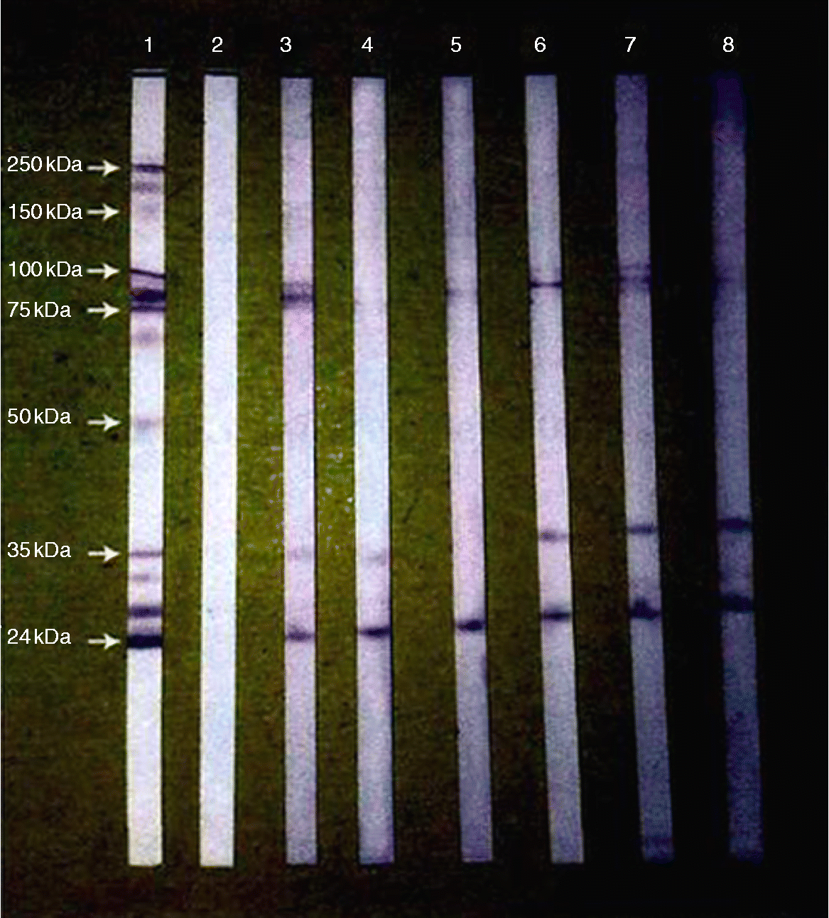
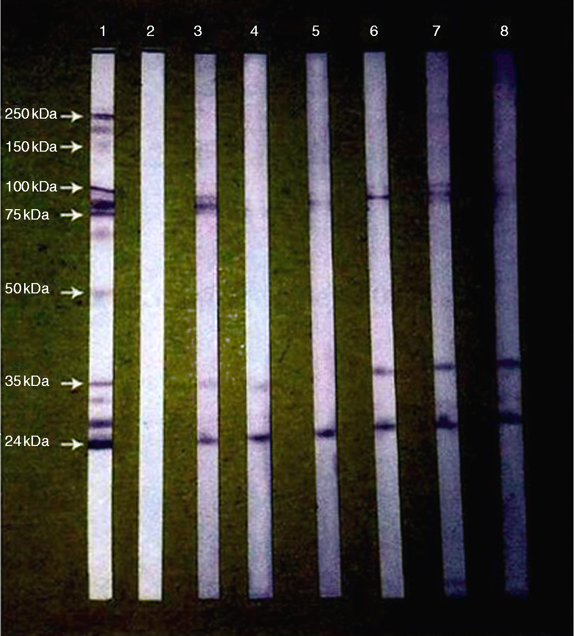
A total of 100 hypereosinophilic individuals were studied, 60 male and 40 female with a mean age of 35.14 years (range 1–92 years) and mean eosinophil count of 16.42% of white blood cells (range 10–65% of white blood cells). Seroprevalence antibodies against Toxocara with ELISA were obtained in 19 (19%) individuals, of whom 12 (63.15%) were female and 7 (36.85%) male. The median age of Toxocara seropositive patients was 48 years. Positive sera with ELISA were subsequently confirmed by Western blot (fig. l). All Western blot strips distinguished two or more low molecular weight bands in the range of 24–35 kDa that are specific for Toxocara IgG, and in 11 strips there were also high molecular weight bands in the range of 75–100 kDa,which are less specific for Toxocara. Antibodies against Toxocara were found in 1% of the control group but were not confirmed by Western blot.
Western blot analyses. Lane 1: positive control; lane 2: serum testing negative by enzyme-linked immunosorbent assay (ELISA); lanes 3–8: sera testing positive by ELISA. (See online for a colour version of this figure)
Discussion
The present study investigated the prevalence of toxocariasis in hypereosinophilic individuals in Ahwaz city, south-western Iran, an infection previously reported elsewhere in different countries at between 1 and 86%, as determined by ELISA (Thompson et al., Reference Thompson, Bundy, Cooper and Schantz1986; Portus et al., Reference Portus, Riera and Prats1989). Another study, in Nigeria by Ajayi et al. (Reference Ajayi, Duhlinska, Agwale and Njoku2000), also using ELISA, found that people residing in tropical regions infected by other worms may have a cross-reaction with anti-Toxocara. Thus, in such conditions, the Western blot method has been used. In the present study, hypereosinophilic individuals showed 19% anti-Toxocara antibody with ELISA. This was confirmed by the Western blot method and corresponded with previous studies by Park et al. (Reference Park, Park, Park, Lee, Huh, Kong and Magnaval2000), Fan et al. (Reference Fan, Hung, Du, Liao and Su2004) and Logar et al. (Reference Logar, Soba, Kraut and Kranjc2004). However, this is not supported by the study performed by Kim et al. (Reference Kim, Huh and Chung2008), in which five sera with anti-Toxocara antibody were not confirmed by Western blot method. In the present study, all specimens examined with the Western blot method showed bands of 24–35 kDa as described by Park et al. (Reference Park, Park, Park, Lee, Huh, Kong and Magnaval2000) and Logar et al. (Reference Logar, Soba, Kraut and Kranjc2004). The bands observed most often were in the range of 24–30 kDa, so low molecular weights are more frequently observed in Toxocara Western blots. Very few specimens had bands in the range of 75–100 kDa, as described by Park et al. (Reference Park, Park, Park, Lee, Huh, Kong and Magnaval2000), Fan et al. (Reference Fan, Hung, Du, Liao and Su2004) and Logar et al. (Reference Logar, Soba, Kraut and Kranjc2004), so bands with high molecular weights have little sensitivity for Toxocara diagnosis.
In the present study, there were significant differences between the frequency of infection with age (P < 0.05). The highest prevalence of infection occurred in adult males aged over 20 years, as seen in Japan (Akao & Ohata, Reference Akao and Ohata2007), France (Gueglio et al., Reference Gueglio, de Gentile, Nguyen, Achard, Chabasse and Marjolet2005), Nigeria (Ajayi et al., Reference Ajayi, Duhlinska, Agwale and Njoku2000) and Slovenia (Logar et al., Reference Logar, Soba, Kraut and Kranjc2004). A high prevalence of infection among the adults probably indicates past infection by Toxocara agents, because larvae can survive inside the body for 10 years (Morimatsu et al., Reference Morimatsu, Akao, Akiyoshi, Kawazu, Okabe and Aizawa2006). Also, infection is not related to age, as risk factors affect all age groups. Glickman & Schantz (Reference Glickman and Schantz1981) reported family toxocariasis in Japan, in which some of the infected individuals were aged 71 and 45 years, supporting this idea well. However, other studies have shown higher infection in children (Herrmann et al., 1985; Sadjjadi et al., Reference Sadjjadi, Khosravi, Mehrabani and Oryan2000; Baboolal & Rawlins, Reference Baboolal and Rawlins2002).
In the present study, there was a significant difference between the frequency of infection with sex (P < 0.05), and higher infection was seen in females, which resembled the results of the study by Magnaval & Baixench (Reference Magnaval, Baixench, Lewis and Maizel1993). The higher infection with Toxocara in women may be due to working in the kitchen and being exposed to contaminated vegetables and fruits and other infected sources. The seroprevalence of toxocariasis in Trinidad (Baboolal & Rawlins, 2002) indicated that a higher rate of infection in boys was related to their behaviour in the playground. In the present study, a significant difference was observed between those with hypereosinophilia and healthy individuals (P < 0.05), which corresponded with studies from Argentina and Korea (Alonso et al., Reference Alonso, Bojanich, Chamorro and Gorodner2000; Choi et al., Reference Choi, Suh, Jung, Song, Suh, Huh, Nahm and Park2003), but studies from Brazil did not show a significant difference between infection with and without hypereosinophilia (Anaruma Filho et al., Reference Anaruma Filho, Chieffi, Correa, Camargo, Silveria, Aranha and Riberio2002). We conclude that the prevalence of toxocariasis in hypereosinophilic individuals is high in Ahwaz, and it necessitates examination of the hypereosinophilic individuals for toxocariasis, not just in Ahwaz but in every region in which even very minute traces of eosinophilia are seen. This may undoubtedly be the best way to keep this enigmatic parasite at bay.
Acknowledgements
We would like to express our appreciation to the Vice Chancellor of research affairs at the Tropical Diseases Research Centre for financial support; Mr S. Rashtipour for his help and, finally, many thanks to Miss E. Nasiri for her kindness and help.