




No CrossRef data available.
Article contents
Transthoracic three-dimensional echocardiography prior to closure of atrial septal defects in children
Published online by Cambridge University Press: 18 April 2005
Abstract
Aims: Our aims were to use transthoracic three-dimensional echocardiography to assess the morphology of atrial septal defects in children prior to closure, and to compare the three-dimensional echocardiographic data with transcatheter and surgical findings. Methods and results:We used transthoracic three-dimensional echocardiography in 62 consecutive patients, aged from 2 to 18 years, with atrial septal defects, measuring the maximal diameter and the extent of the rims. Subsequent to the study, we referred 42 patients for transcatheter closure, the rims being measured at greater than 4 mm. We found a good correlation between the maximal diameter of the defect as measured at transthoracic three-dimensional echocardiography and using a balloon (y = 3.45 − 0.73x; r = 0.78; p < 0.0001), the mean difference between the measurements being 2.4 ± 2.8 mm. Successful closure with the Amplatzer septal occluder, having a mean size of 22 ± 4 mm, was achieved in 95% of the patients. Of the original cohort, 20 patients were referred for surgical closure. In these patients, the inferior rim had been deemed insufficient in 5, the postero-superior rim in 6, and the postero-inferior rim in 9. Complete agreement was found when the deficiency of the rim as judged using transthoracic three-dimensional echocardiography was compared with intraoperative findings. The correlation between measurements of the deficiency of the rim achieved by transthoracic three-dimensional echocardiography and at surgery was excellent (y = 0.2 + 0.98x; r = 0.93; p < 0.0001), the mean difference between the measurements being no more than 0.6 ± 0.4 mm. Conclusions: Transthoracic three-dimensional echocardiography proved accurate in measuring the maximal diameter and rims of atrial septal defects within the oval fossa. This non-invasive method will be valuable in selecting children for transcatheter or surgical closure of such defects.
Keywords
Information
- Type
- Original Article
- Information
- Copyright
- © 2003 Cambridge University Press
Accurate anatomical information is needed when selecting patients for transcatheter or surgical closure of an atrial septal defect within the oval fossa, including its maximal diameter and the amount of circumferential tissues forming its rims.1–3 Cross-sectional echocardiography alone is deficient in defining the criterions for successful closure. Since the defect is visualised from multiple orthogonal planes, its maximal diameter is widely under-estimated.4–7 The transcatheter approach measures the stretched diameter, but similarly cannot be used in isolation for selection of patients, since it does not provide information regarding the extent of the rims.8
Three-dimensional echocardiography, in contrast, provided unique en face views of the atrial septum. Previous studies have shown the capability of this technique to provide information with regard to the shape, the maximal diameter, and the extent of the rims surrounding the defect.9–11 Three-dimensional echocardiographic reconstructions to date, however, have been obtained from the transesophageal approach. This is an obvious limitation for routine use in children, since it requires general anaesthesia. The development of new software for three-dimensional reconstruction now allows the use of the transthoracic approach for surface imaging.12, 13 Our aims in this study, therefore, were to use this new technology to assess children with an atrial septal defect prior to closure.
Methods
Population studied
We examined 62 consecutive children referred for closure of an atrial septal defect using transthoracic three-dimensional echocardiography. There were 22 males and 40 females, the mean age being 8.5 ± 5 years, with a range from 2 to 18 years. The mean weight was 30 ± 15 kg, with a range from 10 to 85 kg. All patients had significant left-to-right atrial shunting with right ventricular overload as assessed by transthoracic cross-sectional echocardiography. The mean delay between the transthoracic three-dimensional echocardiographic study and the planned transcatheter or surgical closure of the defect was 4 ± 1 months, with a range from 1 to 6 months.
Acquisition of transthoracic three-dimensional echocardiographic reconstructions
Transthoracic echocardiography was performed with 3,5 MHz transducer connected to Acuson Sequoia C256 platform (Acuson, Montain View, CA, USA). The cross-sectional images were acquired with a small rotational device adapted for use in children (TomTec, Munich, Germany). Electrocardiographic gating by measurements of thoracic impedance ensured optimal temporal registration of the images, which were acquired from the subcostal position in patients weighing less than 20 kg, or from the precordial position in the heavier patients. The software obtains 36 sequential cross-sections for every 5° of sweep, each during a complete cardiac cycle, and stores them in the memory of the computer. We performed an average of 2 acquisitions for each patient, the one with minimal motion artefact being chosen for three-dimensional echocardiographic measurements. The mean time required for acquisition was 45 seconds, with a range from 30 to 90 seconds, depending on the heart rate of the patient.
Analysis of the three-dimensional images
From the conical data volume, we reconstructed specific cut planes to image en face the atrial septal defect and its rims as seen from the right atrium. We measured the maximal diameter of the defect in each patient when the septal deficiency was maximal in size during the cardiac cycle.11 We then measured the extent of the rims surrounding the septal deficiency, taking the antero-inferior rim as the distance from the tricuspid valve, the postero-superior rim as the distance from the superior caval vein, and the postero-inferior rim as the distance from the inferior caval vein. The mean time required to acquire the three-dimensional echocardiographic reconstructions and make the measurements was 15 minutes per patient. The criterions used to select patients for transcatheter closure were adapted to the geometry of the device to be used for closure.14 In all patients, we used the Amplatzer atrial septal occluder, which is a circular double disk frame with a conjoint waist made of windings of nitinol. We required rims of greater than 4 mm to be present all around the septal deficiency, apart from the antero-superior rim, before proceeding to attempt interventional closure. Patients with rims of no more than 4 mm as measured from the three-dimensional echocardiographic reconstructions were referred for surgical closure (Fig. 1).

Figure 1. Transthoracic three-dimensional echocardiographic views from the right atrium. The left panel shows the centrally located deficiency within the atrial septum (arrow). The large rims, greater than 4 mm, all round the defect allowed transcatheter closure with the Amplatzer septal occluder. The right panel shows a large atrial septal defect (arrow). The restricted rim towards the tricuspid valve, less than 4 mm, precluded interventional closure. TV: tricuspid valve; ICV: inferior caval vein; SCV: superior caval vein.
Transcatheter closure
We measured the stretched diameter of the defects by profiling a balloon (PTA-OS NuMed, Inc) in left anterior oblique view, inflating the balloon with diluted contrast until a waist appears.15 The size of the Amplatzer septal occluder was then adjusted to the stretched diameter of the defect. After placement of the defect, residual shunting was assessed by cross-sectional transesophageal echocardiography.
Surgical closure
The atrial septal defect was exposed through a right atrial incision. The extent of the rims deemed deficient by three-dimensional echocardiography was measured intraoperatively on an empty flaccid heart, and the defect was closed using a pericardial patch. Absence of residual shunting was confirmed by cross-sectional transthoracic echocardiography.
Statistical analysis
Data are expressed as mean values with standard deviations. Blinded observers made the transthoracic three-dimensional echocardiographic measurements. Comparisons between the transcatheter and three-dimensional echocardiography methods for measuring the size of the septal deficiency were done by linear regression analysis. The agreement between methods was evaluated using the approach of Bland and Altman.16 Interobserver and intraobserver variability for measurement of maximal diameter by transthoracic three-dimensional echocardiography was determined in all studies. Measurements for the purposes of comparison were made on separate three-dimensional echocardiographic reconstructions.
Results
Transthoracic three-dimensional echocardiographic reconstructions allowed optimal imaging of the atrial septal defect in all patients. The mean maximal diameter of the deficiency within the oval fossa as measured by transthoracic three-dimensional echocardiography was 20.5 ± 4.8 mm, with a range from 12 to 35 mm. Intraobserver variability for the three-dimensional echocardiographic measurements was 3.4%. The absolute mean difference was 0.2 mm for the maximal diameter of the defect. Interobserver variability for three-dimensional echocardiographic measurements was 4.1%; with the absolute mean difference being 0.3 mm for the maximal diameter.
Transcatheter closure
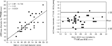
We referred 42 patients for transcatheter closure. The comparisons between the measurements made by transthoracic three-dimensional echocardiography and those using the sizing balloon are shown in Table 1. The correlation between the measurements was good (y = 3.45 − 0.73x; r = 0.78; p < 0.0001), with the mean difference between the average values for the maximal diameter obtained by the two methods being 2.4 ± 2.8 mm (Fig. 2). Of the 42 patients, 40 (95%) underwent successful transcatheter closure with no residual shunt. The mean size of the Amplatzer septal occluder was 22 ± 4 mm, with a range from 15 to 30 mm. In two patients with floppy septums, the stretched diameters as measured with the balloon were half as great again as predicted by the three-dimensional echocardiographic reconstructions. In these patients, therefore, the transcatheter procedure was abandoned, and patients referred for surgical closure.
Table 1. Patients selected by transthoracic three-dimensional echocardiography for transcatheter closure.


Figure 2. Comparisons of measurements made with transthoracic three-dimensional echocardiography (TT3DE) compared to the stretched diameter assessed using the balloon (BSD). The left panel shows the correlations between the measurements, while the right panel shows the differences of each pair of measurements as assessed using the method of Bland and Altman.
Surgical closure
We referred 20 patients directly for surgical closure. In these, the mean maximal diameter of the defect measured by transthoracic three-dimensional echocardiography was 23.8 ± 5.2 mm, with a range from 15 to 35 mm. From the transthoracic three-dimensional echocardiographic views, the antero-inferior rim was found to be deficient in 5 patients, the postero-superior rim in 6 patients, and the postero-inferior rim in 9 patients. We then found complete agreement on these assessments when comparing the three-dimensional echocardiographic and the intraoperative findings (Fig. 3). In the 2 patients in whom we had abandoned the transcatheter procedure, the surgeon found a floppy septum with multiple holes. All patients underwent uncomplicated surgical closure with no residual shunt.

Figure 3. Transthoracic three-dimensional echocardiographic and surgical views from the right atrium. The left panel shows a large round atrial septal defect (arrow). The size of the rim separating the defect from the inferior caval vein, measured at 3 mm, indicated surgical closure. The right panel shows the view obtained during the surgical correction, confirming the deficient postero-inferior rim. RPV: right pulmonary vein; ICV: inferior caval vein; SCV: superior caval vein.
Discussion
The success of transcatheter closure of defects within the oval fossa is strongly related with the anatomy of the defect. Pre-selection of patients is a challenge for the paediatric cardiologist, who needs an accurate and non-invasive method. Our study has shown that transthoracic three-dimensional echocardiography is now able to measure the maximal diameter of the septal deficiency, along with the extents of the rims surrounding the atrial septal defect. By using such transthoracic three-dimensional measurements prior to attempted closure, we were able to select over two-thirds of our patients for transcatheter treatment, and all but one-twentieth of these underwent successful closure. To achieve successful closure, and to choose a device of appropriate size, it is essential to measure these two crucial parameters, namely the extent of the rims round the defect, and its maximal diameter.
The rims surrounding the septal deficiency
Should the rims be insufficient, then the device is prone to migration.1–3 The minimal size of these rims depends on the geometry of the device. The umbrellas of the Amplatzer septal occluder extend from the waist for 7 mm all around the defect.14 Since the cross-sectional echocardiographer approaches the atrial septum from multiple orthogonal planes, mental three-dimensional constructs are needed to comprehend the relation between the defect and the surrounding structures. Transthoracic three-dimensional echocardiography allows surface imaging of the atrial septum. The extent of the septal deficiency, and the size of the rims throughout the circumference of the defect, is simultaneously and directly imaged from the three-dimensional echocardiographic views. Based on the transthoracic three-dimensional echocardiographic measurements of the rims, we excluded almost one-third of the patients from attempted transcatheter closure. The operative findings in these patients endorsed completely the three-dimensional echocardiographic data. The agreement on the localisation of the deficient rims was complete, while the mean difference between the echocardiographic and surgical measurements was less than 1 mm. It was the rim towards the inferior caval vein that was deficient in nearly half the cases. As did Magni et al.,17 we encountered difficulties in visualising the entire length of the postero-inferior rim using transesophageal three-dimensional echocardiography17. In contrast, transthoracic three-dimensional echocardiography depicted very well the inferior rims, as shown in Figures 4 and 5. Because of the geometric profile of the Amplazter septal occluder, we chose to ignore the extent of the antero-superior rim, which charts the distance from the aorta.1–3

Figure 4. Transthoracic three-dimensional echocardiographic views from the right atrium. The left panel shows an oval septal deficiency (arrow). The rim from the tricuspid valve is well seen. The right panel shows the same patient after transcatheter closure. The Amplatzer septal occluder has a concave shape in the right atrial cavity, and does not obstruct flow through the inferior caval vein. ICV: inferior caval vein.

Figure 5. Transthoracic three-dimensional echocardiographic views from the right atrium. There is a large and round deficiency within the oval fossa. The narrowness of the rim separating the defect from the tricuspid valve, less than 3 mm, contra-indicated transcatheter closure. The inferior caval vein, and the Eustachian valve floating in the right atrial cavity, are well seen.
The maximal diameter of the septal deficiency
Three-dimensional echocardiography mimics the view of the beating heart as seen through an atriotomy, and allows description of the septal deficiency throughout the cardiac cycle.18 One striking finding was the great variability in the shape of the atrial septal defect, this observation emphasising how such alterations in the shape of the defect might alter the accuracy of calculations made using cross-sectional echocardiography. In those defects that are circular, cross-sectional images certainly cut the defect in its maximal diameter, but this is no longer true in those defects having complex shapes. Thus, in patients with an elongated oval-shaped defect, cross-sectional echocardiography could underestimate significantly the maximal diameter. Moreover, we found that the surface area of the septal deficiency changed significantly during the cardiac cycle, being maximal in late ventricular systole, and minimal in late left ventricular diastole.19 Such variation in surface area through the cardiac cycle increases the difficulty of measuring the maximal diameter by selecting the appropriate cross-sectional frame. It is the sizing balloon, nonetheless, which is still used most frequently to determine the extent of the septal deficiency.1–3, 15 The relation between the stretched diameter measured with the balloon and the maximal diameter of the septal deficiency is controversial. We found a mean difference of 2 mm between our three-dimensional measurements and the diameters determined using the balloon. This explained the slight oversize of the Amplatzer septal occluder used compared to the maximal diameters predicted from the three-dimensional reconstructions, these being 22 and 19.2 mm, respectively. In particular, we found that in our patients with floppy septum, the three-dimensional reconstructions define different properties of the defect compared to balloon sizing. The three-dimensional echocardiographic views demonstrate the major axis, incorporating information about the shape of the atrial septal defect. Balloon sizing provides information about the degree of physical stretch, a feature that cannot be predicted by echocardiography.
Limitations
Our measurements were obtained only from reconstructions that recreated the right atrial views obtained by the surgeon. While transesophageal three-dimensional echocardiography offers reconstructions from both sides of the atrial septum, our transthoracic three-dimensional approach did not allow good visualisation of the atrial septal defect from the left atrium. This is explained by the smaller cavity of the left compared to the dilated right atrium, and the greater distance from the probe, permitting poorer resolution for the left atrial reconstructions. The absence of obstruction to pulmonary venous flow, nonetheless, was assessed by cross-sectional transesophageal echocardiography after deployment of the device. The resolution of the transthoracic three-dimensional reconstructions was insufficient in most of patients to visualise the Amplatzer septal occluder. Three-dimensional color Doppler reconstructions may help to further define small defects or residual shunting.20, 21 The ability to reconstruct directly from the digital images, without transformation into video signals, would also improve the quality of the three-dimensional images, which is still very dependent on the gain settings.22, 23
In conclusion, we have shown that transthoracic three-dimensional echocardiography is accurate in measuring the maximal diameter of defects within the oval fossa, and the extent of the rims surrounding them. This non-invasive technique should prove invaluable in selecting children for interventional closure. The use of the sizing balloon during closure provides necessary additional information, such as the resistance of the septum. Sizing and placement of the device should take in account both the three-dimensional echocardiographic and transcatheter findings.
References
Berger F, Ewert P, Bjornstad PG, et al. Transcatheter closure as standard treatment for most interatrial defects: experience in 200 patients with the Amplatzer Septal Occluder. Cardiol Young 1999; 9: 468–473.Google Scholar
Losay J, Petit J, Lambert V, et al. Percutaneous with Amplatzer device is a safe and efficient alternative to surgery in adults with large atrial septal defects. Am Heart J 2001; 142: 544–548.Google Scholar
Chan KC, Godman MJ, Walsh K, Wilson N, Redington A, Gibbs JL. Transcatheter closure of atrial septal defect and interatrial communications with a new self expanding nitinol double disc device (Amplatzer septal occluder): multicentre UK experience. Heart 1999; 82: 300–306.Google Scholar
Mehta R, Helmcke F, Nanda NC, Pinheiro L, Samdarshi TE, Shah VK. Uses and limitations of transthoracic echocardiography in the assessment of atrial septal defect in adults. Am J Cardiol 1991; 67: 288–294.Google Scholar
Godart F, Rey C, Francart C, Jarrar M, Vaksmann G. Two-dimensional echocardiographic and color Doppler measurements of atrial septal defect, and comparison with the balloon-stretched diameter. Am J Cardiol 1993; 72: 1095–1097.Google Scholar
Faletra F, Scarpini S, Moreo A, et al. Color Doppler echocardiographic assessment of atrial septal defect size: correlation with surgical measurements. J Am Soc Echocardiogr 1991; 4: 429–434.Google Scholar
Hellenbrand WE, Fahey JT, McFowan FX, Weltin GG, Kleinman CS. Transesophageal echocardiographic guidance of transcatheter closure of atrial septal defect. Am J Cardiol 1990; 66: 207–213.Google Scholar
Rao PS, Langhough R, Beekman RH, Lloyd TR, Sideris EB. Echocardiographic estimation of balloon-stretched diameter of secundum atrial septal defect for transcatheter occlusion. Am Heart J 1992; 124: 172–175.Google Scholar
Acar P, Laskari C, Rhodes J, Pandian NG, Warner K, Marx G. Determinants of mitral regurgitation after atrioventricular septal defect surgery: a three-dimensional echocardiographic study. Am J Cardiol 1999; 83: 745–749.Google Scholar
Magni G, Cao QL, Sugeng L, et al. Volume-rendered three-dimensional echocardiographic determination of the size, shape, and position of atrial septal defects: validation in an in vitro model. Am Heart J 1996; 132: 376–381.Google Scholar
Acar P, Saliba Z, Bonhoeffer P, et al. Influence of atrial septal defect anatomy in patient selection and assessment of closure by the CardioSEAL device: a three-dimensional transesophageal echocardiography. Eur Heart J 2000; 21: 573–581.Google Scholar
Lange A, Walayat M, Turnbull CM, et al. Assessment of atrial septal defect morphology by transthoracic three dimensional echocardiography using standard grey scale and Doppler myocardial imaging techniques: comparison with magnetic resonance imaging and intraoperative findings. Heart 1997; 78: 382–389.Google Scholar
Acar P, Maunoury C, Antonietti T, Bonnet D, Sidi D, Kachaner J. Left ventricular ejection fraction in children measured by three-dimensional echocardiography using a new transthoracic integrated 3D-probe. Eur Heart J 1998; 19: 1583–1588.Google Scholar
Acar P, Saliba Z, Bonhoeffer P, Sidi D, Kachaner J. Assessment of the geometric profile of the Amplatzer and Cardioseal occluders by three-dimensional echocardiograpy. Heart 2001; 85: 451–453.Google Scholar
Gu X, Han YM, Berry J, Urness M, Amplatz K. A new technique for sizing of atrial septal defects. Catheter Cardiovasc Interv 1999; 46: 51–57.Google Scholar
Bland JM, Altman DG. Statistical methods for assessing agreement between two methods of clinical measurement. Lancet 1986; 1: 307–310.Google Scholar
Magni G, Hijazi Z, Pandian NG, et al. Two- and three-dimensional transesophageal echocardiography in patient selection and assessment of atrial septal defect closure by the new DAS-Angel wings device. Circulation 1997; 96: 1722–1728.Google Scholar
Marx GR, Fulton DR, Pandian NG, et al. Delineation of site, relative size and dynamic geometry of atrial septal defect by real-time three-dimensional echocardiography. J Am Coll Cardiol 1995; 25: 482–490.Google Scholar
Franke A, Kühl HP, Rulands D, et al. Quantitative analysis of the morphology of secundum-type atrial septal defects and their dynamic change using transesophageal three-dimensional echocardiography. Circulation 1997; 96 (suppl II): II-323–327.Google Scholar
Acar P, Jones M, Shiota T, et al. Quantitative assessment of chronic aortic regurgitation using three-dimensional echocardiographic reconstruction: comparison with electromagnetic flow-meter measurements. J Am Soc Echocardiogr 1999; 12: 138–148.Google Scholar
De Simone R, Glombitza G, Vahl CF, Albers J, Meinzer HP, Hagl S. Three-dimensional color Doppler: a clinical study in patients with mitral regurgitation. J Am Coll Cardiol 1999; 33: 1646–1654.Google Scholar
Chauvel C, Bogino E, Clerc P, et al. Usefulness of three-dimensional echocardiography for the evaluation of mitral valve prolapse: an intraoperative study. J Heart Valve Dis 2000; 9: 341–349.Google Scholar
Acar P, Saliba Z, Sidi D, Kachaner J. New insight into the single ventricular function after total cavopulmonary connection: a three-dimensional echocardiographic study. Cardiol Young 2000; 10: 83–89.Google Scholar

Transthoracic three-dimensional echocardiographic views from the right atrium. The left panel shows the centrally located deficiency within the atrial septum (arrow). The large rims, greater than 4 mm, all round the defect allowed transcatheter closure with the Amplatzer septal occluder. The right panel shows a large atrial septal defect (arrow). The restricted rim towards the tricuspid valve, less than 4 mm, precluded interventional closure. TV: tricuspid valve; ICV: inferior caval vein; SCV: superior caval vein.
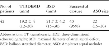
Table 1.
Comparisons of measurements made with transthoracic three-dimensional echocardiography (TT3DE) compared to the stretched diameter assessed using the balloon (BSD). The left panel shows the correlations between the measurements, while the right panel shows the differences of each pair of measurements as assessed using the method of Bland and Altman.

Transthoracic three-dimensional echocardiographic and surgical views from the right atrium. The left panel shows a large round atrial septal defect (arrow). The size of the rim separating the defect from the inferior caval vein, measured at 3 mm, indicated surgical closure. The right panel shows the view obtained during the surgical correction, confirming the deficient postero-inferior rim. RPV: right pulmonary vein; ICV: inferior caval vein; SCV: superior caval vein.

Transthoracic three-dimensional echocardiographic views from the right atrium. The left panel shows an oval septal deficiency (arrow). The rim from the tricuspid valve is well seen. The right panel shows the same patient after transcatheter closure. The Amplatzer septal occluder has a concave shape in the right atrial cavity, and does not obstruct flow through the inferior caval vein. ICV: inferior caval vein.

Transthoracic three-dimensional echocardiographic views from the right atrium. There is a large and round deficiency within the oval fossa. The narrowness of the rim separating the defect from the tricuspid valve, less than 3 mm, contra-indicated transcatheter closure. The inferior caval vein, and the Eustachian valve floating in the right atrial cavity, are well seen.
 You have
Access
- 19
- Cited by