Introduction
Ossifying fibromyxoid tumour was first described by Enzinger et al. in 1989.Reference Enzinger, Weiss and Liang1 It is a rare soft tissue tumour which typically occurs in the subcutaneous tissue of the upper and lower extremities. It predominantly affects adult males and usually follows a benign course. It is characterised histologically by lobules of small, round cells arranged in a cord or nestlike pattern within a myxoid matrix, surrounded partially by metaplastic bone.Reference Enzinger, Weiss and Liang1 The nature of this tumour is under much debate, with neuronal and chondroid origins being favoured by most.Reference Fisher, Hedges and Weiss2 Excision is considered the treatment of choice.
Ossifying fibromyxoid tumours are uncommon in the head and neck region. Enzinger et al., in their original analysis of 59 patients, found eight cases in this region.Reference Enzinger, Weiss and Liang1 Williams et al. reported nine cases in the head and neck, seven in the subcutaneous tissue and two intraorally.Reference Williams, Ellis, Meis and Heffner3 In a study of 70 cases of ossifying fibromyxoid tumour of soft tissue by Folpe and Weiss, only eight occurred in the head and neck.Reference Folpe and Weiss4 More recently, ossifying fibromyxoid tumour has been reported to occur in the nasal septum and ethmoid sinus.Reference Blum, Back, Naim, Hormann and Riedal5, Reference Al-Mazrou, Mansoor, Payne and Richardson6 To our knowledge, the current case represents the first report of an ossifying fibromyxoid tumour involving the sphenoid sinus.
Case report
A 59-year-old man with Parkinson's disease had undergone preparatory assessment for deep brain stimulation. Computed tomography (CT) scanning, revealed an incidental finding of a mass lesion in the sphenoid sinus. The presence of this lesion prohibited him from proceeding with deep brain stimulation to treat his Parkinson's disease, and he was therefore referred to our endoscopic skull base service for further assessment.
The patient had no symptoms of sino-nasal disease, and endoscopic nasal examination was unremarkable. He was taking medication for Parkinson's disease but was otherwise in good health.
Transnasal biopsies of the sphenoid sinus lesion had already been attempted in another centre on two separate occasions, but both had yielded diagnostically inconclusive samples.
Computed tomography imaging of the paranasal sinuses revealed a 3 cm, enhancing, soft tissue mass arising anteriorly within the sphenoid sinus with a bony capsule posteriorly. The sphenoid intersinus septum appeared to be eroded by the mass. The margins of the lesion were partly calcified, and there was a mucocele lying posteriorly within the sphenoid locule. The mass had expanded the sphenoid sinus particularly along the right lateral wall, with limited extension into the posterior ethmoidal air cells. There was no evidence of intracranial or intra-orbital extension; however, there was bony erosion of the anterior cranial fossa (Figure 1).
High definition (a) coronal, (b) axial and (c) parasagittal computed tomography scans (bone window). R = right; L = left; P = posterior

Following discussion at the skull base multidisciplinary team meeting, a decision was made to attempt an endoscopic transnasal approach to enable surgical resection of the lesion and reconstruction.
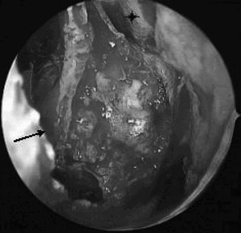
The surgical team included an otorhinolaryngologist and a neurosurgeon. The surgical approach involved a standard right spheno-ethmoidectomy conducted by performing an uncinectomy, middle meatal antrostomy, anterior ethmoidectomy and opening of the posterior ethmoid. A large swelling was encountered in the posterior aspect of the nasal cavity, bowing the nasal septum and extending into the left posterior ethmoid and sphenoid. A posterior nasal septectomy was performed with the septum lowered to allow complete exposure of the anterior face of the sphenoid (Figure 2). The tumour had a firm, rubbery consistency. It appeared to arise from the roof of the right spheno-ethmoid, extending extensively into the sphenoid. It had a firm, bony capsule posteriorly and extended up to the skull base superiorly. Posteriorly, the sphenoid sinus beyond the tumour was normal. No pituitary breach was encountered. The optic nerves and carotid arteries were non-dehiscent.
Peri-operative photograph demonstrating the tumour bulge, with bony capsule, through the anterior face of the sphenoid. Also shown are the posterior septectomy edge (black arrow) and the left superior turbinate (black star).

A submucosal circumferential dissection of the tumour was carried out, with central debulking to allow dissection of the capsule away from the dura of the anterior skull base. The dissection was extended posteriorly and laterally to allow the tumour to be delivered inferiorly. Frozen sections were performed, showing a low grade tumour. Haemostasis was achieved with bipolar diathermy and bone wax. DuraSeal™ (Confluent Surgical, Waltham, USA) was placed into the sphenoid cavity, and DuraForm™ (Codman, Raynam, USA) was laid in the right lateral wall of the sphenoid.
Despite the patient's debilitating Parkinson's disease, he was mobile within a few hours after the procedure. He was discharged from hospital after two days, having been prescribed a one week course of oral antibiotics and six weeks of nasal douching (using SinuRinse™; Confluent Surgical, Waltham, USA).
Follow up at four weeks, and repeat CT scanning at eight weeks, showed no evidence of residual tumour. The patient recovered well, and went on to undergo deep brain stimulation for his Parkinson's disease.
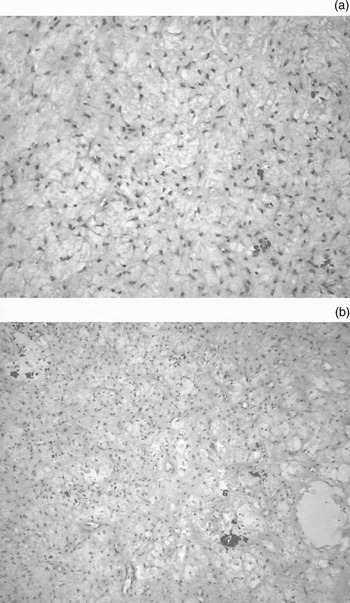
Histopathological analysis showed a tumour of low cell density, composed of small, spindle-shaped or stellate cells with small, irregular nuclei set in a fibromyxoid stroma (Figure 3). There was no tumour necrosis, mitotic figures were not identified and the cell proliferation index was very low. Immunohistochemical staining revealed cells positive for α smooth muscle actin and antimyosin heavy chains smooth muscle. Cells were negative for: the cytokeratins MNF116 and CAM5.2; desmin; cluster of differentiation 31, 34 and 117 glycoproteins; S100 protein; and epithelial membrane antigen. In terms of both morphology and immunohistochemical profile, the appearances were of a benign or low grade ossifying fibromyxoid tumour with possible myofibroblastic features.
(a) High power and (b) medium power photomicrographs of the tumour, demonstrating low cellularity within a myxoid stroma, with a vascular background. The tumour cells are arranged haphazardly and have small, bland nuclei (H&E; ×400, ×200).

Discussion
Ossifying fibromyxoid tumour is an unusual neoplasm that rarely occurs in the head and neck region. Its origin remains the subject of much debate. The original description by Enzinger et al. suggested cartilaginous or neural origins.Reference Enzinger, Weiss and Liang1 The immunohistochemical analyses of more recent cases have certainly substantiated this theory. Positivity to S100 protein and glial fibrillary acidic protein, and the presence of an external lamina, are common to both ossifying fibromyxoid tumour and peripheral nerve sheath differentiation tumours.Reference Fisher, Hedges and Weiss2, Reference Al-Mazrou, Mansoor, Payne and Richardson6 Irregular cell borders with short processes and intracellular microfilaments suggest a cartilaginous differentiation.Reference Folpe and Weiss4
The introduction of cytogenetic analysis may prove to be more fruitful in defining the origins of ossifying fibromyxoid tumour. In fact, cytogenetic analysis by Sovani et al. found chromosomal abnormalities suggestive of an osteochondroblastic lineage, and Nishio and colleagues' cytogenetic findings suggested ossifying fibromyxoid tumour to be a distinct disease entity altogether.Reference Sovani, Velagaleti, Filipowicz, Gatalica and Knisely7, Reference Nishio, Iwaski, Ohjimi, Ishiguro, Isayama and Naito8 Further cytogenetic studies are needed to determine common chromosomal abnormalities.
Ossifying fibromyxoid tumour classically follows a benign course; however, atypical and malignant variants have been described.Reference Kilpatrick, Ward, Mozes, Miettinen, Fukunaga and Fletcher9 Folpe and Weiss's study of 70 cases reported a broad histological spectrum of ossifying fibromyxoid tumours, including tumours displaying features of high nuclear grade or high cellularity and mitotic activity, which should be regarded as sarcomas with significant metastatic potential, i.e. ‘malignant ossifying fibromyxoid tumour’.Reference Folpe and Weiss4
The main differential diagnoses of ossifying fibromyxoid tumour include schwannoma, myxoid chondrosarcoma, smooth muscle tumours, osteosarcoma and benign fibro-osseous tumours, with the latter being particularly important to consider in the paranasal sinuses.Reference Al-Mazrou, Mansoor, Payne and Richardson6 Histopathological characteristics which distinguish ossifying fibromyxoid tumour from other tumours include the presence of a rim of lamellar bone, the arrangement of cells in nests and cords, short cytoplasmic processes, a discontinuous external lamina, cytoplasmic intermediate filaments, and scant organelles.Reference Schofield, Krausz, Stamp, Fletcher, Fisher and Azzopardi10
Whilst the histopathological features of our patient's tumour were not completely classical, in that it lacked a bony component and exhibited S100-negativity, this does not rule out a diagnosis of ossifying fibromyxoid tumour. These tumours do not always conform to classical types, as is often the case for unusual stromal lesions. Whilst the peri-operative findings clearly identified a bony capsule around the tumour, histological assessment of the classical bony component can be difficult if the tumour is sited in an area where there is already significant normal bone, as is the case in the paranasal sinuses. It is also worth noting that the classical bony component is not always present in these tumours; indeed, non-ossifying variants have been described in the literature.Reference Williams, Ellis, Meis and Heffner3, Reference Thompson, Castillo, Reddick, Smith and Shockley11 Furthermore, up to 30 per cent of ossifying fibromyxoid tumour can be S100-negative.Reference Oda, Yasui, Ito and Tahara12
Ossifying fibromyxoid tumour typically presents as a small, painless, well defined mass in the deep subcutaneous tissue, which is often attached to underlying fascia, muscle or tendon. Local excision is the treatment of choice, with reported recurrence rates ranging from 17 to 25 per cent.Reference Enzinger, Weiss and Liang1, Reference Folpe and Weiss4 Metastasis is uncommon, occurring in only 5 per cent of tumours.Reference Folpe and Weiss4 Ossifying fibromyxoid tumours commonly occur in the extremities and involvement of the head and neck region is rare, accounting for around 13 per cent of cases.Reference Folpe and Weiss4 To our knowledge, the current case represents the first report of ossifying fibromyxoid tumour involving the sphenoid sinus. These tumours should be considered in the differential diagnosis of sino-nasal tumours.
• This paper gives the first description of ossifying fibromyxoid tumour in the sphenoid sinus
• This neoplasm has distinctive histopathological features which aid diagnosis
• This case reinforces the evolving role of the endoscopic transnasal approach in the management of inflammatory and neoplastic pathology of the skull base
The management strategy in this case reinforces the evolving role of the endoscopic transnasal approach as the new standard for treatment of pathological conditions of the skull base.Reference Carrau, Kassam, Snyderman, Duvvuri, Mintz and Gardner13 The primary advantage of an endoscope, compared with other methods, is improved visualisation, which in turn results in improved access to poorly accessible areas, and may facilitate tumour resection and avoidance of complications due to poor visualisation. Other potential benefits of endoscopic surgery include improved cosmesis and decreased morbidity from tissue trauma and manipulation of vessels and nerves. The consequences of decreased morbidity are faster recovery, shortened hospitalisation, and decreased cost of medical care. A natural extension of endoscopic sinus surgery has been the application of endoscopic techniques to the surgical treatment of pathological conditions, including benign (as in our patient) and malignant sino-nasal malignancies that secondarily involve the skull base.Reference Snyderman, Carrau, Kassam, Zanation, Prevedello and Gardner14 This change has largely been driven by the development of endoscopic technology, and increasingly also by consumer demand. As the limits of such surgery are tested, the possibilities of endoscopic transnasal skull base surgery are constantly being expanded, as evidenced by the use of such terms as endoscopic transnasal craniotomy. Crucially, however, this has only become possible through the development of the multidisciplinary skull base team formed by close collaboration between multiple surgical specialties, including otorhinolaryngology, neurosurgery, maxillofacial surgery, and plastic and reconstructive surgery.