Congenital coronary artery fistulas connect one of the coronary branches with one of the cardiac chambers or the adjoining vessels.Reference Said 1 , Reference Jama, Barsoum, Bjarnason, Holmes and Rihal 2 Although rare, complications like myocardial ischaemia, congestive heart failure, fistula rupture, and endocarditis have been described. In such a rare disease with an incidence of 0.1–0.2%,Reference Taverne, de Jong, Witsenburg and Bogers 3 endocarditis has been described in 3–12% of the cases.Reference Said 1 Most of the cases described are of adult patients. To our recent knowledge, there are only two case reports of children aged 16 years or under with coronary artery fistulas complicated by infective endocarditis: a 7-year-old girl with a connection from the left main coronary artery to the right atrium, which was closed surgically,Reference Mishra, Barik and Patnaik 4 and a 4-year-old girl with a fistula between the right coronary artery and the right atrium, which was also operated upon.Reference Rein, Yatsiv and Simcha 5
We report the case of a 9-year-old boy with a fistula connecting the right coronary artery with the junction of the superior caval vein and the right atrium diagnosed during infective endocarditis.
Case report
A 9-year-old boy presented with a 3-week history of fever up to 40°C and general malaise. There was no other organ infection or dental treatment before this presentation. He lost 0.5 kg of weight and complained of joint pain. On physical examination he had normal heart sounds, but a 4/6 systolic–diastolic murmur best heard at the left upper sternal border. Inflammatory markers were elevated with a C-reactive protein level of 45 mg/l and an erythrocyte sedimentation rate of 55 in the 1st hour. Blood cultures showed Streptococcus pneumoniae. The electrocardiogram showed sinus rhythm with a ventricular rate of 90 beats per minute, normal time intervals, and no signs of ischaemia or hypertrophy.
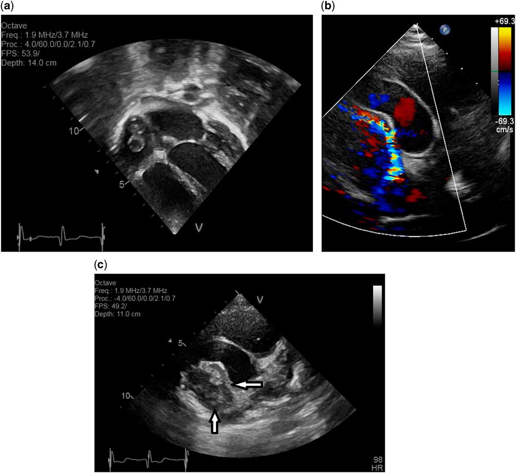
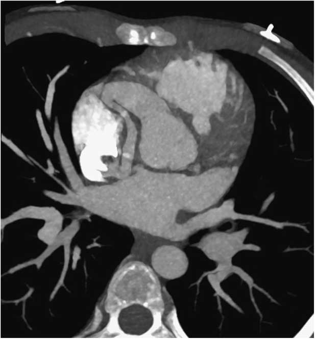
Echocardiography showed vegetations with a diameter of ca. 1 cm close to the connection of the superior caval vein to the right atrium (Fig 1a). The cardiac valves were free of vegetations. The left coronary artery had normal dimensions, but the right coronary artery was dilated with a narrow connection to the junction of the superior caval vein and the right atrium (Fig 1b and c). This was confirmed using computed tomography (Fig 2). The diagnosis of a right coronary fistula was first made at the time of presentation. Beforehand, there was no suggestion of congenital heart disease.
Echocardiography: ( a ) vegetations at the connection of the superior caval vein and the right atrium. Modified four-chamber view. ( b ) Color Doppler flow through the fistula from the right coronary artery to the junction of the superior caval vein and right atrium. Short-axis view. ( c ) Same view as ( b ). Vegetations seen directly connected to the distal end of the fistula, indicated by the horizontal arrow. Note the thin connection of the right atrial vegetations, indicated with a vertical arrow, to the end of the fistula.

Computed tomography showing the dilated proximal right coronary artery with a narrow connection to the junction of the superior caval vein and the right atrium.

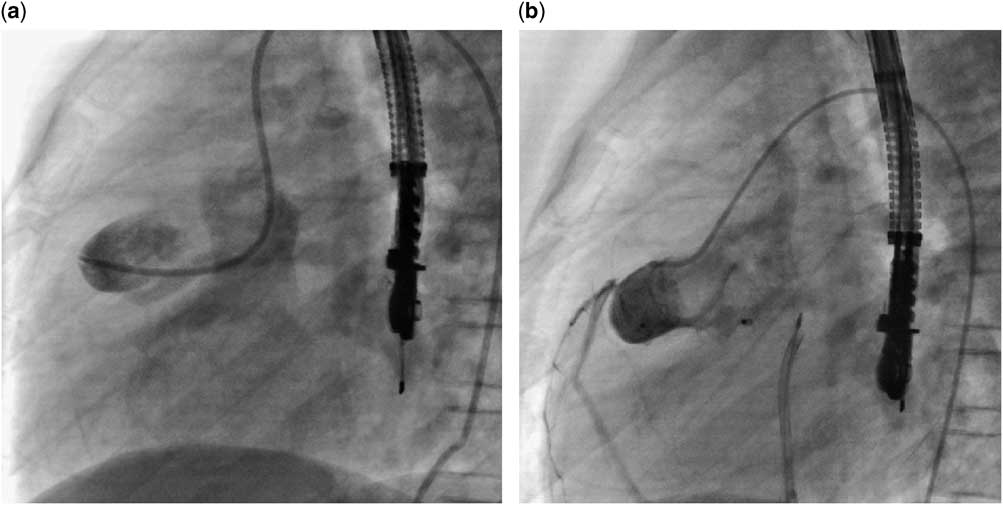
He was treated with ceftriaxone intravenously for 6 weeks. On echocardiography, it was observed that the vegetations disappeared. After 4 further months without any recurrent symptoms, the fistula was closed interventionally with an Amplatzer vascular plug II (St. Jude Medical, St. Paul, Minnesota, United States of America). Direct access from the venous side was possible, but no stable position of the delivery catheter could be achieved; hence, an arteriovenous guidewire circuit was created and the 6 F Launcher delivery catheter (Medtronic, Santa Rosa, California, United States of America) was positioned from the venous side through the fistula in the ascending aorta. The first disk of the device was deployed and the catheter–device ensemble pulled back into the fistula. The body of the device and second disk were deployed within the narrow part of the fistula under transoesophageal echocardiographic guidance and check-angiography in order not to interfere with the right coronary flow. The device was released in a good position (Fig 3a and b). The patient was started on dual antiplatelet medication with aspirin and dipyridamole.
Angiography: ( a ) fistula before closing. ( b ) Amplatzer vascular plug II closing the fistula.

No further episodes of fever occurred over the following 3 months. Prophylaxis for endocarditis was recommended.
The electrocardiogram remained stable without ischaemic signs. Echocardiography showed good biventricular function and good flow into the dilated right coronary. No residual shunt could be seen on color Doppler.
Discussion
Congenital coronary artery fistulas are rare. Infective endocarditis of these occurs in 3–12% of cases.Reference Said 1 Although congenital fistulas are present in childhood, the symptoms encountered are mainly congestive heart failure in fistulas with large shunt volumes or coronary steal.Reference Jama, Barsoum, Bjarnason, Holmes and Rihal 2 Infective endocarditis is exceedingly rare in children before puberty. A PubMed search found two such cases, as mentioned in the introduction.Reference Mishra, Barik and Patnaik 4 , Reference Rein, Yatsiv and Simcha 5 Both cases were treated surgically. In the 7-year-old girl with a connection from the left main coronary artery to the right atrium, the endocarditis involved the tricuspid valve,Reference Mishra, Barik and Patnaik 4 and in the 4-year-old girl with a fistula between the right coronary artery and the right atrium the aortic valve was involved.Reference Rein, Yatsiv and Simcha 5 Interestingly, both these fistulas drained to the right heart, as in our case. On the other hand, ca. 90% of all coronary artery fistulas connect to right heart structures.Reference Gerede, Acibuca and Uzun 6 We chose interventional treatment 13 weeks after effective antibiotic treatment without recurrence, as the anatomy seemed quite favourable, and we considered the risk from an intervention to be lower than that from surgical treatment.
Electrocardiographical changes may occur if coronary steal is present. In our case, the effective shunt volume was probably too small to cause ischaemia. On the other hand, the narrow connection to the junction of the right atrium and the superior caval vein may have led to high shear forces leading to minor lesions of the vascular surface, which then allowed a vegetation to form at this place. The jet was not directed towards the tricuspid valve. It is possible that, in the cases reported previously in the literature, the endocarditis initially involved the fistula itself and, secondarily, the valves.
Our case is the first description of an infective endocarditis not of one of the valves, but of the fistula itself, in a boy aged 9 years, which was treated interventionally once no inflammatory signs were seen for months. The fistula was not big enough to cause heart failure, and ischaemia was not seen in any of the electrocardiograms.
It is noteworthy that the history of our patient before endocarditis was empty. Fever of unknown origin maybe caused by endocarditis. In our patient the newly appreciated cardiac murmur lead to the investigations establishing the diagnosis.
It seems that small coronary fistulas can close spontaneously.Reference Cotton 7 Treatment options are surgery or intervention.Reference Said 1 – Reference Taverne, de Jong, Witsenburg and Bogers 3 In our case we closed the fistula interventionally to prevent further endocarditis that may be triggered by the jet lesion.
Conclusion
In children, infective endocarditis of a coronary artery fistula is exceedingly rare. Treatment consists of antibiotic medication and closure of the fistula, which can be performed transcutaneously in selected cases.
Acknowledgements
None.
Financial Support
This research received no specific grant from any funding agency, commercial, or not-for-profit sectors.
Conflicts of Interest
None.