Introduction
Cetacean populations are vulnerable to a variety of natural threats, such as predator attacks, intra- or inter-specific interactions (Scott et al., Reference Scott, Mann, Watson-Capps, Sargeant and Connor2005; Steiger et al., Reference Steiger, Calambokidis, Straley, Herman, Cerchio, Salden, Urbán-R, Jacobsen, Von Ziegesar, Balcomb, Gabriele, Dahlheim, Uchida, Ford, Ladrón de Guevara, Yamaguchi and Barlow2008; Dwyer & Visser, Reference Dwyer and Visser2011; Marley et al., Reference Marley, Cheney and Thompson2013), and human-derived pressures. Coastal and continental shelf areas are susceptible to human impacts (Halpern et al., Reference Halpern, Diamond, Gaines, Gelcich, Gleason, Jennings, Lester, Mace, McCook and McLeod2012); fishery interactions, prey depletion caused by overfishing, noise and chemical pollution, vessel strikes, and plastics or marine debris (Rejinders et al., Reference Rejinders, Donovan, Aguilar and Bjørge1999; Bearzi et al., Reference Bearzi, Politi, Agazzi and Azzellino2006; Read et al., Reference Read, Drinker and Northridge2006; Van Waerebeek et al., Reference Van Waerebeek, Baker, Félix, Gedamke, Iñiguez, Sanino, Secchi, Sutaria, Van Helden and Wang2007; Moore & Barco, Reference Moore and Barco2013; Dwyer et al., Reference Dwyer, Kozmian-Ledward and Stockin2014; Luksenburg, Reference Luksenburg2014; Sierra et al., Reference Sierra, Zucca, Arbelo, García-Álvarez, Andrada, Déniz and Fernández2014; Fossi et al., Reference Fossi, Baini, Panti, Baulch, Fossi and Panti2018; Marsili et al., Reference Marsili, Jiménez, Borrell, Fossi and Panti2018; Murphy et al., Reference Murphy, Law, Deaville, Barnett, Perkins, Brownlow, Penrose, Davison, Barber, Jepson, Fossi and Panti2018) are the most reported threats worldwide for cetaceans.
Small cetaceans have been used as bioindicators of bioaccumulative pollutants (Aguilar, Reference Aguilar1984; Borrell et al., Reference Borrell, Aguilar, Cantos, Lockyer, Heide-Jørgensen and Jensen2004; Borrell & Aguilar, Reference Borrell and Aguilar2007; Lemos et al., Reference Lemos, De Moura, Hauser-Davis, De Campos and Siciliano2013) and are suitable as indicators for monitoring other natural or anthropogenic threats, specifically in areas heavily impacted by human activities. Examination of the injuries in wild cetaceans using photo-ID techniques (Evans & Hammond, Reference Evans and Hammond2004; Van Bressem et al., Reference Van Bressem, Van Waerebeek, Reyes, Félix, Echegaray, Siciliano, Di Beneditto, Flach, Viddi, Avila, Bolaños, Castineira, Montes, Crespo, Flores, Haase, Mendonça de Souza, Laeta and Bernadete Fragoso2007, Reference Van Bressem, Flach, Reyes, Echegaray, Santos, Viddi, Félix, Lodi and Van Waerebeek2015; Kiszka et al., Reference Kiszka, Pelourdeau and Ridoux2008; Murdoch et al., Reference Murdoch, Reif, Mazzoil, McCulloch, Fair and Bossart2008; Tetley et al., Reference Tetley, Kiszka and Hoyt2012; Bessesen et al., Reference Bessesen, Oviedo, Hart, Herra-Miranda, Pacheco-Polanco, Baker, Saborío-Rodríguez, Bermúdez-Villapol and Acevedo-Gutiérrez2014; Sanino et al., Reference Sanino, Van Bressem, Van Waerebeek and Pozo2014; Hupman et al., Reference Hupman, Pawley, Lea, Grimes, Voswinkel, Roe and Stockin2017) might provide good information on direct impact of potential threats on these animals (Hupman et al., Reference Hupman, Pawley, Lea, Grimes, Voswinkel, Roe and Stockin2017). Vessel strikes, fishing entanglement and by-catch, or inter- and intra-species interactions can heavily impact small cetacean populations in coastal areas (Parsons & Jefferson, Reference Parsons and Jefferson2000). Depending on the grade of impact, these interactions do not always result in mortality, leading to deformities, cuts or amputation of appendages among other outcomes (Read, Reference Read2008; Bechdel et al., Reference Bechdel, Mazzoil, Murdoch, Howells, Reif, McCulloch, Schaefer and Bossart2009).
Examining the nicks and scar markings on cetaceans could help determine the causes of lesions, whether caused by physical interactions with other species, such as sharks (Biton et al., Reference Biton Porsmoguer, Bănaru, Boudouresque, Dekeyser, Viricel and Merchán2015; Wilkinson et al., Reference Wilkinson, Wells, Pine and Borkhataria2017; Smith et al., Reference Smith, Allen, Bejder and Brown2018), or by social or aggressive interactions with other cetaceans (Corkeron et al., Reference Corkeron, Morris and Bryden1987; Wedekin et al., Reference Wedekin, Daura-Jorge and Simões-Lopes2004; Murphy et al., Reference Murphy, Collet and Rogan2005; Scott et al., Reference Scott, Mann, Watson-Capps, Sargeant and Connor2005; Volker, Reference Volker2016; Lee et al., Reference Lee, Wallen, Krzyszczyk and Mann2019).
According to the literature, the species most affected by natural and human threats are the bottlenose dolphins (Tursiops truncatus), the Indo-Pacific humpback dolphin (Sousa chinensis), and the Irrawaddy dolphin (Orcaella brevirostris) (Bloom & Jager, Reference Bloom and Jager1994; Bechdel et al., Reference Bechdel, Mazzoil, Murdoch, Howells, Reif, McCulloch, Schaefer and Bossart2009; Donaldson et al., Reference Donaldson, Finn and Calver2010; Maldini et al., Reference Maldini, Riggin, Cecchetti and Cotter2010; Melero et al., Reference Melero, Rubio-Guerri, Crespo, Arbelo, Vela, García-Párraga, Sierra, Domínguez and Sánchez-Vizcaíno2011; Félix et al., Reference Félix, Centeno, Romero, Zavala and Vásconez2018). At least 82% of odontocetes species have been reported entangled in fishing gear, while 18 species of small cetaceans have been observed to have collided with different types of vessels (Van Waerebeek et al., Reference Van Waerebeek, Baker, Félix, Gedamke, Iñiguez, Sanino, Secchi, Sutaria, Van Helden and Wang2007). There is less information about injuries in short-beaked common dolphin species, which have mostly been reported from strandings, by-catch records, or fishermen interviews (Dixon, Reference Dixon1984; Van Bressem et al., Reference Van Bressem, Van Waerebeek, Jepson, Raga, Duignan, Nielsen, Di Beneditto, Siciliano, Ramos, Kant, Peddemors, Kinoshita, Ross, López-Fernández, Evans, Crespo and Barrett2001, Reference Van Bressem, Van Waerebeek, Reyes, Félix, Echegaray, Siciliano, Di Beneditto, Flach, Viddi, Avila, Bolaños, Castineira, Montes, Crespo, Flores, Haase, Mendonça de Souza, Laeta and Bernadete Fragoso2007; López et al., Reference López, Pierce, Santos, Gracia and Guerra2003; Silva & Sequeira, Reference Silva and Sequeira2003; Fernández-Contreras et al., Reference Fernández-Contreras, Cardona, Lockyer and Aguilar2010; Martinez & Stockin, Reference Martinez and Stockin2013; Goetz et al., Reference Goetz, Read, Santos, Pita and Pierce2014). Only three references were identified where the lesions on free-ranging common dolphins were examined using photo-ID (Bamford & Robinson, Reference Bamford and Robinson2015; Hupman et al., Reference Hupman, Pawley, Lea, Grimes, Voswinkel, Roe and Stockin2017).
Our study was developed in a bay that comprises the waters between Gibraltar and Algeciras (WBGA), Andalusia, southern Spain, geographically located in the north-eastern end of the Strait of Gibraltar. The bay, facing south-eastward, is 8–10 km wide and 10–12 km long, with a surface area of ~73 km2. A submarine canyon intersects the WBGA, connecting the shallow continental waters close to the shoreline of the bay with the basin of the Strait of Gibraltar, reaching depths of 1476.38 ft in the southern end (Sammartino et al., Reference Sammartino, García Lafuente, Sánchez Garrido, De los Santos, Álvarez Fanjul, Naranjo, Bruno and Calero2014).
The short-beaked common dolphins (Delphinus delphis) are considered resident in these waters (Espada et al., Reference Espada, Martín, Haasova, Olaya-Ponzone and García-Gómez2018), turning the area into a critical hotspot for this species; they use the bay for breeding, birthing newborns during the summer months and raising their calves year round (Ruiz-Giráldez et al., Reference Ruiz-Giráldez, Montero, Gálvez and García-Gómez2005; Giménez et al., Reference Giménez, Gauffier, Verborgh, Esteban, Jiménez-Torres, De Stephanis, Gauffier and Verborgh2011; Espada et al., Reference Espada, Martín, Haasova, Olaya-Ponzone and García-Gómez2018). Local upwelling processes have been observed in the southern end of the bay (Gómez-Jakobsen et al., Reference Gómez-Jakobsen, Mercado, Cortés, Yebra and Salles2019) bringing deep nutrients towards the surface, providing vast amounts of fish and molluscs (Ruiz-Giráldez et al., Reference Ruiz-Giráldez, Montero, Gálvez and García-Gómez2005) for a main feeding ground for common (Giménez et al., Reference Giménez, Gauffier, Verborgh, Esteban, Jiménez-Torres, De Stephanis, Gauffier and Verborgh2011), striped and bottlenose dolphins.
In the Mediterranean Sea, conservation problems regarding D. delphis have been recognized since the 1970s. In 2003, the Mediterranean common dolphin ‘subpopulation’ was listed as endangered in the IUCN Red List of Threatened Animals, based on criterion A2 which refers to a 50% decline in abundance over the last three generations. The causes of this decline ‘may not have ceased or may not be understood or may not be reversible’ (Bearzi et al., Reference Bearzi, Reeves, Notarbartolo di Sciara, Politi, Cañadas, Frantzi and Mussi2003) (http://www.redlist.org).
The local population of common dolphins can be seriously affected by the industrialization of the area; industrial complexes are formed by petrochemical installations, the ports of Algeciras and Gibraltar, oil bunkering activities, and busy shipping lanes including containers, oil tankers and passenger liners. There is also tremendous impact from recreational activities, including whale watching, as well as sport and commercial fishing activities (Espada et al., Reference Espada, Martín, Haasova, Olaya-Ponzone and García-Gómez2018).
There is a great knowledge gap regarding the evolution of wounds in individuals of D. delphis, but in this study, thanks to the level of residence of these dolphins in WBGA and to the frequency with which observations have been made in the area, it has been possible to obtain data, using photo ID, that provide new information on the wounds found in this species. We sought to identify individuals with lacerations, to characterize sequences of ‘before – during (recent injury) – after’ with respect to the occurrence of these lacerations, and to identify associations with a type of injury and its severity. This work may inform future studies and contribute to the periodical monitoring of individuals in this area, especially with regard to locating new wounded specimens, and aid in predicting post-laceration outcomes. This will facilitate implementation of action protocols that allow the rescue of badly injured dolphins so that they can be treated in captivity. This type of information will also allow statistical evaluation of the degree of laceration threat and whether threat decreases or increases depending on the mitigation and conservation measures that could be implemented. A study proposal has already been made in this regard (Espada et al., Reference Espada, Martín, Haasova, Olaya-Ponzone and García-Gómez2018).
In this study, five cases of specimens with external lacerations have been detected; all cases have been monitored since the initial stage of the accident, confirming their anterior presence in the WBGA and establishing healing times posteriori. Although it cannot be confirmed, the possible sources of the lacerations have been noted and the degree of injury associated with their scarring has been described where possible. Surviving specimens can be subject to immunosuppression and a greater vulnerability to infection in the case of certain types of external wounds. In this sense, newborns are particularly sensitive since they obtain temporary immunological protection from maternal antibodies, and the immune system of many marine mammalian species is not fully developed at birth (Ross et al., Reference Ross, de Swart, Visser, Vedder, Murk, Bowen and Osterhaus1994).
Materials and methods
The study site included the waters between Gibraltar (UK) and Algeciras (Spain) where a dedicated dolphin-watching catamaran (14 m of length) runs non-systematic trips promoting responsible approaches to cetaceans on a nearly daily basis from Gibraltar. Between 2013 and 2017, 90,391 photographs were taken during a total of 1402 sightings. GPS position, number of individuals, general behaviour, wind direction and speed (Beaufort scale), sea state (Douglas scale), and water temperature were recorded for each sighting. Each time an individual with fresh injuries was detected, photographs were taken using two Nikon DSLR cameras with a 300 mm lens in order to identify the individual dolphins using standard photo-ID methods (Würsig & Jefferson, Reference Würsig and Jefferson1990). The lesions were also characterized and to assess the progression of healing of external wounds during the study period.
To be able to identify the causes of such injuries, an exhaustive examination of the wound was conducted. Depending on the shape, depth, area of the body, deformities, mutilations, and other factors it is possible to determine what interacted with the individual to create the wound. For example:
Horizontal and clean wounds: these could be related to interactions with fisheries (Wells et al., Reference Wells, Hofmann and Moors1998; Baird & Gorgone, Reference Baird and Gorgone2005; Kiszka et al., Reference Kiszka, Pelourdeau and Ridoux2008). When a fishing line wraps around a dorsal fin or the body, it can cause knife-shaped cuts, unique or opposite. Linear marks embedded in the skin can also represent tangles (Wells et al., Reference Wells, Hofmann and Moors1998; Read & Murray, Reference Read and Murray2000). These types of cuts can cause partial or total mutilations, as well as the collapse of the dorsal fin.
Single or multiple parallel deep cuts, or V-shaped cuts: these types of wounds are most likely to be caused by helix blows (Read & Murray, Reference Read and Murray2000; Elwen & Leeney, Reference Elwen and Leeney2010; Byard et al., Reference Byard, Winskog, Machado and Boardman2012; Dwyer et al., Reference Dwyer, Kozmian-Ledward and Stockin2014; Luksenburg, Reference Luksenburg2014).
Deep and unclean wounds: half-moon and spaced tooth marks (Wilkinson et al., Reference Wilkinson, Wells, Pine and Borkhataria2017) may be the result of shark attacks (Heithaus & Dill, Reference Heithaus and Dill2002; MacLeod et al., Reference MacLeod, MacLeod, Learmonth, Jepson, Reid, Deaville and Pierce2007) or fatal interactions between species (Visser et al., Reference Visser, Zaeschmar, Halliday, Abraham, Ball, Bradley, Daly, Hatwell, Johnson, Johnson, Kay, Maessen, McKay, Petres, Turner, Umuroa and Pace2010), that do not match the natural marks caused by dolphins' interactions (Heithaus, Reference Heithaus2001).
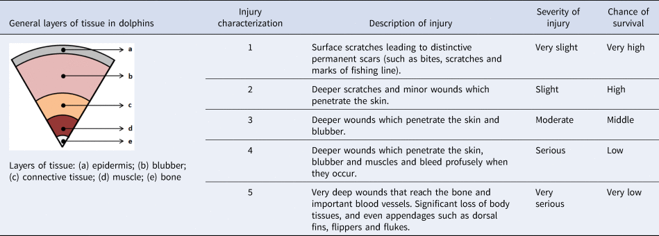
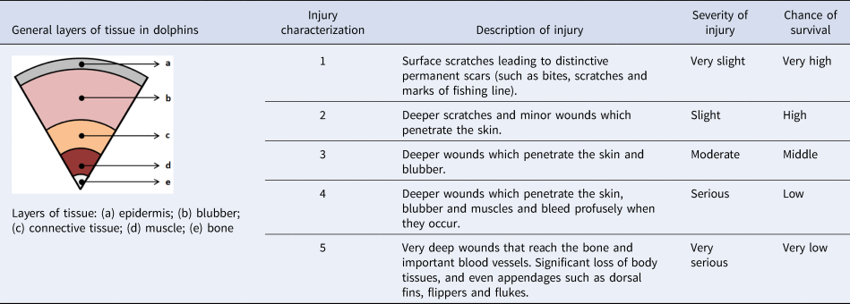
The injuries were classified into five different categories according to the type and severity of the wound. The classification was inspired by Lockyer & Morris (Reference Lockyer and Morris1990) and the categories are listed in Table 1. The severity of the injury is related to the depth of the wound. When the wound runs deeper into the body of the animal (from the epidermis to the bone), it is considered more severe. For less severe injuries, the wound coverage is taken into consideration. According to the data collected in this study, the probability of survival is reduced when the severity of the injury increases. In addition, the affected area of the body was also taken into consideration: head, back, peduncle and dorsal fin. Five individuals (D. delphis) with external injuries were identified using photo-identification methods combined with field observations. These individuals have been named as Dd-1 through Dd-5. ArcGIS 10 software was used to generate maps that included the specimen's location and compare it to the most frequent itineraries of large ships that transit the bay. The location of injured animals was also overlaid with a map published by Espada et al. (2018) which represented the accumulation of recreational activities associated with dolphin groups during the summer months, which in some occasions included newborns.
Table of severity of injuries. Depending on the depth and the damage of tissue the injuries are categorized from 1 (Surface wounds) to 5 (Very deep wound). This is related to the chance of survival which decreases as the severity of the wound increases
Results
Between 2013–2017, 79% of 1961 sightings were groups of common dolphins. For this study, only common dolphins which presented fresh wounds were selected for tracking of healing and mortality over time. Five injured common dolphins showing fresh wounds were observed. The healing process of four specimens was documented until their recovery (Dd-1, Dd-2, Dd-3 and Dd-4); one specimen wasn't observed again subsequent to the first detection (Dd-5).
Specimen Dd-1
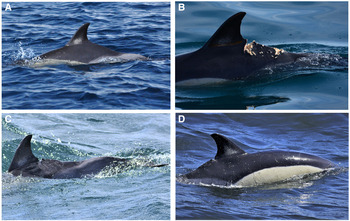
Specimen Dd-1 was recognizable due to a triangular creamy patch on both sides of its dorsal fin, centred on its base, and its particular small and irregular nicks along the trailing edge. There was a visible dorsal hump, resulting in a prominent concavity on its tail stock that showed no scars or symptoms of external injuries, suggesting a vertebral column anomaly (Weir & Wang, Reference Weir and Wang2016). Photographic evidence was collected between May 2013 to February 2017 for this individual. Analyses verified that the individual did not show any visible wounds until 22 September 2015 (Figure 1A).
Specimen Dd-1. (A) Female common dolphin (D. delphis) in the BA, 14.10.2014, showing a prominent concavity on its tail stock and no visible scars or symptoms of external injuries; (B) recaptured on 22.11.2015 showing a severe wound on its back, behind the dorsal fin; (C) and (D) animal recaptured in 2016 showing clear signs of healing, 21.1.2016 and 02.2.2016 respectively.

The individual was recaptured during 20 and 22 November 2015 (Figure 1B), and displayed two parallel fresh wounds in the dorsal ridge. The wounds were widely spaced, coinciding with a large bite gap and a crescent-shaped wound located behind the dorsal fin ~5 cm deep and 2 cm wide, penetrating through the skin and blubber. Pale, pink-coloured tissue was identified in the bed of the wound. A smaller and more superficial wound was located in the posterior tailstock, only penetrating the blubber. Muscle tissue and blood were not explicitly observed, but sloughing of soft epidermal tissue was detected during the healing procedure. Natural causes, such as inter-species interactions or attempted predation, may explain these injuries. On 21 and 22 January 2016, the animal was photographed again with superficial light grey pigmented scars and a separation gap of ~1.5 cm. This individual was photographed again that year in February and consecutively during the next 5 months, showing a completely healed wound and a remaining gap on the dorsal ridge of the peduncle (Figure 1C and D). These wounds may have resulted from natural causes, as small irregular and uncleaned wounds were observed surrounding the main cuts. A propeller hit is also a suspected cause of injury due to the parallel orientation of the cuts.
Specimen Dd-2
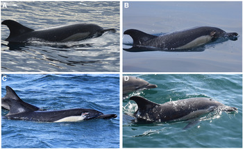
Specimen Dd-2 was a female that was identifiable due to the round creamy patch located at the base and a triangular-shaped nick on the tip in the superior trailing edge of its falcate dorsal fin. Photographic records of this animal were collected between January and March 2016. Prior to 21 January, the animal showed no injuries (Figure 2A).
Specimen Dd-2. (A) Female common dolphin (D. delphis) showing no injuries, 21.1.2016; (B) individual recaptured with open wound on its rostrum, just above the right eye, 29.1.2016; (C) healing wound showing granulation tissue and depigmented edges, 23.2.2016; (D) complete closure was detected on 13.3.2016, with only a non-pigmented scar remaining.

On 29 January, this animal was recaptured with a deep open wound on the right side of its head/rostrum with visible blubber and muscle tissue. The shape of the cut suggests that it may be due to natural causes; due to the unclean aspect around the main cut a propeller hit must also be taken into consideration. The margins of the wound were swollen and granulation tissue was observed along with soft sloughing tissue in the bed of the wound (Figure 2B). On the 23 and 24 February, the animal showed clear signs of healing and the depth of the wound had considerably decreased, filling up with granulation tissue and showing contraction of the margins of the wound (Figure 2C). A few days later, on 9 and 13 March, the individual was photographed again and the wound was almost completely healed (Figure 2D).
Specimen Dd-3
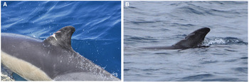
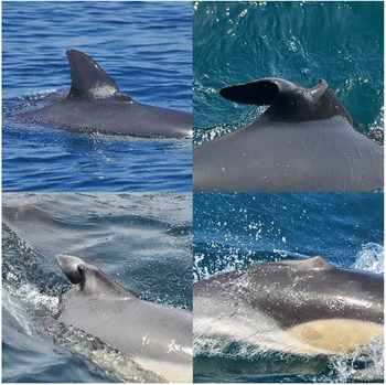
Specimen Dd-3 was identifiable by the natural creamy and irregular stripe-shaped patch and a small unpigmented spot on the left side of its dorsal fin. Rake markings were also detected on the tip, inflicted by intra-species interactions (Marley et al., Reference Marley, Cheney and Thompson2013). The individual was photographed between July and September 2015. On 21 July, the animal showed two linear shaped wounds in the leading edge of its dorsal fin, exhibiting sloughing and pink-coloured tissue. These wounds were almost healed during photographic recapture on 14 September, showing only non-pigmented scars on its skin (Figure 3A and B, respectively). According to Luksenburg (Reference Luksenburg2014), epidermal lacerations similar to those that were recorded (Figure 3), especially indentations around the head and fins, might be the result of entanglement in fishing lines or nets. Other common dolphins with similar healed injuries on their dorsal fin have been observed repeatedly in the area. The linear cuts on the dorsal fin of different individuals follows the same patterns (Figure 4). Different grades of penetration on the medial area of the leading edge of the dorsal fin can produce a superficial cut as the case presented, penetrating only a few centimetres (Figure 4A–C) or can mutilate the whole dorsal fin (Figure 4D).
Specimen Dd-3. (A) Common dolphin (D. delphis) sighted in the WBGA, 21.7.2015. Two cuts on the leading edge of its dorsal fin showing sloughing and pink colouration tissue; (B) individual recaptured on 14.9.2015 showing only non-pigmented scars on its skin.

Four different cases of common dolphins with similar injuries at different grades. (A) Small incision on the base of the leading edge of the dorsal fin. (B) and (C) Different degrees of severe incisions on the leading edge of the dorsal fin partially collapsed. (D) Total mutilation apparently produced by the same type of laceration.

Specimen Dd-4
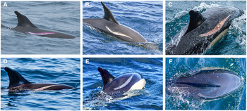
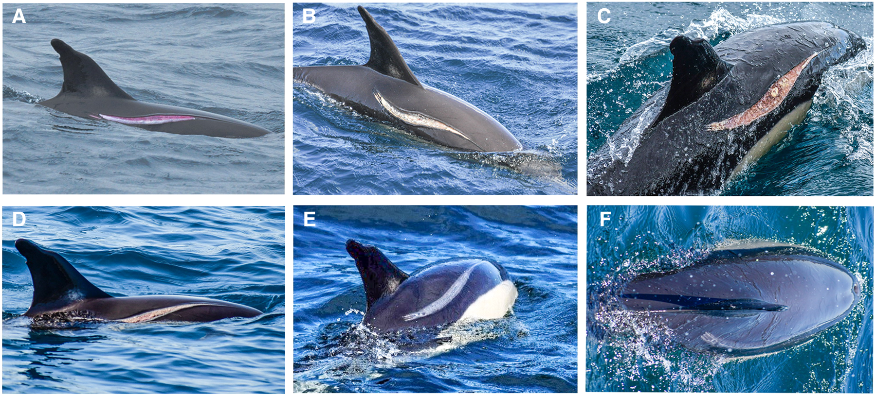
Specimen Dd-4 was an adult female, ~1.70 m in length, exhibiting good health. The animal was easily observed due to its opposed cuts with partial loss of tissue on the top leading and trailing edge of her dorsal fin, likely produced by fishing interactions (Wells et al., Reference Wells, Hofmann and Moors1998, Reference Wells, Allen, Hofmann, Bassos-Hull, Fauquier, Barros, Delynn, Sutton, Socha and Scott2008; Read & Murray, Reference Read and Murray2000). On 26 January 2017, the animal was observed showing a fresh single slash that covered the top right flank. The skin was superficially penetrated in the cranial area, but the blubber was seriously cut through with pale pink muscle layer and connective tissue exposed below on the dorsal side (Figure 5A). The wound did not bleed and was very fresh when the individual was encountered, suggesting that the accident likely occurred in the previous 24–48 h (Bloom & Jager, Reference Bloom and Jager1994; Elwen & Leeney, Reference Elwen and Leeney2010). On 28 January 2017, the wound edges showed a light grey colouration and were raised and swollen, while the wound itself showed grey-brownish colouration and soft epidermal sloughing tissue (Figure 5B). A creamy-coloured tissue was observed in the deepest bed of the wound. On 4 February 2017, the wound initially began to close up cranially, displaying a thin line followed by a buffer zone with white-brownish degenerating tissue, the same that was observed in the dorsal extreme of the wound. Fibroplasia was also detected, which extended over the pink granulation tissue, forming in the bed of the wound (Figure 5C). There was also a creamy-yellow degenerating tissue isolated in an elliptical shape in the centre of the wound, suggesting a possible infection. The lack of blubber under the dorsal fin exhibited a concave shape, and there were signs of blubber movement from the surrounding tissue that migrated over the exposed wound surface for the subsequent healing process (Zasloff, Reference Zasloff2011). On 2 March 2017, the previously concave wound had a convex and corrugated appearance and was filled with granulation tissue. During this sighting, it wasn't possible to photograph the dorsal extreme of the cut, but cranially, a darker line covered the healed extreme of the skin while caudally depigmented fibrous scar tissue squeezed the wound margins together (Figure 5D). By 1 April 2017, the slash was completely closed and a light grey discoloured band remained, along with a caudally depigmented area (Figure 4E). The band became more pigmented, maintaining only a slim pale grey line in the middle and divided in three small lines caudally on 8 May 2017 (Figure 5F).
Specimen Dd-4. (A) Female common dolphin (D. delphis) showed a fresh slash on its right flank, 26.1.2017; (B) wound with grey-brownish colouration and soft epidermal sloughing tissue, 28.1.2017; (C) buffer zone with a white-brownish degenerating tissue and pink granulation tissue forming in the bed of the wound, 4.2.2017; (D) wound with convex and corrugated effect, filled with granulation tissue and contracting the wound, 2.3.2017; (E) the cut was completely closed showing a light grey discoloured band, 1.4.2017; (F) slim pale grey line in the middle and divided in three small lines caudally, 8.5.2017.
Specimen Dd-5
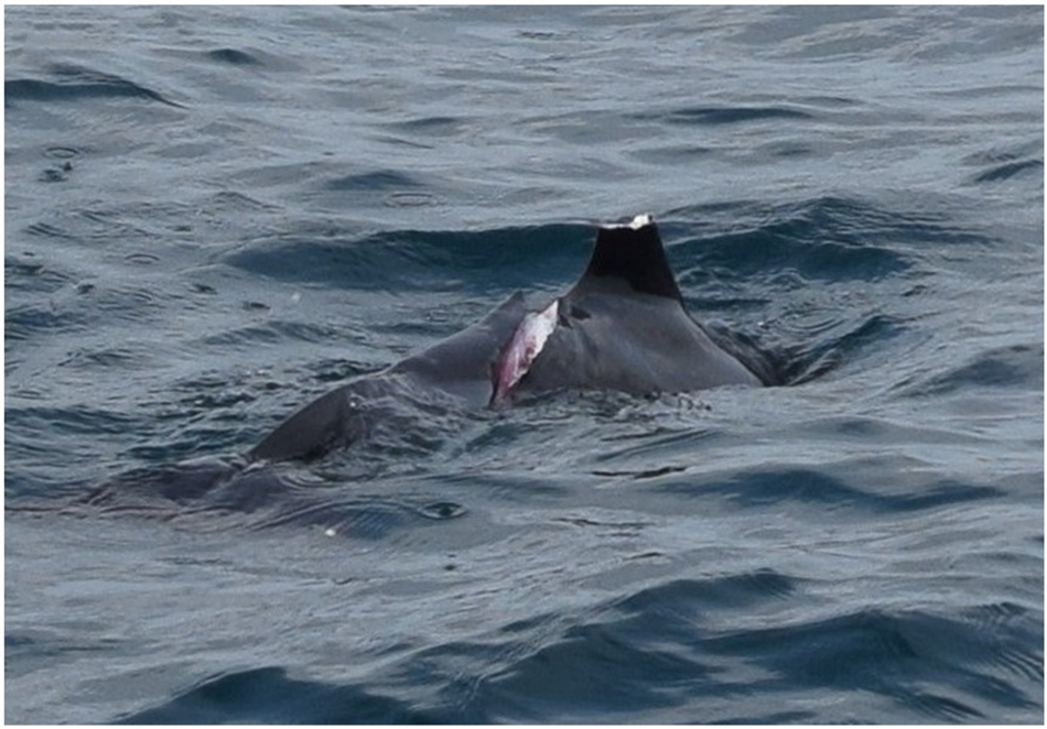
On 31 January 2017, a subadult was photographed with one large open deep cut on the peduncle running diagonally on the right side, immediately caudal to the dorsal fin (Figure 6).
Specimen Dd-5. A subadult common dolphin (D. delphis) photographed on 31.1.2017 showing one large open deep cut on the peduncle running diagonally on the right side, immediately caudal to the dorsal fin, which is partially mutilated.
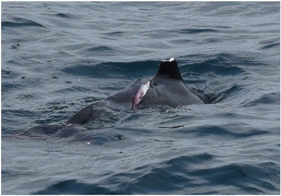
The caudal vertebrae looked damaged and the deep cut penetrated the skin, blubber and muscle. The borders of the wound were clean, not showing deformity or discolouration. Bleeding processes were not detected during the sighting. In addition, a second wound was observed on a partially mutilated dorsal fin. Both wounds were fresh, suggesting that the dolphin was injured within the previous 24 h. Although not conclusive, a propeller strike was likely the cause of the wound. Considering the animal's swimming restrictions, as well as its incapacity to follow its group (the animal was observed alone), we consider this type of wound to likely have been fatal and the animal was not observed again in the area.
Unlike the rest of the individuals described in this paper, Dd-5 was not observed again. Due to the severity of its wounds, among which included the mutilation of part of the dorsal fin, it was impossible to identify the individual.
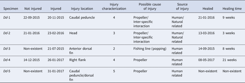
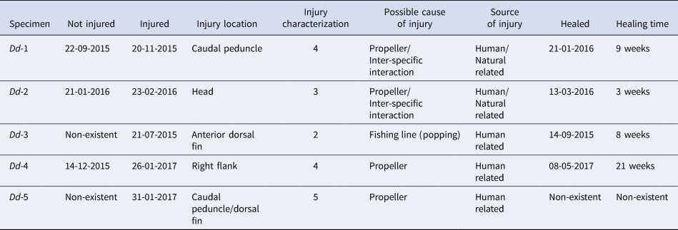
The five monitored specimens' history and healing times (animal photographed not injured–injured–healed) are compiled in Table 2. The injuries of Dd-1, Dd-2, Dd-3, Dd-4 and Dd-5 are classified as 4, 3, 2, 4 and 5, respectively based on the classifications of Lockyer & Morris (Reference Lockyer and Morris1990) (Table 1). The fact that Dd-5 wasn't located again suggests the death of the animal. For all tracked individuals, this is possibly the only individual that suffered death due to the severity of its injuries.
History of the injured specimens. Injury location, cause of injuries, dates and pictures were registered before and after the injury; these were then monitored during the healing process. Injury characterization was based on type and severity level of the wound, influenced by Lockyer & Morris (Reference Lockyer and Morris1990) (Table 1)
Discussion
Photographic records and analysis techniques constitute a powerful tool to distinguish one individual from another in wild range cetaceans (Chu & Nieukirk, Reference Chu and Nieukirk1988; Gowans & Whitehead, Reference Gowans and Whitehead2001; Rowe & Dawson, Reference Rowe and Dawson2009), even over long periods of time (Hammond, Reference Hammond1986; de Boer et al., Reference de Boer, Clark, Leopold, Simmonds and Reijnders2013). Furthermore, the remaining scars of past injuries, as well as mutilations or deep, long-term lasting wounds (Würsig & Jefferson, Reference Würsig and Jefferson1990), are also valuable as a tool to identify individuals (Scott et al., Reference Scott, Mann, Watson-Capps, Sargeant and Connor2005). Examination of such wounds could help potentially determine the human or natural source of the injury.
While it is usually difficult to assess the evolution of a wound over a period of time in wild animals (Dwyer et al., Reference Dwyer, Kozmian-Ledward and Stockin2014), injuries in dolphins and whales, both in natural and controlled situations, heal fully and quickly (Bruce-Allen & Geraci, Reference Bruce-Allen and Geraci1985; Corkeron et al., Reference Corkeron, Morris and Bryden1987; Lockyer & Morris, Reference Lockyer and Morris1990; Bloom & Jager, Reference Bloom and Jager1994). Even serious injuries that expose deep muscle tissue heal almost completely within the first 5 months (Bossley & Woolfall, Reference Bossley and Woolfall2014). Nevertheless, the healing period depends on the severity of the injury (Corkeron et al., Reference Corkeron, Morris and Bryden1987; Bloom & Jager, Reference Bloom and Jager1994; Visser, Reference Visser1999).
Zasloff (Reference Zasloff2011) proposed that blubber plays an important role in wound healing, as it contains antimicrobial properties and antibiotic compounds. This could explain how wild dolphins survive severe injuries in soft tissues without antibiotics or clinical care. The same author also suggested that dolphins do not exhibit typical responses to pain while recovering from injuries, feeding and behaving naturally even when seriously injured. Injury-healing rates in dolphins may be related to water temperature and salinity levels (Wilson et al., Reference Wilson, Arnold, Bearzi, Fortuna, Gaspar, Ingram, Liret, Pribanic, Read, Ridoux, Schneider, Urian, Wells, Wood, Thompson and Hammond1999). Healing appears to occur faster in warmer and saltier waters (S. Ridgway 2010, personal communication), whereas low water temperatures can decrease blood flow to the skin, thus limiting the cell regeneration rate of the epidermis (Feltz & Fay, Reference Feltz and Fay1966). Waters with poor physio-chemical conditions can also slow down the healing process. According to Bloom & Jager (Reference Bloom and Jager1994), prolonged exposure to untreated wastewater with high bacterial levels can delay the healing process and facilitate the development of secondary infections. Belugas (Delphinapterus leucas Pallas, 1776) heal five times faster than bottlenose dolphins (Tursiops truncatus) due to the thickness of their epidermis (Geraci & Bruce-Allen, Reference Geraci and Bruce-Allen1987).
Animals suffering soft tissue injuries caused by boat propellers usually survive, and even when the distal ends of their tails have been amputated, those individuals have successfully reproduced (Wells et al., Reference Wells, Allen, Hofmann, Bassos-Hull, Fauquier, Barros, Delynn, Sutton, Socha and Scott2008). One specific example of an injury caused by propellers in bottlenose dolphins illustrated that a severe head cut on a specimen healed in 3 months (Lockyer & Morris, Reference Lockyer and Morris1990). Another case of a similar injury took 65 days for scarring and ~100 days to heal completely. However, it should be noted that human-impacted environments, such as WBGA, are not an ideal area for recovering from severe wounds as injured individuals can suffer immunosuppression and increased susceptibility to infection (Daszak et al., Reference Daszak, Cunningham and Hyatt2001; Ross, Reference Ross2002; Van Bressem et al., Reference Van Bressem, Raga, Di Guardo, Jepson, Duignan, Siebert, Barrett, Santos, Moreno, Siciliano, Aguilar and Van Waerebeek2009; Desforges et al., Reference Desforges, Sonne, Levin, Siebert, De Guise and Dietz2016; Gui et al., Reference Gui, Èl, Zhang, Tu, Chen, Feng, Liu, Mai and Wu2018; Rubio-Guerri et al., Reference Rubio-Guerri, Jiménez, Melero, Díaz-Delgado, Sierra, Arbelo, Bellière, Crespo-Picazo, García-Párraga, Esperón and Sánchez-Vizcaíno2018).
Among the five individuals observed, two showed injuries presumably related to natural causes such as attempted predation, inter-species interactions with other cetaceans, or propeller hits (Dd-1, Dd-2); one was presumably related to fishing activities (Dd-3) and the final two showed injuries likely produced by propellers (Dd-4, Dd-5).
Boat propeller strikes can produce curved or straight parallel spaced slashes or cuts (Van Waerebeek et al., Reference Van Waerebeek, Baker, Félix, Gedamke, Iñiguez, Sanino, Secchi, Sutaria, Van Helden and Wang2007). They generally occur on the back of the dolphins since they do not expose their bellies to the surface for prolonged periods of time (Morgan & Patton, Reference Morgan and Patton1990). The coverage and the severity of the injury are directly related to the size of the propeller, the speed and bearing of the vessel, as well as the position and activity of the animal. Boat propeller injuries, if not fatal, are usually severe (Shane, Reference Shane1977; Sergeant, Reference Sergeant, Geraci and St Aubin1979). Dd-4 and Dd-5 presumably suffered propeller hits but at different degrees; while on Dd-4 the propeller hit only cut through the skin and blubber, Dd-5 had the propeller blade penetrate muscle and bone. The results indicated that Dd-4 survived its encounter while Dd-5 did not. Propeller hits could be related to the injuries presented by Dd-1 and Dd-2, but these two individuals survived.
The dorsal fin of one individual (Dd-3) was probably damaged by a fishing line. As we have shown in Figure 4, some animals show straight cuts with similar patterns, normally on the leading edge of the dorsal fin. One hypothesis suggests that the increase in this type of injury is related to sport fishing for bluefin tuna (Thunnus thynnus) in the area as the tuna population is recovering in the Mediterranean Sea (Van Beveren et al., Reference Van Beveren, Fromentin, Bonhommeau, Nieblas, Metral, Brisset, Jusup, Bauer, Brosset and Saraux2017). It has been observed that fishermen in the area use a technique called ‘popping’, where an artificial lure or ‘popper’ is presented to feeding tuna aggregations via casting out and reeling in a braided fishing line, using short bursts to pull the lure across the shoals of fish. During the summer in 2017, Espada et al. (Reference Espada, Martín, Haasova, Olaya-Ponzone and García-Gómez2018) carried out a study in the area, where 11% of the activities related to cetaceans belonged to fishermen casting directly on the dolphin-tuna feeding aggregates (Clua & Grosvalet, Reference Clua and Grosvalet2001; Cruz et al., Reference Cruz, Machete, Menezes, Rogan and Silva2018). It should also be noted that the ‘popping technique’ uses braided lines (Spectra twine type) which have been proved to be significantly more abrasive and to cut deeper than monofilament lines (Barco et al., Reference Barco, D'Eri, Woodward, Winn and Rotstein2010).
Cetaceans are considered prey of sharks (Leatherwood et al., Reference Leatherwood, Perrin, Garvie and LaGrange1973; Ebert et al., Reference Ebert, Compagno and Cowley1992; Long & Jones, Reference Long, Jones, Klimley and Ainley1996), which can attack live dolphins or predate on their carcasses. There are no records of shark attack reports on cetaceans in the WBGA but the presence of sharks in the Strait of Gibraltar is well known (Buencuerpo, Reference Buencuerpo1998). Only about six species of sharks including the great white shark (of more than 450 described species) are known to prey on small odontocetes (Long & Jones, Reference Long, Jones, Klimley and Ainley1996). In the Mediterranean, several shark attacks have been cited to cetaceans (Long & Jones, Reference Long, Jones, Klimley and Ainley1996; Celona et al., Reference Celona, De Maddalena and Comparetto2006), but only De Maddalena (Reference De Maddalena1999, Reference De Maddalena2000; unpublished data; see De Maddalena & Heim, Reference De Maddalena and Heim2012) reported attack of white shark (Carcharodon carcharias) on common dolphins. However, the white shark is exceptionally present in the Strait of Gibraltar (García-Gómez & Magariño, Reference García-Gómez and Magariño2010). Also, inter-species predatory interactions between cetaceans have been also reported (Pitman et al., Reference Pitman, Ballance, Mesnick and Chivers2001; Visser et al., Reference Visser, Zaeschmar, Halliday, Abraham, Ball, Bradley, Daly, Hatwell, Johnson, Johnson, Kay, Maessen, McKay, Petres, Turner, Umuroa and Pace2010). Dd-1 and Dd-2 showed unclean and punctured irregular-shaped crescent wounds that might also be related to shark attacks or inter-species interactions.
Environmental and management implications
The WBGA is considered to be a very busy shipping lane where container ships, ferries and oil tankers transit the area on a daily basis, along with internal shipping commercial activities.
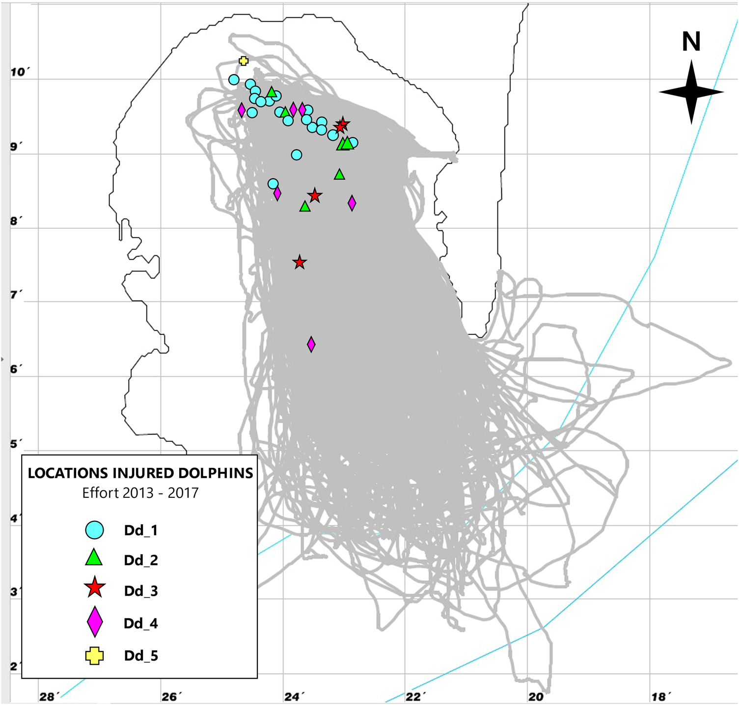
During the healing process, the tracked individuals were detected in the shallowest area of the bay overlapping with recreational and commercial whale watching and sport fishing activities, which were associated with groups of common dolphins, including newborns (see Espada et al., Reference Espada, Martín, Haasova, Olaya-Ponzone and García-Gómez2018) (Figure 7).
Map representing the total sightings of each injured animal in relation to effort. This may suggest that injured animals preferred to stay in shallow waters to heal their wounds.

Small cetaceans are rarely hit by propellers (David, Reference David and Notarbartolo di Sciara2002) but in situations of high concentrations of boats in a small and shallow area, the risk of collision might increase (Wells & Scott, Reference Wells and Scott1997). The WBGA becomes very busy in the summer, when there are intense whale watching commercial and recreational activities, increasing the concentration of boats (Espada et al., Reference Espada, Martín, Haasova, Olaya-Ponzone and García-Gómez2018) in a small area, close to the ports and marinas.
It is important to consider the impact of unregulated recreational and commercial whale watching along with sport and commercial fishing activities in the area, as the WBGA serves as an important breeding and feeding hotspot for the endangered common dolphin Mediterranean subpopulation (Ruiz-Giráldez et al., Reference Ruiz-Giráldez, Montero, Gálvez and García-Gómez2005; Giménez et al., Reference Giménez, Gauffier, Verborgh, Esteban, Jiménez-Torres, De Stephanis, Gauffier and Verborgh2011).
Conservational perspectives
Considering the results of this study, and due to the high maritime traffic and accumulation of boats in the area, there should be an overall assessment that monitors the noise output of individual commercial ships to gain a more detailed understanding of how the common dolphin population is affected by acoustic pollution and maritime traffic recurrence.
Conservation measures should be applied in order to mitigate the stress, injuries and death of dolphins. Monitoring these variables would establish a controlled framework for recreational and commercial dolphin watching activities, which is currently hindered by ineffective law enforcement and controls on water quality and marine litter. Fishing methods for bluefin tuna must be revised, as dolphins are frequently used as signs to find the aggregations of fish, and the ‘popping’ technique should be controlled by penalizing fishermen who clearly cast on dolphin–tuna feeding aggregation groups.
These methods should be banned not only because of the injuries produced by their braided lines, propellers or hull collisions but also for the stress inflicted on the cetacean groups. Mitigation measures have been applied and navigation is forbidden when human activities interfere with breeding, reproduction and feeding areas in other parts of the world. There are alternative traffic routes and maritime activities to avoid overlap with important areas for cetaceans (Nowacek et al., Reference Nowacek, Wells and Solow2001). One suggested mitigation measure includes issuing speed guidance for vessels to require slow, safe speed standards (Constantine et al., Reference Constantine, Johnson, Riekkola, Jervis, Kozmian-Ledward, Dennis, Torres and Aguilar de Soto2015), as well as posting dedicated observers and establishing marine mammal protected areas and sanctuaries.
The results of this work highlight the value of whale watching operators who voluntarily develop their activities in a responsible manner; these operators are a powerful asset to enforcement and conservation. Due to their fixed itineraries and recurrences they also help researchers to collect observation data, photographic records and long-term information to track injured animals. Operators can obtain great learning experience from researchers in order to minimize their impact on dolphin populations. However, whale watching activities can become a major threat to cetaceans in the area when they are unregulated or rules are unenforced (Parsons, Reference Parsons2012).
This study contributes new cases of common dolphins surviving injury, providing information on their healing processes and time and rates of survival. These data also aid in the development of injury catalogues for common dolphins and help determine chances of healing, healing times and survival rates of animals living in environments with drastic human impact. In the long term, these injury patterns might help to determine the human and natural interactions impacting dolphins in the area.
Human threats have increased in the WBGA over the past few years and common dolphins are now facing several challenges. The protection of this breeding and feeding area is crucial for the survival of this endangered species, as maritime human activities are rapidly and substantially jeopardizing this important hotspot. This issue therefore needs to be quickly addressed with determination and delicacy.
Acknowledgements
We thank the entire team of Dolphin Adventure for donating their whale watching platforms in the waters of the Bay of Algeciras. Thanks to Luisa Haasova for her collaboration in data collection, Alessia Scuderi for her work and determination and Darren Fa (University of Gibraltar) and Clive Crisp (Government of Gibraltar) for their unconditional support with the investigations. Thanks to David Hanrahan and José Manuel Guerra for his collaboration in part of the translation of some content to English.
Financial support
This paper has been financed under the research project number 18/0014 (FIUS) and benefited from infrastructures acquired in the framework of the research project number 1889 (FIUS), co-financed by the Port Authority of Seville, the Aquarium of Seville and the University of Seville.