Introduction
Traumatic brain injury (TBI) is a leading cause of death and disability in youth. In the United States, at least 700,000 children and adolescents seek medical care for TBI annually, resulting in approximately 50,000 hospitalizations and 6000 deaths (Faul, Xu, Wald, & Coronado, Reference Faul, Xu, Wald and Coronado2010). Previous research has clearly documented that survivors of childhood TBI demonstrate a variety of cognitive, emotional, and behavioral deficits, especially following more severe injuries (Yeates, Reference Yeates2010).
In contrast, the social outcomes of childhood TBI remain largely uncharacterized and poorly understood. Despite the importance of social competence as a predictor of numerous other outcomes, including psychological adjustment, academic performance, and health status (Cacioppo et al., Reference Cacioppo, Hawkley, Crawford, Ernst, Burleson, Kowalewski and Berntson2002; Rubin, Bukowski, & Parker, Reference Rubin, Bukowski and Parker2006), little is known about the nature, basis, and consequences of social problems among children with TBI. Nevertheless, poor social functioning is likely to play a major role in the reductions in quality of life reported following childhood TBI (Di Battista, Soo, Catroppa, & Anderson, Reference Di Battista, Soo, Catroppa and Anderson2012).
Several different lines of research suggest that children with TBI are vulnerable to poor social outcomes. First, children with chronic health conditions affecting the central nervous system, such as epilepsy and cerebral palsy, are consistently reported to be less socially competent than peers (Martinez, Carter, & Legato, Reference Martinez, Carter and Legato2011). Second, neuroimaging research has revealed an anterior–posterior gradient in the brain abnormalities associated with TBI. Larger and more numerous lesions, as well as volumetric reductions, characterize frontal and anterior temporal regions (Bigler, Reference Bigler2007; Wilde et al., Reference Wilde, Bigler, Haider, Chu, Levin, Li and Hunter2005, Reference Wilde, Bigler, Hunter, Fearing, Scheibel, Newsome and Levin2006, Reference Wilde, Hunter, Newsome, Scheibel, Bigler, Johnson and Levin2007), many of which have been implicated as the neural substrates of social information-processing and the regulation of social behavior (Adolphs, Reference Adolphs2001). Third, the few existing studies of social outcomes in childhood TBI have shown that children with severe TBI show deficits in social information processing and social adjustment and that their poor social outcomes persist over time (Rosema, Crowe, & Anderson, Reference Rosema, Crowe and Anderson2012; Yeates et al., Reference Yeates, Bigler, Dennis, Gerhardt, Rubin, Stancin and Vannatta2007).
Previous studies of social outcomes in childhood TBI have focused largely on social adjustment in a broad sense, assessed primarily via parent reports. Social difficulties have been documented using standardized checklists, as well as clinical and qualitative interviews, but significant limitations characterize this literature (Yeates et al., Reference Yeates, Bigler, Dennis, Gerhardt, Rubin, Stancin and Vannatta2007). Most research has relied on broad measures of behavioral symptoms, such as the Child Behavior Checklist (Achenbach & Rescorla, Reference Achenbach and Rescorla2001), or adaptive functioning, such as the Vineland Adaptive Behavior Scales (Sparrow, Cicchetti, & Balla, Reference Sparrow, Cicchetti and Balla2005), that use a limited number of items to assess social adjustment and provide little detail about children's social interactions with peers. Although ratings of social adjustment are usually provided by parents, children's social interactions with peers occur largely outside the home, so parent and peer perceptions of social adjustment may not match.
Classroom-based peer nominations and ratings are widely used by developmental researchers to assess social behavior, peer acceptance, and friendships (Masten, Reference Masten2005; Parker & Asher, Reference Parker and Asher1987; Rubin, Shulz Begle, & McDonald, Reference Rubin, Shulz Begle and McDonald2012), and these techniques have been successfully applied with multiple pediatric illness populations (Reiter-Purtill, Waller, & Noll, Reference Reiter-Purtill, Waller and Noll2009). The perspectives of numerous classmates who interact daily at school are combined and yield scores that are reliable, stable, and predictive of concurrent and future academic, emotional, and behavioral outcomes (Masten, Reference Masten2005; Rubin et al., Reference Rubin, Shulz Begle and McDonald2012), all of which continue to be concerns for children with TBI even after they reach adulthood (Anderson, Brown, Newitt, & Hole, Reference Anderson, Brown, Newitt and Hole2009).
The primary goal of our study was to compare school-aged children with TBI to children with orthopedic injuries (OI) on measures of social behavior, acceptance, and friendship derived from the children's classmates. We believe this to be the first study of childhood TBI to use classroom-based peer nominations and ratings for this purpose. Based on previous studies of peer relationships in children with other central nervous system (CNS) disorders (Noll et al., Reference Noll, Reiter-Purtill, Moore, Schorry, Lovell, Vannatta and Gerhardt2007; Vannatta, Gerhardt, Wells, & Noll, Reference Vannatta, Gerhardt, Wells and Noll2007), we hypothesized that children with TBI, especially those with severe injuries, would be described by peers as demonstrating less leadership and popularity, more shy and withdrawn behavior, and more rejection and victimization than children with OI. We also hypothesized that children with TBI would be less accepted or liked by peers and have fewer mutual friendships with classmates. We expected that the magnitude of these differences would be most pronounced among children with severe TBI.
A secondary goal of the study was to determine whether differences in peer relationships among children with TBI were related to underlying variations in brain structure as measured by magnetic resonance imaging (MRI). Using voxel-based morphometry (VBM; Ashburner & Friston, Reference Ashburner and Friston2000), we explored how volumetric measures derived from MRI varied as a function of differences in social behavior and friendship status. We hypothesized that those aspects of peer relationships on which the TBI and OI groups differed would correlate with volumetric reductions in the so-called “social brain” network, which includes a distributed set of regions that gradually become integrated during the course of social development (Adolphs, Reference Adolphs2001; Johnson et al., Reference Johnson, Griffin, Csibra, Halit, Farroni, De Haan and Richards2005).
Methods
Participants
Participants were drawn from a larger pool of children taking part in a multi-site study examining social outcomes in children with traumatic brain injuries. The children were recruited from children's hospitals at three metropolitan sites, including the Hospital for Sick Children in Toronto (Canada), Nationwide Children's Hospital in Columbus (US), and Rainbow Babies and Children's Hospital and MetroHealth Medical Center in Cleveland (US). Eligible participants included children hospitalized for either a traumatic brain injury (TBI) or orthopedic injury (OI) who were from 8 to 13 years of age at the time of their participation and injured between 12 and 63 months before participation. All children were injured after 3 years of age and the vast majority after 4 years. The TBI group was restricted to children with complicated mild to severe TBI. Severe TBI was defined based on a lowest post-resuscitation Glasgow Coma Scale (GCS) score of 8 or less, moderate TBI based on a GCS score from 9 to 12, and complicated mild TBI based on a GCS score of 13 to 15 in association with trauma-related abnormalities on neuroimaging at the time of hospitalization. A depressed skull fracture was sufficient to warrant inclusion in the complicated mild TBI group, but intracranial abnormalities were required in all other cases; only three children were considered eligible for the complicated mild TBI group based solely on a depressed skull fracture. For the purposes of the current study, children with complicated mild and moderate TBI were combined into a single group. The OI group consisted of children who sustained fractures not associated with any loss of consciousness or other risks or indications of brain injury (e.g., skull or facial fractures).
All children were subject to the following exclusion criteria: (a) history of more than one serious injury requiring medical treatment; (b) premorbid neurological disorder or mental retardation; (c) any injury determined to be a result of child abuse or assault; (d) a history of severe psychiatric disorder requiring hospitalization before the injury; (e) any sensory or motor impairment that prevented valid administration of study measures; (f) not fluent in English; and (g) any medical contraindication to MRI. Children in full-time special education classrooms were excluded because the reliability and validity of classroom data for such classrooms is not established. Children with a history of premorbid learning or attention problems were not excluded from the study. Parents reported that six children had a premorbid diagnosis of ADHD and four had a premorbid diagnosis of learning disability. The groups did not differ in the proportion of children with these premorbid conditions.
Among children eligible to participate and approached about the study, 82 (47%) of those with TBI and 61 (26%) of those with OI agreed to enroll. The participation rate was significantly higher for TBI than OI participants. However, participants and non-participants in both groups did not differ in age at injury, age at initial contact about the study, sex, race, or census tract measures of socioeconomic status (SES) that included mean family income, percentage of minority heads of household, and percentage of households below the poverty line. Participants and non-participants also did not differ on measures of injury severity (i.e., mean length of stay, median Glasgow Coma Scale score for children with TBI).
The current study was restricted to the 87 of 143 (61%) children from the larger study for whom classroom data were obtained (n = 15 of 25 with severe TBI, n = 40 of 57 with complicated mild/moderate TBI, and n = 32 of 61 with OI). Completion of classroom data collection did not differ significantly as a function of injury group, sex, race, SES (measured using a standardized composite based on parental education, parent occupational status, and census tract mean family income), age at injury, or age at assessment. Classroom data were provided by 1598 children in 87 mainstream classrooms. On average, 18.4 students (SD = 4.7; range = 7–30) participated in each class. Parental consent was obtained for 82% of available classmates and 96% were present on the day of data collection; thus, 79% of classmates participated.
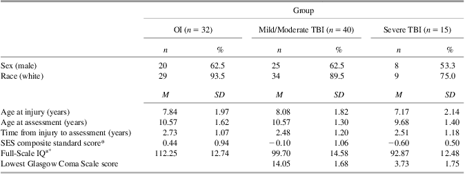
Demographic and injury characteristics for the three injury groups are shown in Table 1. The groups did not differ in sex, race, age at injury, age at assessment, or time from injury to assessment. The groups differed significantly in Full Scale IQ and SES, with the severe TBI group having the lowest mean IQ and SES, although IQ scores for all groups were within the average to high-average range. The groups also differed in the distribution of mechanism of injury, with injuries arising from motorized vehicles being most common among the severe TBI group (67% for severe TBI, 30% for mild/moderate TBI, 6% for OI) and those arising from sports and recreational events being most common among the OI group (20% for severe TBI, 40% for mild/moderate TBI, 75% for OI). Falls accounted for the remaining injuries. Group differences in SES were no longer significant when injury mechanism was taken into account. These findings are consistent with epidemiological studies showing that the risk of TBI, particularly those linked to motorized vehicles, is highest for children of lower SES and minority status (Brown, Reference Brown2010; Howard, Joseph, & Natale, Reference Howard, Joseph and Natale2005; Langlois, Rutland-Brown, & Thomas, Reference Langlois, Rutland-Brown and Thomas2005; Parslow, Morris, Tasker, Forsyth, & Hawley, Reference Parslow, Morris, Tasker, Forsyth and Hawley2005; Yates, Williams, Harris, Round, & Jenkins, Reference Yates, Williams, Harris, Round and Jenkins2006). For that reason, we did not treat SES as a covariate in primary data analyses, because the SES differences appeared to be intrinsic to the injury groups. When a covariate is an attribute of a disorder, or is intrinsic to the condition, “adjusting” for differences in the covariate is not meaningful and can be potentially misleading (Dennis et al., Reference Dennis, Francis, Cirino, Schachar, Barnes and Fletcher2009).
Group demographics and injury characteristics
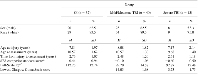
Note. OI = orthopedic injury, TBI = traumatic brain injury, SES = socioeconomic status.
aIQ measured using the two-subtest version of Wechsler Abbreviated Scale of Intelligence.
*Groups differ significantly, p < .05.
Procedure
Institutional review boards approved all study procedures before recruitment, and informed parental consent and child assent were obtained before participation. School principals received written information about the study, followed by a telephone call to obtain their permission to contact participants’ teachers. A teacher meeting was held to explain the study. Teachers distributed and collected parental consent forms from students. To protect the confidentiality of participants, the work was described to students as a study of friendships without mentioning traumatic brain injuries or identifying the participating child. Questionnaires were administered during a single group session in the primary classroom for elementary school students or a required academic subject (e.g., English) for students in middle school. Classroom data was not collected during the first 2 months of the school year, to ensure that children were familiar with one another before completing ratings. The groups did not differ in the average number of months that had passed during the school year before classroom collection (M = 6.30; SD = 2.09).
Classroom Measures
Extended Class Play (ECP)
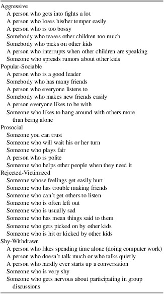
Participants completed an extended version of the Revised Class Play (Masten, Morison, & Pellegrini, Reference Masten, Morison and Pellegrini1985) to assess social behavior with peers (Rubin, Wojslawowicz, et al., Reference Rubin, Wojslawowicz, Burgess, Rose-Krasnor and Booth-LaForce2006; Wojslawowicz Bowker, Rubin, Burgess, Rose-Krasnor, & Booth-LaForce, Reference Wojslawowicz Bowker, Rubin, Burgess, Rose-Krasnor and Booth-LaForce2006). Administration of the ECP requires students to imagine that they are the director of a play and to “cast” one boy and one girl from the class into 31 hypothetical “roles” (see Table 2). Popular-Sociable, Prosocial, Aggressive, Rejected-Victimized, and Shy-Withdrawn subscales have been identified through factor analysis (Rubin, Wojslawowicz, et al., Reference Rubin, Wojslawowicz, Burgess, Rose-Krasnor and Booth-LaForce2006; Wojslawowicz Bowker et al., Reference Wojslawowicz Bowker, Rubin, Burgess, Rose-Krasnor and Booth-LaForce2006). Tallies of nominations received from classmates for each role were standardized within sex in each class to adjust for unequal class size, composition, and participation rates and then summed to create dimension scores. The summed scores were standardized (M = 0; SD = 1) within sex in each class to create subscales. The scores reflect nominations relative to same-sex peers.
Extended Class Play subscale items

Note. Copyright © 1985 by the American Psychological Association. Item content adapted from Table 1 appears here with permission. The official citation that should be used in referencing this material is Masten, A.S., Morison, P., & Pellegrini, D.S. (1985) A revised class play method of peer assessment. Developmental Psychology, 21(3), 523, 533. Doi: 10.1037/0012-1649.21.3.523. No further reproduction or distribution is permitted without written permission from the American Psychological Association.
Peer acceptance ratings
Students completed 5-point ratings of how much they liked each classmate (Asher, Singleton, Tinsley, & Hymel, Reference Asher, Singleton, Tinsley and Hymel1979). Mean acceptance ratings were standardized within sex for each class.
Best friend nominations
Students recorded names of their three “best friends” from the class. Two scores were computed, one for total friendship nominations and one for mutual friendship nominations, which reflects the number of friendship nominations each child received from classmates who also identified the child as a friend (Bukowski & Hoza, Reference Bukowski and Hoza1989). Both scores were standardized within sex for each class. We also classified children based on whether they had no mutual friendships versus at least one mutual friendship in their classrooms.
MRI and VBM
Magnetic field strength was 1.5 Tesla for all studies. The Toronto and Columbus sites used GE Signa Excite scanners and the Cleveland site used a Siemens Symphony scanner. All sites acquired the following sequences on each participant: thin slice, volume acquisition T1-weighted ultrafast three-dimensional gradient echo, commonly referred to as MPRAGE or FSPGR (depending on scanner manufacturer); a dual-echo proton density (PD)/T2 weighted sequence; FLAIR; and GRE. Standard phantoms were used for quality control and to calibrate scans across sites.
FreeSurfer (surfer.nmr.mgh.harvard.edu) was used to derive whole brain volumetric measurements, using methods described by Bigler et al. (Reference Bigler, Abildskov, Wilde, McCauley, Li, Merkley, Fearing and Levin2010). Volumes obtained included total white matter (WM), total gray matter (GM), total ventricular volume, and total cerebrospinal fluid space (CSF), along with total brain volume (TBV), summing for right and left hemisphere structures. Also, a ventricle-to-brain ratio (VBR) was obtained by using the FreeSurfer computation of total ventricular volume divided by TBV and multiplied by 100 to provide whole number values (Tate, Khedraki, Neeley, Ryser, & Bigler, Reference Tate, Khedraki, Neeley, Ryser and Bigler2011).
For voxel based morphometry (VBM), the major preprocessing and analysis steps were done in SPM8 (Wellcome Trust Centre for Neuroimaging, London, UK; available at: http://www.fil.ion.ucl.ac.uk/spm) using the VBM8 toolbox (available at: http://dbm.neuro.uni-jena.de/vbm). The images were (1) corrected for bias-field inhomogeneities, (2) registered using a linear (i.e., 12-parameter affine) and a nonlinear transformation, and (3) stripped of non-brain tissue in the T1-weighted images. The SPM8 unified segmentation package (Ashburner & Friston, Reference Ashburner and Friston2005) was used to initialize a VBM8 algorithm-based classification of GM, WM, and CSF, following the methods outlined by Ziegler et al. (2012).
WM population templates were generated using the entire image dataset, using the Diffeomorphic Anatomical Registration through Exponentiated Lie Algebra (DARTEL) technique (Ashburner, Reference Ashburner2007). Affine registration of the WM DARTEL templates to the tissue probability maps in Montreal Neurological Institute (MNI) space was performed (see http://www.mni.mcgill.ca/), along with non-linear warping of WM images to the DARTEL WM template in MNI space with a 1.5 mm3 resolution (as recommended for the DARTEL procedure). WM volume at each voxel was obtained through modulation, which was performed by multiplying the WM concentration map by the Jacobian determinants derived from the non-linear spatial normalization step followed by smoothing of WM volume images with a full width at half maximum kernel of 8 mm. Following all of these spatial pre-processing methods, the smoothed, modulated, and normalized WM volume maps were used for statistical analysis.
For the automated image analyses, useable data were available for only 58 cases (severe TBI = 10, complicated mild/moderate TBI = 28, OI = 22). The availability of useable imaging data did not vary by group. Reasons for exclusion were poor scan quality as a result of movement or artifact (either post-surgical for children with TBI or from dental braces) or incomplete imaging sequences due to claustrophobia, equipment failure, or participant non-compliance. Additionally, two children with severe TBI had such significant parenchymal distortion as a result of their injuries that it precluded automated image analysis.
Statistical Analyses
Peer relationship data
Group comparisons on the continuous measures of social behavior, peer acceptance, and friendship were conducted using univariate analyses of variance. Planned contrasts compared each of the TBI groups to the OI group. The proportion of children in each group who did or did not have a mutual friendship was compared using a χ2 analysis. Within the TBI sample, children with and without mutual friendships were compared on the indices of social behavior and peer acceptance using t tests. Effect sizes for continuous variables were estimated using η2, which reflects the proportion of variance accounted for by the three injury groups, or d, which reflects the difference in standard deviation units between any two groups. Effect sizes for proportions were estimated using odds ratios.
Neuroimaging data
Overall measures of brain volume (total GM, WM, CSF, and VBR) were first correlated with variables that demonstrated significant group differences, to determine if peer relationships were affected by diffuse atrophy. Correlations were computed within the OI and TBI groups separately (with complicated mild/moderate and severe TBI groups combined) and across the entire sample. Voxel-based partial correlation analyses then were carried out in SPM to test relationships between WM volumes and classroom variables using all available participants. Age, sex, and MRI acquisition site were treated as covariates, to reduce error variance and to control for variations between scanners across sites. We first identified significant individual voxels using an uncorrected threshold of T of 2.5 (p < .008, Friston, Holmes, Poline, Price, & Frith, Reference Friston, Holmes, Poline, Price and Frith1996). We next identified clusters of voxels that were significant based on a threshold of p < .01 after family-wise error correction. We interpreted only those clusters that surpassed a size threshold of 250 voxels, which represented the expected number of voxels per cluster based on random field theory, as computed by SPM8.
Results
Group Differences in Peer Relationships
Social behavior
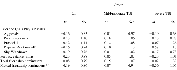
Group mean scores on the five subscales of the ECP are presented in Table 3. The three injury groups differed significantly on the Rejected-Victimized subscale of the ECP, F(2,83) = 3.70, p = .033, η2 = .08; they did not differ on the Popular-Sociable, Prosocial, Aggressive, or Shy-Withdrawn subscales. Planned contrasts showed that the severe TBI group had higher scores on the Rejected-Victimized subscale than the OI group, p = .010, d = .80. The severe TBI group did not differ from the OI group on other subscales, despite medium effect sizes for Popular-Sociable (d = −.46, lower scores for severe TBI group) and Shy-Withdrawn (d = .41, higher scores for TBI group). The complicated mild/moderate TBI group did not differ from the OI group on any subscales from the ECP.
Group mean ratings for social behavior, acceptance, and friendship
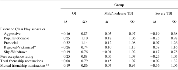
*Groups differ significantly, p < .05; Severe TBI < OI, p < .01.
**Severe TBI < OI, p < .07.
OI = orthopedic injury; TBI = traumatic brain injury.
Peer acceptance
Group scores for the peer acceptance ratings are presented in Table 3. The three injury groups did not differ significantly in how they were rated. Additionally, neither TBI group differed significantly from the OI group, despite a medium effect size reflecting lower scores in the severe TBI group (d = −.48).
Friendship
Group scores for the number of best friend nominations and mutual best friend nominations are presented in Table 3. The groups did not differ on either measure. Neither TBI group differed from the OI group on the number of best friend nominations, but planned contrasts indicated that the severe TBI group showed a trend toward fewer mutual best friend nominations than the OI group, p = .065, d = −.58.
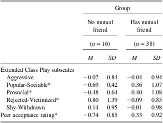
The groups differed significantly in the proportion of children who had at least one mutual friendship with a classmate, χ2(2) = 9.84, p = .007. Children with severe TBI were significantly less likely to have a mutual friend in their classrooms than children with OI (47% vs. 88%, odds ratio = 0.125, 95% CI = 0.03, 0.54). Children with moderate TBI did not differ from those in the OI group in the likelihood of having a mutual friend (80% vs. 88%). Within the TBI group, as seen in Table 4, children without a mutual friend were rated significantly lower than those with a mutual friend on the Popular-Sociable and Prosocial subscales from the ECP, t(51) = −3.90, p = .000, d = −1.01, and t(51) = −2.51, p = .009, d = −.72, respectively, and significantly higher on the Rejected-Victimized subscale, t(51) = 2.86, p = .006, d = 1.05. They did not differ on the Aggressive or Shy-Withdrawn subscales. Children with TBI without a mutual friend also had significantly lower peer acceptance ratings than those with mutual friends, t(52) = −3.98, p = .000, d = −1.16. Children in the TBI group with mutual friends displayed acceptance ratings and ECP scores that were similar to those of children with OI.
Mean ratings for children with TBI with and without mutual friendships in classroom
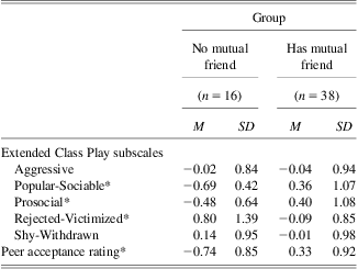
*Groups differ significantly, p < .01.
TBI = Traumatic brain injury.
Neuroimaging and Peer Relationships
Analyses of the neuroimaging correlates of peer relationships were restricted to the Rejected-Victimized scale and mutual best friend nominations, which were the variables with the most pronounced group differences. Neither variable correlated significantly with total WM, GM, CSF, or VBR, either within the TBI and OI groups separately or in the combined group.
In contrast, VBM identified one significant cluster of voxels (p = .005 after family-wise error correction) that predicted the number of mutual best friend nominations (see Table 5). After controlling for age, sex, and MRI acquisition site, mutual friendship was positively correlated with WM volume within a cluster that involved regions within the parietal and frontal lobes, as well as limbic system, largely within the right hemisphere. The specific regions within the cluster included the precuneous, inferior parietal lobule, posterior cingulate gyrus, cingulum bundle, insula, posterior corpus callosum, and paracentral lobule (see Figure 1). VBM did not identify any significant clusters correlated with the Rejected-Victimized scores.
Results of VBM analysis showing positive correlations with mutual friendship ratings.

VBM = voxel-based morphometry.
Voxel cluster in which WM volume was positively correlated with mutual friendship ratings on VBM (right hemisphere on right side). Regions within the cluster included the precuneous; inferior parietal lobule; posterior cingulate gyrus; cingulum bundle; insula; posterior corpus callosum; and paracentral lobule. Individual voxels identified with uncorrected threshold of T = 2.5 (p < .008); significant clusters identified with family-wise error corrected threshold of p < .01 and cluster size threshold of 250 voxels per cluster. Brain template is an averaged image based on T1 images from children with orthopedic injuries. Scale represents T values for individual voxels.

Discussion
According to their classmates, children with severe TBI demonstrate problematic peer relationships characterized by higher levels of peer rejection and victimization and a lower likelihood of having a mutual friend in the classroom. Both of these characteristics put children with severe TBI at substantial long-term risk, because rejection and victimization by peers and a lack of reciprocal friendships are both associated with an increased likelihood of poorer psychological adjustment, academic performance, and health status (Cacioppo et al., Reference Cacioppo, Hawkley, Crawford, Ernst, Burleson, Kowalewski and Berntson2002; Rubin et al., Reference Rubin, Shulz Begle and McDonald2012). In this regard, the poor peer relationships of children with severe TBI may help explain or may exacerbate the wide range of poor outcomes associated with TBI during childhood (Yeates, Reference Yeates2010). Poor peer relationships also are likely to contribute the poorer overall quality of life experienced by children with severe TBI, both shortly after their injury and continuing into adulthood (Anderson et al., Reference Anderson, Brown, Newitt and Hole2009; Di Battista et al., Reference Di Battista, Soo, Catroppa and Anderson2012), because of the cascading effects of social problems on other long-term outcomes, such as psychopathology (Burt, Obradovic, Long, & Masten, Reference Burt, Obradovic, Long and Masten2008).
Classmates did not simply report global changes in the peer relationships of children with TBI. As a group, children with TBI did not differ significantly from children with OI in terms of popularity and sociability, prosocial or aggressive behavior, or shy and withdrawn behavior. Nor did they differ in overall peer acceptance. Thus, the findings suggest some specificity in the nature of the peer difficulties experienced by children with TBI. On the other hand, the severe TBI group was described by classmates as demonstrating notably less popularity and sociability, more shy and withdrawn behavior, and lower peer acceptance ratings based on effect sizes, although these differences did not reach statistical significance. Taking the latter findings into account, the profile of differences is not dissimilar to that displayed by other groups of children with CNS disorders (Noll et al., Reference Noll, Reiter-Purtill, Moore, Schorry, Lovell, Vannatta and Gerhardt2007; Vannatta et al., Reference Vannatta, Gerhardt, Wells and Noll2007).
Group differences in peer relationships were related to injury severity. Only children with severe TBI differed significantly from children with OI; the complicated mild/moderate TBI group did not demonstrate any significant differences from the OI group, although they have shown deficits in social information processing in previous analyses based on the same sample (e.g., Dennis, Agostino, et al., Reference Dennis, Agostino, Taylor, Bigler, Vannatta, Gerhardt and Yeates2012; Dennis, Simic, et al., Reference Dennis, Simic, Taylor, Bigler, Rubin, Vannatta and Yeates2012). Historically, social outcomes after childhood TBI have not been consistently related to injury severity, with some studies finding a relationship and others not (Rosema et al., Reference Rosema, Crowe and Anderson2012). The inconsistency likely reflects multiple factors, including the nature of the outcome measure under study. The current findings add to the existing literature by suggesting that injury severity is related specifically to the impact of TBI on peer relationships.
The current findings also highlight the importance of mutual friendship as a predictor of peer relationships. Among children with TBI, those without mutual friends were described by peers as less popular and sociable, less prosocial, more rejected-victimized, and less liked than those with mutual friends, who were rated similarly to children with OI. This finding is in accord with an extensive literature on the critical function that friendship plays in children's social development (Rubin et al., Reference Rubin, Shulz Begle and McDonald2012). One of the more compelling findings of the current study is that less than 50% of the children with severe TBI had a mutual friend in their classrooms, compared to nearly 90% of the children with OI or a mild-moderate TBI. This finding suggests that interventions designed specifically to promote friendship may be an important vehicle for promoting better social outcomes following childhood TBI (Frankel, Reference Frankel2005). Clinical trials are needed to adapt and test the efficacy of such interventions specifically for children with TBI.
The differences in peer relationships between children with severe TBI and those with OI also were related to underlying variations in brain volume. Interestingly, global measures of brain atrophy were not predictive of either rejection/victimization or mutual friendship scores. However, VBM revealed specific brain regions in which WM volumes were positively correlated with mutual friendships. These included both major fiber pathways (e.g., corpus callosum, cingulum bundle) and other structures (e.g., posterior cingulate gyrus, precuneous, paracentral lobule, inferior parietal lobule) implicated in social information-processing and thought to be part of the social brain network (Adolphs, Reference Adolphs2001; Johnson et al., Reference Johnson, Griffin, Csibra, Halit, Farroni, De Haan and Richards2005). Interestingly, these regions also overlap substantially with the so-called “default node network,” which has an intriguing relationship to the social brain (Mars et al., Reference Mars, Neubert, Noonan, Sallet, Toni and Rushworth2012). Notably, the brain regions that correlated with reciprocal friendship involved primarily non-frontal regions. Posterior brain regions have been identified as key components of networks for social cognition (Hein & Singer, Reference Hein and Singer2008). Although damage to frontal regions has been linked to difficulties with behavioral regulation and associated social problems following childhood TBI (Levin et al., Reference Levin, Zhang, Dennis, Ewing-Cobbs, Schachar, Max and Hunter2004), several studies have now linked damage to or dysfunction in non-frontal structures to deficits in social information-processing in children with TBI that may also help to account for their poor social outcomes (Hanten, Levin, Newsome, & Scheibel, Reference Hanten, Levin, Newsome and Scheibel2012).
A concern could be raised about comparability across different sites and scanners in the VBM analyses. Ideally, comparability would be addressed by running separate analyses by site, but the sample size in the current study was too small for that approach. However, prior studies using VBM with larger samples have demonstrated that multi-site acquisitions result in reliable findings (Bendfeldt et al., Reference Bendfeldt, Hofstetter, Kuster, Traud, Mueller-Lenke, Naegelin and Borgwardt2012; Schnack et al., Reference Schnack, van Haren, Brouwer, van Baal, Picchioni, Weisbrod and Hulshoff Pol2010; Segall et al., Reference Segall, Turner, van Erp, White, Bockholt, Gollub and Calhoun2009). In the current investigation, the authors worked closely with the MR physicists and technologists at each site to duplicate the imaging sequences across the different platforms, and both human and machine phantoms were used to ensure reliability during the study. In the VBM analyses, moreover, site was treated as a covariate. The cluster that remained significant with FWE correction involved contiguous white matter in regions where multi-site comparisons typically have their best agreement.
Sample size was a more general limitation in the current study, especially in the severe TBI group. Unfortunately, classroom data could not be obtained for all children in the larger parent study, and some of those with classroom data did not have useable neuroimaging data. Although children with and without classroom data did not differ on demographic or injury characteristics, the restricted sample did reduce the statistical power of analyses. Another limitation is that the effects of SES and TBI severity on the assessed outcomes cannot be entirely disentangled; however, the lack of evidence for recruitment bias, and the absence of group differences in SES when injury mechanism is taken into account, indicate that the group difference in SES is intrinsically related to TBI severity and is not a function of biased sampling. Moreover, although we did not include SES as a covariate in our primary analyses because it would be potentially misleading to do so, supplementary analyses showed that group differences remained significant when SES was covaried. A final limitation was that information regarding the children's pre-injury peer relationships was not available. We could have obtained retrospective ratings of social adjustment from parents, but the study was conducted at least 1 year post-injury, so retrospective ratings would be subject to recall bias.
Future research should address the mechanisms that give rise to the difficulties in peer relationships reported here. For instance, studies are needed that examine various aspects of social information processing, such as mentalizing and the social communication of affect (Dennis, Agostino, et al., Reference Dennis, Agostino, Taylor, Bigler, Vannatta, Gerhardt and Yeates2012; Dennis, Simic, et al., Reference Dennis, Simic, Taylor, Bigler, Rubin, Vannatta and Yeates2012), and observed social interactions as predictors of the quality of peer relationships among children with TBI. Few previous studies have examined the relationship between social information processing and social adjustment following TBI (cf. Yeates et al., Reference Yeates, Swift, Taylor, Wade, Drotar, Stancin and Minich2004), and to the best of our knowledge, none has involved peer ratings. Similarly, no published studies have conducted direct behavioral observations of the peer interactions of children with TBI, although such observations are likely to prove critical in determining exactly which behaviors of children with TBI elicit peer rejection and victimization and inhibit friendship formation. The identification of deficits in social information processing and social interaction that impair peer relationships will be important to the development of intervention programs designed to prevent or ameliorate poor social outcomes following childhood TBI.
Acknowledgments
The preparation of this study was supported in part by grant 5 R01 HD048946 from the National Institute of Child Health and Human Development to the first author. The authors do not have any conflicts of interest to declare.