Introduction
The food-borne zoonotic nematode Angiostrongylus cantonensis has been recognized as the most common cause of eosinophilic meningitis and/or eosinophilic meningoencephalitis in humans (Cross and Chen, Reference Cross, Chen, Black and Seed2007). Rodents are the definitive host while molluscs are the intermediate host. Humans are accidental host where transmission is through eating infected undercooked snails, or other infected paratenic hosts such as freshwater prawns, crabs, frogs or monitor lizards as well as poorly cleaned contaminated vegetables (Cross and Chen, Reference Cross, Chen, Black and Seed2007; Wang et al., Reference Wang, Lai, Zhu, Chen and Lun2008, Reference Wang, Wu, Wei, Owen and Lun2012; Eamsobhana, Reference Eamsobhana2015). Typical clinical manifestations of central nervous system (CNS) angiostrongyliasis include severe headache, neck stiffness/nuchal rigidity and paresthesias (Wang et al., Reference Wang, Lai, Zhu, Chen and Lun2008; Graeff-Teixeira et al., Reference Graeff-Teixeira, da Silva and Yoshimura2009).
A. cantonensis is endemic in Asia and the Pacific islands where most cases of human infection have occurred. In recent years, the parasite has spread from its recognized endemic areas to the American continent including the USA, Brazil and Caribbean islands, and the incidence of human infection has increased rapidly (Wang et al., Reference Wang, Lai, Zhu, Chen and Lun2008, Reference Wang, Wu, Wei, Owen and Lun2012; Eamsobhana, Reference Eamsobhana2015; Barratt et al., Reference Barratt, Chan, Sandaradura, Malik, Spielman, Lee, Marriott, Harkness, Ellis and Stark2016). Angiostrongyliasis is of an increasing public health risk as globalization contributes to the geographical spread and more international travellers encounter the disease. The rapid global spread of A. cantonensis and the emerging occurrence of the infection pose challenges in clinical and laboratory diagnosis.
Suspected cases of A. cantonensis infection can only be confirmed by isolation of the nematodes in cerebrospinal fluid (CSF) or eye chamber of the infected individuals, but this finding is rare. A presumptive diagnosis is primarily based on clinical characters, specific eating habits, peripheral blood eosinophilia and serological testing (Wang et al., Reference Wang, Lai, Zhu, Chen and Lun2008, Reference Wang, Wu, Wei, Owen and Lun2012; Eamsobhana, Reference Eamsobhana2015). Currently, the immunological diagnosis of human infection to detect specific A. cantonensis antibodies/antigens remains problematic because there are no commercially available validated tests. Most laboratories in endemic areas still use domestically prepared tests such as the enzyme-linked immunosorbent assay (ELISA) or immunoblotting. The utilization of immunodiagnostic tests based on enzyme immunoassay format requires long reaction time and involves multiple biochemical reagent steps. Therefore, it has been confined to laboratories equipped with tools and devices for performing the analyses. Development of a more reliable user-friendly test to support the clinical diagnosis of eosinophilic meningitis/meningoencephalitis due to A. cantonensis is still needed towards the improvement of laboratory diagnosis for large-scale field application and in clinical laboratories.
The 31-kDa glycoprotein antigen of A. cantonensis has been used in immunoblotting as a diagnostic marker for differential diagnosis of human angiostrongyliasis with very high sensitivity and specificity (Eamsobhana and Yong, Reference Eamsobhana and Yong2009; Eamsobhana, Reference Eamsobhana2015). Recently, a more rapid non-enzymatic, dot-immunogold filtration assay (DIGFA), or flow-through immunoassay, using purified 31-kDa antigen to detect specific antibody against A. cantonensis has been developed and applied to detect specific anti-A. cantonensis antibodies from angiostrongyliasis patients, with diagnostic sensitivity and specificity of 100% (Eamsobhana et al., Reference Eamsobhana, Gan, Ma, Wang, Wanachiwanawin and Yong2014). The test has been proven to be useful for the parasite endemic regions in Thailand (Eamsobhana et al., Reference Eamsobhana, Prasartvit, Gan and Yong2015). Although our current established AcDIGFA for detecting specific A. cantonensis antibodies has the advantages of simplicity, rapidity and without the use of specific/expensive equipment, it cannot confirm/detect active/acute A. cantonensis infection which is very much required for the patient management. False-negative results for specific antibodies detection occur before or at the early seroconversion phase of infection. Therefore, tests that detect circulating antigens of A. cantonensis would be of value in individual diagnosis to distinguish past or current infection. In the present work, a rapid, portable, easy-to-use, and low cost test (based on the vertical flow-through immunoassay), namely ‘AcDIGFAAg Test’, for detection of 31-kDa A. cantonensis specific antigens was established and assessed in the Parasitology Laboratory at Siriraj Hospital, Bangkok.
Materials and methods
Rabbit immune sera against crude somatic extracts and purified 31-kDa glycoprotein of A. cantonensis were produced according to the standard protocol (Eamsobhana, Reference Eamsobhana1994; Eamsobhana et al., Reference Eamsobhana, Yong, Mak and Wattanakulpanich1997, Reference Eamsobhana, Yoolek, Suvouttho and Suvuttho2001). They were aliquoted and kept at −70°C. The hyper-immune sera used in the present study were those collected 2 weeks after the last immunizing dose. Previous immunoblot analysis had shown that the crude A. cantonensis extracts were recognized by both the polyclonal antibodies to A. cantonensis soluble proteins and purified 31-kDa glycoprotein. Anti-A. cantonensis polyclonal antibody reacted with multiple proteins, including the 31-kDa antigenic protein, whereas the anti-31 kDa antibody reacted strongly as a broad, reactive band with a molecular mass of about 31 kDa. Both the anti-A. cantonensis and anti-31 kDa A. cantonensis immune sera were retrieved and purified using Melon IgG Spin Purification Kit (Thermo Scientific, MA) according to the manufacturer's instructions. After purification, the viability of both the purified rabbit antibodies was confirmed to ensure the test performance. From immunoblotting, crude A. cantonensis extracts were recognized by both the purified polyclonal antibodies to crude soluble A. cantonensis proteins and 31-kDa glycoprotein to reveal respective band/s. The purified anti-A. cantonensis IgGs were used as an antigen-capture-antibody, while the purified anti-31 kDa A. cantonensis IgGs were used for colloidal gold-labelling as a detection agent.
From CSF specimens submitted to the Parasitology Laboratory, Department of Parasitology, Faculty of Medicine Siriraj Hospital, Bangkok, Thailand, for routine antibody testing (ELISA and/or immunoblot) of tissue-invading parasites, i.e., Gnathostoma spinigerum, A. cantonensis and Taenia solium metacestodes, 20 CSF samples were used for the present assessment. These CSF specimens were from clinically diagnosed cases with positive immunoblot tests for the presence of a 31-kDa band specific for A. cantonensis (n = 10; designated as CSF1–10) and clinically suspected cases with negative immunoblot tests for A. cantonensis infection (n = 5; CSF11–15). CSF samples (representing other clinically related parasitic infections) with positive immunoblot tests showing a 24-kDa band specific for G. spinigerum (n = 2; CSF16 and CSF17) and with ELISA-positive cases of T. solium neurocysticercosis (n = 3; CSF18–20) were included in the study. The CSF specimens were kept at −70°C after the initial routine immunological investigations. Additionally, a total of 97 sera – 19 samples from clinically diagnosed patients with detectable A. cantonensis-specific antibody in immunoblotting; 43 samples from patients with other parasitic diseases, i.e. gnathostomiasis (n = 13), toxocariasis (n = 2), trichinellosis (n = 2), hookworm infection (n = 4), filariasis (n = 5), cysticercosis (n = 9), paragonimiasis (n = 2), opisthorchiasis (n = 3), and malaria (n = 3); and 35 samples from normal healthy subjects were used for evaluation testing. All these patient sera were from the reference serum stock collected from the left-over clinical samples. This set of archived serum samples kept at −70°C had been retrieved and tested by AcQuickDx to detect the 31-kDa specific antibody against A. cantonensis (Eamsobhana et al., Reference Eamsobhana, Tungtrongchitr, Wanachiwanawin and Yong2018) and was re-tested in this study using AcDIGFAAg to detect the 31-kDa A. cantonensis antigen. The use of stored left-over clinical samples for this study was approved by the Director of Siriraj Hospital, Faculty of Medicine Siriraj Hospital, Mahidol University.
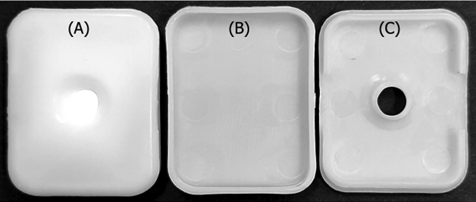
The vertical flow-through test device, designated AcDIGFAAg, to allow rapid antigen–antibody reaction was assembled and used as per the original design of the study group (Institute of Parasitic Diseases, Zhejiang Academy of Medical Sciences, Hangzhou, PR China). The purified polyclonal antibodies against A. cantonensis (1.0 mg mL−1) was dotted (0.5 μL dot−1) onto a 0.45-μm pore size nitrocellulose membrane (NCM) (Bio-Rad Laboratories, Richmond, California). Furthermore, the membrane was blocked with 1% (w/v) bovine serum albumin (BSA) in wash buffer [0.1 m phosphate buffer, pH 6, containing 0.9% (w/v) NaCl and 0.1% (v/v) Tween 20] and incubated at 37°C for 1 h. The dried membrane was laid on the top of water-absorbing pads over the test hole of a flow-through cassette, comprising a rectangular plastic box (30 mm × 40 mm × 6 mm) with a test hole (diameter 10 mm) cut into the centre of the lid (Fig. 1).
The configuration of flow-through DIGFA device. (A) Whole device filled with water-absorbing pads and a porous nitrocellulose membrane coated with antibodies laid on the top; (B) support box; (C) box cover with a hole-opening.

The colloidal gold-labelled anti-31 kDa A. cantonensis polyclonal antibody was prepared according to the standard method by Serve Science Co., Ltd, Bangkok, Thailand. This anti-31 kDa A. cantonensis polyclonal antibody-coated colloidal gold probe was stored at 4°C.
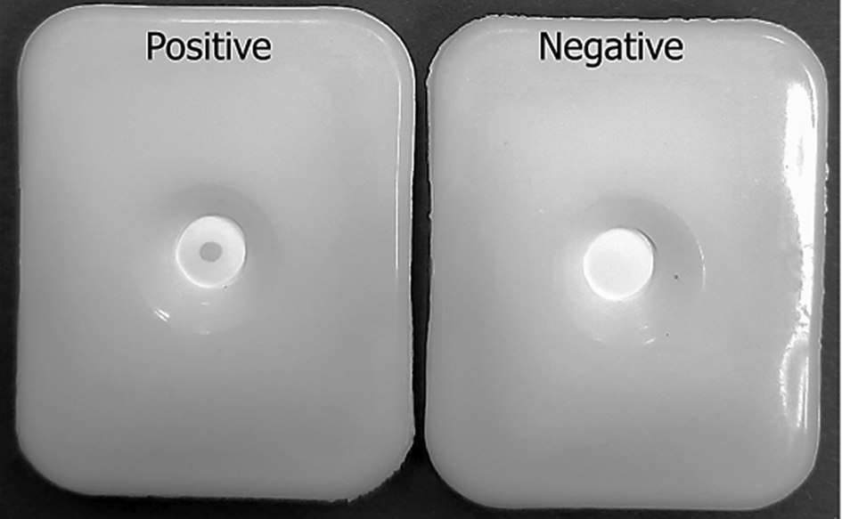
The AcDIGFAAg Test was performed at room temperature. At first, one drop (about 50 μL) of 2% BSA in Tris buffer (140 mm NaCl, 10 mm Tris–HCl, pH 7.4) was applied to the centre of the device test hole onto the NCM in the well to activate the membrane. Then, 25 μL of test CSF/serum was dripped slowly into the test hole until completely infiltrated. The membrane was subsequently washed with a drop of Tris buffer. After washing, one drop of the gold-labelled anti-31 kDa A. cantonensis polyclonal antibody conjugate was added and allowed to be absorbed through the membrane. This was followed by a final wash with a drop of Tris washing buffer. The appearance of a reddish dot in the hole-opening indicated a positive reaction, whereas the absence of such a dot was an indication of a negative result (Fig. 2). Each CSF/serum sample was tested twice to confirm the reproducibility of the results.
AcDIGFAAg Test with positive and negative results.

Results and discussion
Various circulating antigens of A. cantonensis in human angiostrongyliasis sera and CSF (and sera of infected animals) had been demonstrated by sandwich ELISA (Eamsobhana et al., Reference Eamsobhana, Mak and Yong1995, Reference Eamsobhana, Yong, Mak and Wattanakulpanich1997; Chye et al., Reference Chye, Yen and Chen1997; Chen et al., Reference Chen, Wang, Ai, Yan, Peng and Zhang2011), dot-blot ELISA (Eamsobhana et al., Reference Eamsobhana, Yong, Mak and Wattanakulpanich1997), enzyme-linked fluorescent assay (Shih and Chen, Reference Shih and Chen1991), lateral flow immunoassay (Chen et al., Reference Chen, Chen, Chen, Huang, Ai and Zhang2016), ELISA and Western blotting (Liang et al., Reference Liang, Huang, Pan and Tan2005).
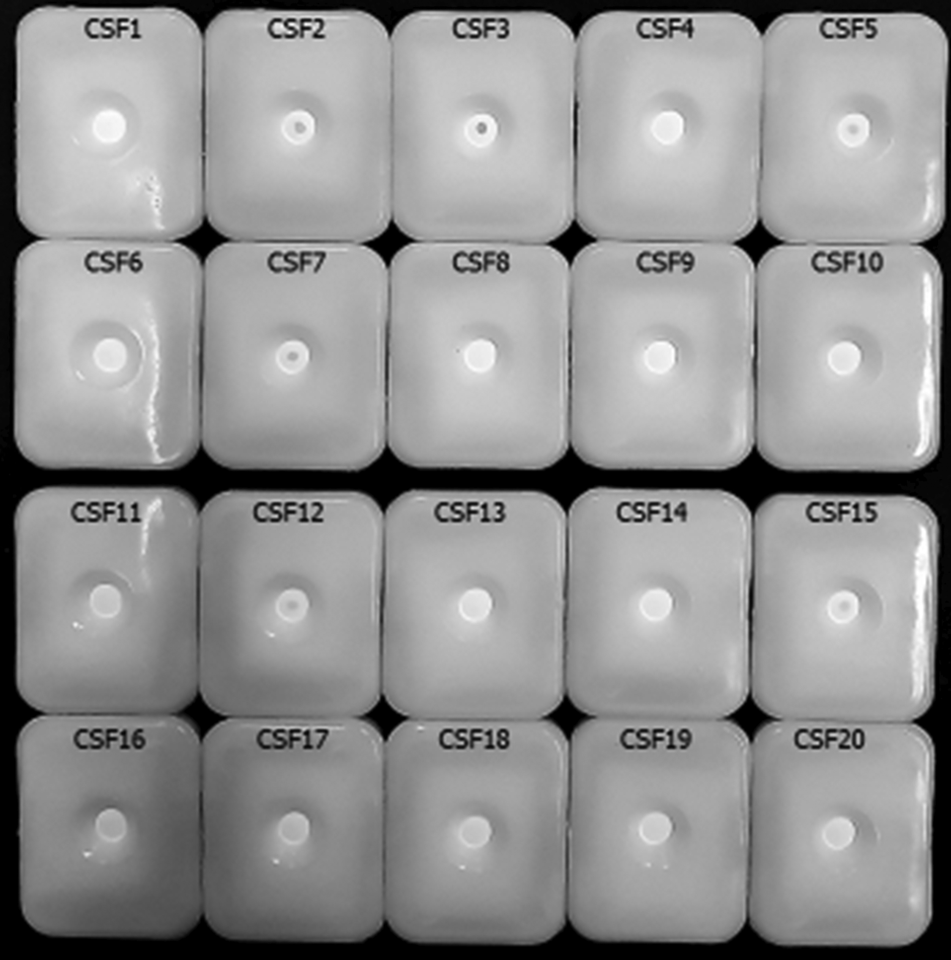
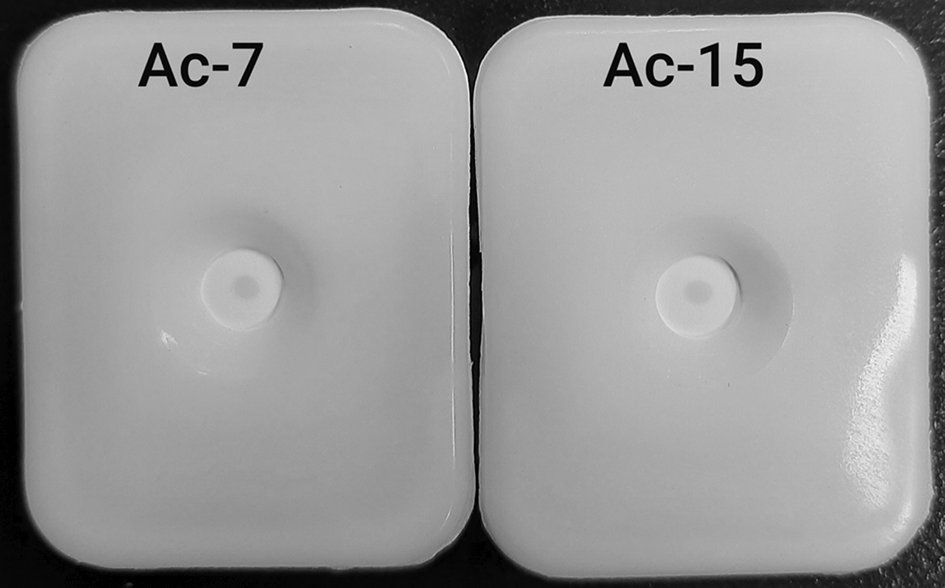
In the present study, positive AcDIGFAAg Test with well-defined, positive pink dots was detected in four (CSF2, CSF3, CSF5 and CSF7) of the 10 CSF samples from patients showing clinical criteria of eosinophilic meningitis and positive results for a 31-kDa A. cantonensis-specific immunoblot band, and two (CSF12 and CSF15) of 5 CSF samples from cases with clinical features of infection, but were negative for A. cantonensis-specific antibodies (Fig. 3). The specificity of the AcDIGFAAg Test was demonstrated by the absence of the positive colour dots in the CSF samples from patients with cerebral gnathostomiasis (CSF16 and CSF17) and neurocysticercosis (CSF18, CSF19 and CSF20). Among 19 patient sera with A. cantonensis infection, two (Ac-7 and Ac-15) showed a positive reaction by AcDIGFAAg with visible pale pink dots (Fig. 4). No positive AcDIGFAAg reaction was observed in the other 17 sera with angiostrongyliasis as well as 78 sera with other parasitic diseases, and the healthy control subjects.
The AcDIGFAAg detection results of 20 CSF samples from clinically diagnosed cases with positive immunoblot test for the presence of a 31-kDa band specific for A. cantonensis (CSF1–10), clinically suspected cases with negative immunoblot test for A. cantonensis infection (CSF11–15), and CSF samples with positive immunoblot test showing a 24-kDa band specific for G. spinigerum (CSF16 and CSF17), and with ELISA-positive cases of T. solium neurocysticercosis (CSF18–20). The pink dot in the centre of the opening indicates a positive sample (CSF2, CSF3, CSF5, CSF7, CSF12 and CSF15), compared with a negative sample (CSF1, CSF4, CSF6, CSF8–11, CSF13, CSF14 and CSF16–20) without a pink dot.

AcDIGFAAg Test results of the two positive serum samples (Ac-7 and Ac-15) for the detection of specific antigens against the 31-kDa antibodies of A. cantonensis.

In recent years, more and more on-site, gold-based immunological techniques based on vertical flow assay (DIGFA) and lateral flow assay (immunochromatographic test (ICT)) are gaining interest in the area of clinical diagnostics for infectious diseases as a rapid, low-tech robust assay for applications at point-of-care settings, and in remote and/or resource-poor environment (Jiang et al., Reference Jiang, Ahmed, Damayantharan, Unal, Butt and Yetisen2019). In comparison with ICT, DIGFA is more economical to prepare to make its use potentially attractive in developing countries. In this work, DIGFA was chosen instead of ICT because the manufacturing of lateral flow chromatographic strip requires expensive equipment; whereas DIGFA device is cheaper and easier to manufacture by hand compared with ICT devices. DIGFA device consists of only a porous membrane (where specific reagents are immobilized) and an absorbent pad, put together into a plastic cassette (Fig. 1).
Theoretically, monoclonal antibodies (MAbs) with defined specificities can be used as immunodiagnostic reagents for the detection of specific parasite antigens during the active phase of the disease. In our previous work, a specific AW-3C2 MAb was evaluated in sandwich and dot-ELISA with 100% specificity to detect specific A. cantonensis circulating antigen in serum samples of CNS angiostrongyliasis patients (Eamsobhana et al., Reference Eamsobhana, Mak and Yong1995, Reference Eamsobhana, Yong, Mak and Wattanakulpanich1997). Despite its high specificity, the use of the AW-3C2 MAb did not help to achieve the required sensitivity in real clinical samples and so the developed tests had little practical importance. Recently, a rapid test based on lateral flow immunoassay using 2 specific MAbs (12D5C12 and 21B7B11) has been described with high diagnostic sensitivity (91.1%) and specificity (100%) to detect circulating antigens of A. cantonensis in sera and CSF of patients with eosinophilic meningoencephalitis (Chen et al., Reference Chen, Chen, Chen, Huang, Ai and Zhang2016). Moreover, the molecular approach that targets genetic sequences of A. cantonensis can also assist an early aetiologic diagnosis. A DNA-based diagnostic technique that relies on polymerase chain reaction (PCR) to amplify and detect specific DNA molecules has been successfully applied to detect A. cantonensis DNA in CSF in 4 out of 10 immunoblot-positive test samples (Eamsobhana et al., Reference Eamsobhana, Wanachiwanawin, Dechkum, Parsartvit and Yong2013). Although detection of parasite DNA in clinical specimens can provide an early detection of the disease, PCR requires bulky and expensive equipment and trained personnel to operate, as well as time-consuming (Verweij and Stensvold, Reference Verweij and Stensvold2014). Currently, driven by a prospect for more rapid, inexpensive, on-site and portable testing, a sandwich DIGFA (designated ‘AcDIGFAAg’) to detect circulating 31-kDa A. cantonensis-specific antigens was developed in the present work for diagnosis of active/current infection.
The specific 31-kDa glycoprotein antigen has been shown earlier to be specific for angiostrongyliasis cantonensis patients (Eamsobhana and Yong, Reference Eamsobhana and Yong2009; Wilkins et al., Reference Wilkins, Qvarnstrom, Whelen, Saucier, da Silva and Eamsobhana2013). It has been suggested to be a potential immunological marker of early infection since this 31-kDa antigen is a component of A. cantonensis excretory/secretory products (Eamsobhana and Dechkum, Reference Eamsobhana and Dechkum2000). Detection of anti-31 kDa A. cantonensis antibody produced in response to the parasite infection could be performed on serum specimens using our earlier developed AcDIGFA Test (Eamsobhana et al., Reference Eamsobhana, Gan, Ma, Wang, Wanachiwanawin and Yong2014, Reference Eamsobhana, Prasartvit, Gan and Yong2015). Nevertheless, in our preliminary test, the same antibody-positive serum set from this group of patients did not have detectable levels of circulating A. cantonensis antigens using the present established AcDIGFAAg Test – all revealed negative test results (data not shown). The explanation for the discordance is likely to be that this batch of samples might be collected during the late phase of the disease, and that the A. cantonensis antigen levels may be masked by circulating antibodies and/or cleared from the circulation. Detection of antigen has the potential to become positive for the active clinical stage of infection earlier than serology or prior to the antibody development.
In this study using a set of patient sera from the reference stock revealed 2 out of 19 angiostrongyliasis serum samples to be positive by AcDIGFAAg Test. This set of archived serum samples had been successfully tested by AcQuickDx to detect the 31-kDa specific antibody against A. cantonensis with 100% sensitivity and 98.72% specificity (Eamsobhana et al., Reference Eamsobhana, Tungtrongchitr, Wanachiwanawin and Yong2018). A tentative explanation of the low sensitivity of AcDIGFAAg in this study could possibly be due to relevant amounts of circulating antigens and antibodies forming antigen–antibody complexes, which may inhibit the detection of antigens in the sera. Nevertheless, all 78 sera from patients with gnathostomiasis, toxocariasis, trichinellosis, hookworm infection, filariasis, cysticercosis, paragonimiasis, opisthorchiasis, malaria, and samples from normal healthy subjects, tested for potential cross-reactions were all AcDIGFAAg negative indicating a high specificity of the AcDIGFAAg Test for detection of circulating A. cantonensis antigens.
CSF is a common type of sample available from patients with suspected cerebral angiostrogyliasis. In our previous report using a genus-specific, standard PCR for the detection of parasite DNA followed by sequencing analysis, we found A. cantonensis DNA in CSF specimens from 4 out of 10 serologically confirmed CNS angiostrongyliasis cases (Eamsobhana et al., Reference Eamsobhana, Wanachiwanawin, Dechkum, Parsartvit and Yong2013). Since the somatic and metabolic antigens of A. cantonensis are more abundant in human CSF than in the serum, the same batch of these clinical CSF samples was retested to assess the validity of the AcDIGFAAg Test. The two methods agreed well for all the positive-PCR samples analysed. Four out of the 10 CSF samples with serologically confirmed angiostrongyliasis were detected to be both positive by the A. cantonensis PCR amplification and AcDIGFAAg Test. The difference is that AcDIGFAAg could confirm 2 additional positive cases from 5 samples with only clinical criteria of eosinophilic meningitis, but were negative for A. cantonensis-specific antibodies, whereas PCR did not reveal any amplified 280-base pairs reactive/diagnostic band in all the 5 patients (Eamsobhana et al., Reference Eamsobhana, Wanachiwanawin, Dechkum, Parsartvit and Yong2013). These 2 positive samples from eosinophilic meningitis patients might be obtained during the active clinical stage of A. cantonensis infection without the production of antibodies. Based on our results, the positivity rate to detect A. cantonensis antigens by AcDIGFAAg was greater than the detection rate of A. cantonensis DNA using a genus-level standard PCR. This is most likely due to a low detection limit of the conventional PCR method. Nevertheless, the present study showed that AcDIGFAAg exhibited a similar diagnostic specificity as the conventional PCR test since no positive test results were observed with CSF samples from other clinically related patients with cerebral gnathostomiasis and neurocysticercosis.
Nonetheless, this study is limited by the fact that neither the AcDIGFAAg Test nor the previously reported PCR–DNA sequencing method (Eamsobhana et al., Reference Eamsobhana, Wanachiwanawin, Dechkum, Parsartvit and Yong2013) could detect circulating A. cantonensis antigens/DNA in CSF samples from all 10 patients with serologically confirmed angiostrongyliasis (with a 31-kDa A. cantonensis-specific immunoblot band). It should be pointed out that, in the present study, only well-defined coloration dot has been considered as positive. The development of very light coloration of the dot by AcDIGFAAg has been considered negative; it may, however, suggest the presence of Angiostrongylus antigens in very low quantity. The initial data (not shown) from our laboratory showed that the minimal readable concentration of the A. cantonensis antigens in phosphate-buffered saline was around 0.01 μg mL−1. Since the method is semi-quantitative, the interpretation of results may be difficult when the antigen concentration of the test sample is close to the cut-off level. Moreover, circulating A. cantonensis antigen has the potential to be present in the acute phase of the disease, prior to the development of antibodies. It is likely that these clinical samples might be obtained in the late phase of infection, and the parasite antigens are degraded and/or cleared from the circulation/CSF. Consequently, additional CSF samples from A. cantonensis-infected patients for whom complete clinical data are available should be evaluated. Likewise, the lack of DNA amplification in 6 out of 10 patients, despite parameters suggesting a high probability of A. cantonensis infection, may represent false-negative results (Eamsobhana et al., Reference Eamsobhana, Wanachiwanawin, Dechkum, Parsartvit and Yong2013). It is possible that the detection rate of parasite DNA in the CSF could be greater using more sensitive PCR-based methods, such as real-time PCR assay (Qvarnstrom et al., Reference Qvarnstrom, Xayavong, da Silva, Park, Whelen, Calimlim, Sciulli, Honda, Higa, Kitsutani, Chea, Heng, Johnson, Graeff-Teixeira, Fox and da Silva2016). Additionally, data from these studies also support a growing body of evidence that to accurately diagnose CNS angiostrongyliasis, the results of at least two standardized tests with different principles should be coincidently positive. If a discrepancy occurs with a positive and a negative test, a third test should be conducted to confirm or rule out infection.
In conclusion, it is apparent that our developed AcDIGFAAg Test holds considerable promise for the rapid detection of active A. cantonensis infection (circulating antigens in the sera) in humans. The assay does not require any sophisticated/specific instruments and can be applied with ease. It is therefore most adaptable for use in the field. More importantly, the availability/use of rapid diagnostic test ‘AcDIGFAAg’ that detects circulating A. cantonensis antigen in the patient sera (acute infection phase), together with the earlier reported ‘AcDIGFA’ (Eamsobhana et al., Reference Eamsobhana, Gan, Ma, Wang, Wanachiwanawin and Yong2014) for A. cantonensis antibody detection (late infection phase) will render a more accurate diagnosis that substantially improves the quality of care and treatment in an infected patient with A. cantonensis. Nonetheless, due to difficulty in obtaining the confirmed cases at an early stage of Angiostrongylus infection, the present study was based on a rather small number of samples from patients. Therefore, further evaluation with a greater panel of clinical CSF as well as serum samples for its diagnostic value is warranted.
Acknowledgements
The authors thank the Director of the Siriraj Hospital, Faculty of Medicine Siriraj Hospital, Mahidol University for permission to publish this research work. We also thank Nuntiya Monkong and Suphaluck Wankhom for assistance in the retrieval of CSF samples kept at the Department of Parasitology, Siriraj Hospital, and Sudarat Boonyong for her help with antibody purification. The constructive comments and valuable suggestions from the editor and two anonymous reviewers have helped to improve this manuscript.
Ethical standards
This study was approved by the Ethics Committee of the Faculty of Medicine Siriraj Hospital, Mahidol University (Si369/2015).
Financial support
This study was supported in part by the Department of Disease Control, Ministry of Public Health, Thailand, and the University of Malaya (YHS: grant H-5620009).
Conflicts of interest
None of the authors have any conflict of interest in this study.