Introduction
The differential diagnosis of childhood stridor includes congenital abnormalities of the upper and lower airway, inflammatory and neoplastic lesions, vocal fold paralysis, foreign bodies, and vascular lesions.Reference Horak, Bodner, Gassner, Schmid, Simma and Grassl1 Herein, we describe the unusual case of an infant whose airway symptoms were the result of a bronchogenic cyst.
The symptoms of bronchogenic cysts are usually due to a mass effect, with those near the carina producing the most symptoms and thus being the most commonly reported.Reference Estrera, Landay and Pass2 Common symptoms include cough, wheezing, dyspnoea, dysphagia, chest pain and stridor.Reference Williams and Johnson3 Symptoms may result from secondary infection, especially where a patent connection to the airway is present.Reference Yerman and Holinger4 However, up to a third remain asymptomatic.Reference Horak, Bodner, Gassner, Schmid, Simma and Grassl1 A combination of rigid airway endoscopy and computed tomography (CT) or magnetic resonance imaging (MRI) can provide information regarding the cyst location and the degree of upper airway narrowing.Reference Lazar, Younis and Bassila5
Once the diagnosis of bronchogenic cyst is suspected, total surgical excision should be undertaken even in an asymptomatic child, to avoid the possible development of cardio-respiratory compromise.Reference Stewart, Cochran, Iglesia, Speights and Ruff6
To our knowledge, the patient presented below represents the first reported case of a bronchogenic cyst presenting with airway obstruction and a cervical mass.
Case report
A six-month-old boy was admitted, via the emergency department of his local hospital, with a neck lump and a provisional diagnosis of croup. He had been born at full term, with no signs of respiratory compromise. His parents reported that he had had a persistent cough for the preceding three months, which was unrelated to feeding but accompanied by inspiratory stridor (during coughing attacks). The parents described one cyanotic episode; this had resolved spontaneously, requiring no intervention. Between coughing attacks, the child maintained a steady respiratory rate, but was noted to have subcostal recession and mild inspiratory stridor.
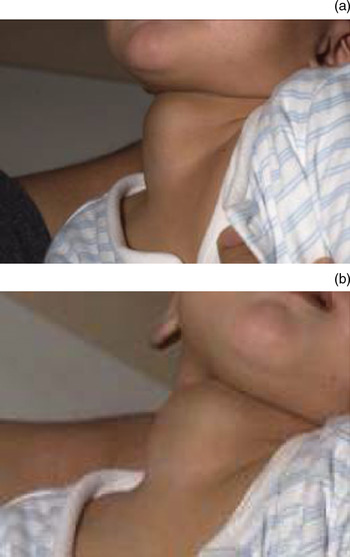
On examination, a 5 cm mass was noted on the right side of the child's lower neck (Figure 1).
Clinical photographs taken pre-operatively.

He was subsequently transferred to our tertiary referral centre.
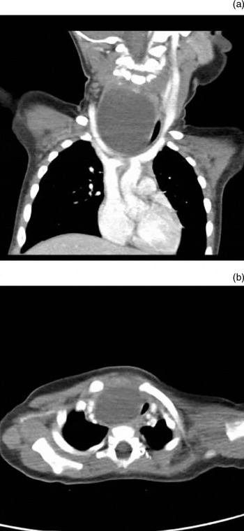
Computed tomography scanning of the neck and thorax demonstrated a 6 × 4.2 cm, unilocular cyst extending from just above the carina to above the thoracic inlet. Significant tracheal narrowing and deviation to the left were noted, along with dramatic splaying of the great vessels from their point of origin. The lesion displaced the sternocleidomastoid muscle, common carotid artery and jugular vein to the right, and the thyroid lobe to the left (Figure 2).
(a) Coronal computed tomography (CT) image showing marked external compression and deviation of the trachea. (b) Axial CT image at the level of the thoracic inlet.

Based on the clinical picture and the radiological findings, a working diagnosis of cystic hygroma was established.
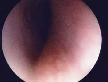
In order to assess the mass effect of the cyst on the airway, and to aid anaesthetic planning, rigid airway endoscopy was performed (Figure 3). This showed that the larynx was displaced to the left, but that active movements of the vocal folds were present and symmetrical. The immediate subglottis was normal, but the airway quickly became laterally compressed, and remained compressed until approximately 2 cm above the carina. The carina and the main bronchi appeared normal. There was significant thoracic extension, which resulted in dramatic splaying of the great vessels from their point of origin.
Pre-operative rigid airway endoscopic view showing significant tracheal compression.

On induction of anaesthesia, significant prolapse of the mass through the thoracic inlet was noted on inspiration.
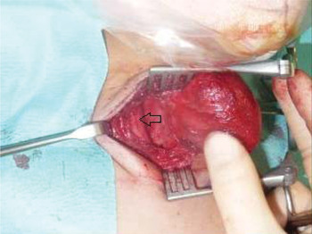
At operation, with thoracic surgical support available, the lesion was approached via a cervical incision. The patient was positioned with the neck extended. Dissection of the lesion capsule allowed delivery of the lesion into the neck (Figure 4). All major neurovascular structures were preserved, and there were no post-operative complications.
Intra-operative photograph showing delivery of the cystic lesion into the neck. The carotid sheath is seen laterally (arrow).

Post-operative rigid airway endoscopy revealed a good calibre airway to the carina, with normal, symmetrical movement of both vocal folds.
Successful extubation was performed 11 hours after the procedure, and feeding was resumed after 24 hours. The child was discharged 9 days post-operatively.
At the time of writing, the child remained well with no evidence of recurrence.
Histological analysis revealed a cystic specimen, comprising a respiratory-type, ciliated, columnar epithelium lined cyst with underlying mucous glands and focal cartilage, along with bundles of smooth muscle. All features were consistent with a diagnosis of bronchogenic cyst.
Discussion
Bronchogenic cysts are remnants of the primitive foregut, which forms between 26 and 40 weeks' gestation.Reference Ombrellaro, Feola, Goldthorn and Shires7 They represent abnormal budding of the foregut, and contain tissue normally found in the trachea and bronchi.Reference Shanmugam, MacArthur and Pollock8
Bronchogenic cysts account for 10 per cent of all mediastinal masses. They tend to occur more often on the right side, and are more frequent in males.Reference Estrera, Landay and Pass2 Bronchogenic cysts are usually unilocular, filled with clear fluid, and lined by pseudostratified, ciliated, columnar epithelium and other elements of the normal respiratory tract (including cartilage, smooth muscle and glandular tissue).Reference Yerman and Holinger4
Two classification systems exist, based on the location of the lesion. Maier performed the first successful surgical cure of a bronchogenic cyst in an infant in 1948, and classified these cysts as paratracheal, carinal, hilar or para-oesophageal.Reference Maier9 An alternative system was suggested by Klin et al., whereby the lesion was classified according to its relation to the trachea, into paratracheal, intraluminal and intramural types.Reference Klin, Springer, Segal, Eshel, Tabachnik and Vinograd10
Bronchogenic cysts have seldom been reported as a cause of infantile stridor.Reference Lazar, Younis and Bassila5, Reference Stewart, Cochran, Iglesia, Speights and Ruff6, Reference Nussenbaum, McClay and Timmons11–Reference Lai, Nguyen, Kim and Campisi14 The available consensus is that surgical excision is the treatment of choice, in order to prevent the morbidity and mortality associated with infection and the mass effect which characterises such cysts.
Lazar et al. described a case of a child in respiratory distress at birth, requiring emergency intubation.Reference Lazar, Younis and Bassila5 Immediately after the infant was weaned from the ventilator and extubated, he developed stridor and respiratory distress and was reintubated. Computed tomography scanning revealed a cystic mass extending from the second thoracic vertebra to the level of the carina. Complete surgical excision was performed. Lazar et al. retrospectively assessed their management of 15 other patients with respiratory distress secondary to bronchogenic cysts. Each was investigated using a combination of chest X-ray, barium swallow and CT scanning. All 15 patients had their cysts excised via a thoracotomy approach. The authors concluded that CT or MRI imaging should be performed in cases of paediatric stridor unresponsive to medical management, and that, once diagnosed, bronchogenic cysts should be promptly excised.
Other single case reports have also been published (Table I).
Case reports detailing bronchogenic cyst treatment

CT = computed tomography; mth = months; F = female; yr = years; M = male; NA = not announced; wk = weeks
Although bronchogenic cysts are a relatively rare cause of infantile stridor, their presence should be considered, and investigations performed where appropriate, as surgical excision provides a reliable cure and reduces the risk of future morbidity. The airway symptoms of bronchogenic cyst may alternatively be caused by tracheobronchomalacia (due to external compression) or cervical cystic hygroma.
We are unable to explain our patient's consistent, documented inspiratory stridor, in the presence of significant tracheal compression which one would expect to cause an expiratory component to the airway noise. The child's stridor was considered to be mild in severity, and this may have hindered detection of any expiratory airway noise.
• Bronchogenic cysts are a rare cause of infantile stridor, and may rarely present initially with a cervical mass
• Bronchogenic cysts are a rare differential diagnosis for cervical cystic lesions
• Surgical excision is the treatment of choice
To our best knowledge, this case represents the first report of a bronchogenic cyst presenting as a neck mass with airway symptoms, and highlights this lesion as a differential diagnosis for a cystic neck mass in an infant.