Abnormalities of the aortic arch present symptomatically in a variety of ways, often because rings are produced that cause tracheal obstruction and respiratory distress.Reference Humphrey, Duncan and Fletcher1 We describe here a very rare form of vascular ring. It was produced by the combination of a left-sided aortic arch, a right-sided descending aorta, and a persistently patent right-sided arterial duct associated with interruption of the aortic arch. An Abbott’s artery was also present, which crossed retro-oesophageally to the right, and connected to the right-sided descending aorta.
Case report
A male infant, aged 10 days, who weighed 3050 grams at birth, was referred to us after an uneventful, full term gestation and normal delivery. He was admitted to the hospital with respiratory distress. On physical examination, we found dyspnoea, retractions, a systolic murmur graded 2 from 6, and peripheral cyanosis. The pulses in the legs and right arm were palpable but weak, but no abnormal sounds were heard on auscultation of the lungs. A chest x-ray revealed moderate cardiomegaly, with increased pulmonary markings. An electrocardiogram showed right ventricular hypertrophy. A transthoracic echocardiogram revealed an unrestrictive subaortic ventricular septal defect, pulmonary hypertension, and severe aortic coarctation.
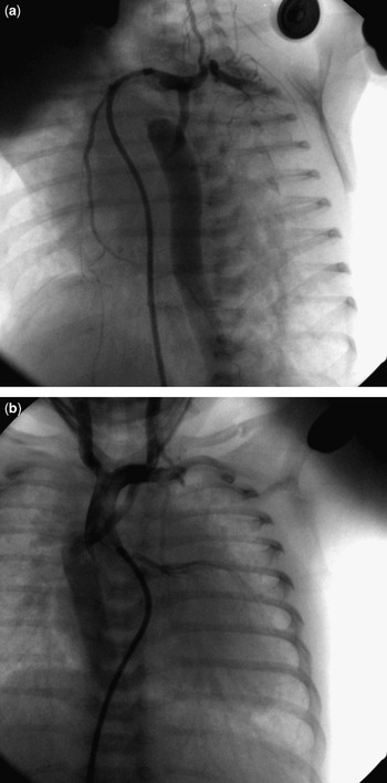
Urgent cardiac catheterization confirmed the presence of the ventricular septal defect, and revealed the aortic arch to be left-sided, but with the aorta descending on the right side. It also showed interruption of the aortic arch with a tiny persistently patent right-sided arterial duct, and Abbott’s artery, which originated from the left subclavian artery and connected to the right-sided descending aorta (Figs 1a and 1b). The right subclavian artery could not be visualized. The patient developed respiratory arrest immediately after cardiac catheterization and required intubation, mechanical respiratory support, and cardiac inotropic support. Because of this, we planned for urgent surgical intervention.
The catheter crosses to the ascending aorta from the right ventricle through a ventricular septal defect. An oblique view is shown of an aortic root angiogram (a). After the injection, it can be seen that the left-sided ascending aorta is interrupted, with connection via Abbott’s artery to the right-sided descending aorta. Contrast also passes in retrograde fashion from the persistently patent right-sided arterial duct. In panel b, we show a frontal view of the aortic root angiogram. After the injection, it can again be seen that the left-sided ascending aorta is interrupted, and is connected with the right-sided descending aorta through Abbott’s artery.

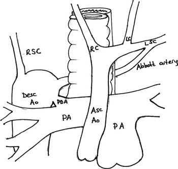
Surgery was performed through a median sternotomy. The aortic arch and its branches, specifically the right common carotid, left common carotid, and left subclavian arteries, were all identified, along with Abbott’s artery, which crossed to the right in retro-oesophageal fashion to connect with the descending aorta on the right. We also found that the right subclavian artery originated from the descending aorta (Fig. 2). Under cardiopulmonary bypass and moderate hypothermia, the patent arterial duct was divided. Abbott’s artery was divided at both its proximal and distal ends. The descending aorta was anastomosed to the ascending aorta in end-to-side fashion, and the pulmonary trunk was banded. Unfortunately, the patient developed thrombocytopenia within the frame of a sepsis syndrome, and died on the eighth day after surgery.
The cartoon shows a schematic view of the vascular ring formed in our patient.

Discussion
Anomalies of the aortic arch and its branches are well recognized to compress the trachea and oesophagus, and are commonly known as vascular rings. All such anomalies, however, do not form true rings, so the term is best applied when the blood vessels encircle and compress the trachea and oesophagus.Reference Roberts, Othersen, Sade, Smith, Tagge and Crawford2
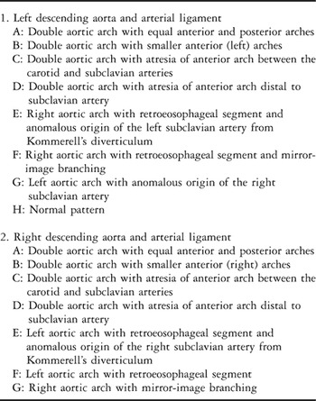
The anomalies are due to defective disappearance on one or other side of the arteries of the fourth and, exceptionally, the third primary branchial arches, and also of the appropriate parts of the paired dorsal aortas.Reference Edwards3 The multiple combinations are readily visualized and understood using the hypothetical scheme of Edwards,Reference Edwards3 consisting of an ascending aorta, right and left aortic arches, a descending aorta on either right or left, and bilateral arterial ducts (Table 1). One of these possibilities is a left-sided aortic arch with retro-oesophageal course to supply as right-sided descending aorta. This can be categorized as group II, type E, in the system of Edwards, and our case fits with this grouping apart from the additional presence of interruption of the aortic arch. The combination of left-sided aortic arch, right-sided descending aorta and right-sided persistently patent arterial duct is rare, and association with interruption of the retro-oesophageal segment of the arch is extremely rare.Reference Planche, Roussin, Paul, Belli and Serraf4, Reference Camilleri, Legault, Boyer, Bailly, Lusson and de Riberolles5 Our case is unique in that we also discovered the presence of Abbott’s artery. This is an anomalous artery that arises from the posterior wall of the aortic arch, or from the subclavian artery within a radius of about 2 centimetres from its origin.Reference Planche, Roussin, Paul, Belli and Serraf4–Reference Lerberg6 The artery was first described by Maude Abbott in 1928.Reference Hamilton and Abbott7 She suggested that the artery might represent the evanescent remnant of the fifth arch, but others suggest that it might be an enlarged bronchial artery arising just proximal to the coarctation.Reference Schuster and Gross8 Since there is no evidence for existence of a fifth arch during normal development, the latter seems a more likely explanation.
Anomalies of the aortic arch (From Edwards, JE.: Anomalies of the derivatives of the aortic arch system. Med Clin North Am 1948; 32: 925–949).

Both the clinical presentation, and the surgical approaches, to complex malformations of the aortic arch vary depending on the specific lesion, and the presence of associated congenital cardiac disease. Clinical manifestations of tracheal and oesophageal compression are wheezing, respiratory distress, stridor, dysphasia, feeding difficulties, cyanosis, vomiting and/or arterial insufficiency.Reference Humphrey, Duncan and Fletcher1, Reference Roberts, Othersen, Sade, Smith, Tagge and Crawford2 Early diagnosis and treatment are important in order to reduce morbidity and mortality due to the possible development of infectious complications.Reference Arciniegas, Hakimi, Hertzler, Farooki and Green9 The methods of diagnosis and treatment are standard in many centers, although preference varies among clinicians. Echocardiography may be useful in diagnosing a vascular ring in infants and children but, in particular cases, this may fail to detect the specific lesion. Computed tomography with or without three-dimensional reconstruction offers technical advantages. Although previously published reports underscore the importance of preoperative cardiac catheterization in evaluating patients suspected of having vascular ring, we believe it is very important correctly to define the anatomy of the aortic arch, its branches, and any associated cardiac defects, and also to choose the best surgical approach. Our patient had a true vascular ring surrounding the trachea and oesophagus, with Abbott’s artery presenting as a small arterial canal linking the ascending aorta to the descending aorta, albeit permitting limited flow.
In the light of all reported experiences, all symptomatic cases, with or without complete rings, need surgical intervention. The type of surgery depends on the vascular anomaly and whether associated conditions are present. In our patient, we performed extensive dissection of the descending aorta and achieved an end-to-side anastomosis of the descending to ascending aorta, having divided the patent arterial duct and Abbott’s artery, and banded the pulmonary trunk, but sadly without a successful outcome.