Introduction
Urinary schistosomiasis caused by the trematode parasite Schistosoma haematobium, is endemic throughout the states of the Nigerian Federation (Awogun, Reference Awogun1990). Freshwater pulmonate snails of the genus Bulinus act as the intermediate hosts for S. haematobium and related species and occur commonly throughout much of Africa and adjacent regions. Knowledge of the geographical distribution of Bulinus species present in Nigeria as well as their frequency of occurrence is key in the design of schistosomiasis treatment and control programmes. Linked to information concerning the level of parasite infection, such data can be valuable in the assessment of likely levels of disease in a particular region and may also highlight areas that might be at greater risk of infection.
There are currently 37 recognized species of Bulinus (Brown, Reference Brown1994) but the specificity of the snail–parasite interaction is such that only certain species are involved in transmission of the parasite. The genus can be divided into four major groups of species, namely, the Bulinus africanus group, the Bulinus forskalii group, the Bulinus reticulatus group and the Bulinus truncatus/tropicus complex. Within each group there are species that act as intermediate hosts for schistosomes in part or all of their geographical range (Brown, Reference Brown1994). Limited morphological distinctiveness within species groups can hinder identification but, in contrast, there is considerable molecular divergence within the mitochondrial cox1 gene (Jones et al., Reference Jones, Rollinson, Mimpfoundi, Ouma, Kariuki and Noble2001; Stothard et al., Reference Stothard, Brémond, Andriamaro, Sellin, Sellin and Rollinson2001) which may be employed to produce clear phylogenetic resolution both between and within these groups. A recent study (Kane et al., Reference Kane, Stothard, Emery and Rollinson2008) took advantage of this situation to generate a phylogeny of the four species groups based on 81 partial cox1 sequences. The latter were lodged as reference material with the European Molecular Biology Laboratory (EMBL) sequence database, allowing the current investigation to perform BLAST searches (Altschul et al., Reference Altschul, Gish, Miller, Myers and Lipman1990) via the National Center for Biotechnology Information and this facilitated initial species identification. Additionally, phylogenetic analysis was performed through the inclusion of representative Nigerian cox1 sequences with those already submitted to EMBL and this provided definitive categorization of the snail species.
Nigeria is a country known to be highly endemic for urinary schistosomiasis, with an estimated 101.28 million persons at risk and 25.83 million people infected (Chitsulo et al., Reference Chitsulo, Engels, Montresor and Savioli2000). Studies among school-aged children in various parts of the country, in both rural and urban environments, have shown that S. haematobium is clearly a problem for this age group, with prevalence ranging from 20 to 40% in typical communities (Okpala, Reference Okpala1961; Amali, Reference Amali1988; Anosike et al., 1992, Reference Anosike, Okafor and Onwuliri2003; Agi, Reference Agi1995; Okoli & Odaibo, Reference Okoli and Odaibo1999; Ekejindu et al., Reference Ekejindu, Ekejindu and Agbai2002; Odaibo et al., Reference Odaibo, Adewunmi and Olorunmola2004; Okoli & Iwuala, Reference Okoli and Iwuala2004; Umar & Parakoyi, Reference Umar and Parakoyi2005) but can be as high as 50–70% in areas where environmental changes occur due to constructions such as man-made dams and quarries (Pugh & Gilles, Reference Pugh and Gilles1978; Edungbola et al., Reference Edungbola, Asaolu, Omonisi and Aiyedun1988; Ogbe & Olojo, Reference Ogbe and Olojo1989; Ofoezie et al., Reference Ofoezie, Imerbore, Balogun, Ogunkoya and Asaolu1991; Nduka et al., Reference Nduka, Ajaero and Nwokel995, Reference Nduka, Etusim, Nwaugo and Oguariri2006; Mafiana et al., Reference Mafiana, Ekpo and Ojo2003; Sarkinfada et al., Reference Sarkinfada, Oyebanji, Sadiq and Ilyasu2009). Focal distribution of freshwater snails belonging to the genus Bulinus has been reported in many parts of the country (Okwuosa & Ukoli, Reference Okwuosa and Ukoli1980; Imevbore et al., Reference Imevbore, Ofoezie and Obot1988; Ofoezie, Reference Ofoezie1999; Owojori et al., Reference Owojori, Asaolu and Ofoezie2006). Ndifon & Ukoli (Reference Ndifon and Ukoli1989) observed the presence of 14 snail species from diverse freshwater habitats in south-western Nigeria: nine pulmonates (Biomphalaria pfeifferi, B. globosus, B. truncatus, Lymnaea natalensis, Physa sp., Aplexa waterloti, B. forskalii, Gyraulus costulatus, Ferrissia sp., Segmentorbis sp.) and five prosobranchs (Lanistes libycus, L. ovum, Pila wernei, Potadoma moerchi and Melanoides tuberculata). They also observed that among the intermediate hosts of S. haematobium, B. globosus was widely distributed and common, while B. truncatus was rare. Nale et al. (2003) reported the presence of B. globosus and B. truncatus in the River Kibanni, at Zaria, north-western Nigeria, while Betterton et al. (Reference Betterton, Ndifon, Bassey, Tan and Oyeyi1988) investigated 165 freshwater habitats throughout Kano state, north-western Nigeria and reported the presence of a number of potential snail intermediate host species, namely Bulinus senegalensis, B. forskali, B. globosus, B. truncatus and Bi. pfeifferi, but stated that the most widespread species was B. senegalensis. Ntonifor & Ajayi (Reference Ntonifor and Ajayi2007) revealed five medically important snail species comprising B. globosus, B. truncatus, B. forskalii, Bi. pfeifferi, and Lymnaea natalensis (which may act as a host for human Fasciola gigantica) present in two local government areas of Bauchi State, north-eastern Nigeria. These snail identification studies were based on morphological identification methods. However, Raahauge & Kristensen (Reference Raahauge and Kristensen2000) observed that differentiation of Bulinus species based on the microsculpture of the shell and the penis to preputium proportion is misleading when one wishes to determine correct species identity.
The aim of this study, therefore, was to identify Bulinus snails involved in the transmission of S. haematobium in Nigeria, through the sequencing of an amplicon for part of the mitochondrial cox1 gene and also polymerase chain reaction-restriction fragment length polymorphism (PCR-RFLP) analysis of the ribosomal its so as to provide information that would serve as a basis for informed predictions and decisions on schistosomiasis control strategies in the country. Sequencing in combination with phylogenetic analysis can be an excellent technique for establishing molecular relationships, but it is expensive, time consuming and requires access to sophisticated apparatus such as an automatic sequencer. For this reason, restriction analysis of the ribosomal its using RsaI was undertaken in the manner outlined by Stothard et al. (Reference Stothard, Hughes and Rollinson1996) and used for the majority of samples. Restriction pattern profiles were capable of distinguishing the various species of Bulinus present and high levels of confidence could be placed on these data when a sample typed by sequencing, BLAST searching and phylogenetic techniques was included on each gel.
Materials and methods
Snail isolates
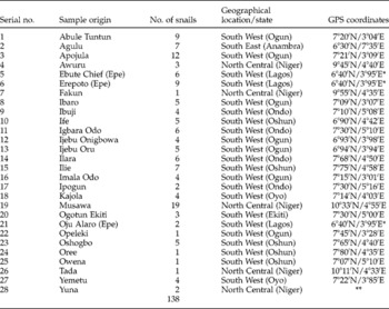
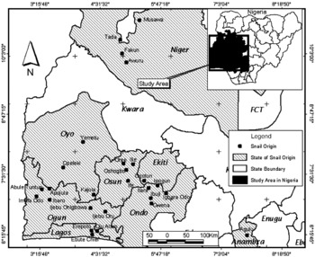
A total of 138 Bulinus snails were collected between May 2007 and June 2009, from 28 sites (table 1) endemic for schistosomiasis in south-west, south-east and north-central Nigeria (fig. 1). All the human–water contact sites in the study areas were identified and searched for snails belonging to the genus Bulinus. The snails were collected using a standard snail scoop, the contents washed and the snails picked manually. Recovered snails were transported to the laboratory in pre-labelled plastic containers, rinsed, sorted and counted. Each snail was identified based on its morphological characteristics using the field guide to African freshwater snails by Kristensen (Reference Kristensen1987). Sequencing of samples was undertaken in conjunction with the Biomedical Parasitology research group at the Natural History Museum in London and methods for DNA extraction and Dra1/its amplification were practised and refined there.
Geographical location/coordinates (where available), description of sample origins and number of snails collected from each site.

* Streets in Epe town, Lagos state. ** Not available.
Map of Nigeria showing the study area.

Genomic DNA extraction from snails
Genomic DNA was extracted from each snail using a modified method of Stothard et al. (Reference Stothard, Hughes and Rollinson1996). Each snail was removed from 70% ethanol and soaked in TE buffer, pH 7.4 (10 mm Tris HCl and 1 mm EDTA) overnight so as to remove the remaining ethanol. Tissue from each of the snails was placed in a sterile 1.5-ml microcentrifuge tube, with 500 μl of CTAB solution (0.2% 2-mercaptoethanol; 2% hexadecyltrimethyl-ammonium bromide (CTAB); 100 mm Tris (hydroxymethyl) amino-methane; 16 mm EDTA; 1.4 m sodium chloride) and the tissue homogenized. Proteinase K solution (10 μl at 20 mg/ml) was added and the digests were incubated at 55°C for 1 h, with occasional gentle mixing. Genomic DNA was extracted from the digests by adding an equal volume of chloroform/isoamyl alcohol (24:1) to each tube. The organic and aqueous layers were gently mixed for 5 min and spun at 13,000 g for 20 min. For each sample, the upper aqueous layer was removed into another sterile microcentrifuge tube and an equal volume of 100% ethanol was added, mixed and the whole incubated at − 20°C overnight in order to enhance DNA precipitation. The precipitate was spun at 13,000 g for 20 min and the pellet washed with 70% ethanol and spun for another 20 min. The supernatant was removed and the pellet was dried at room temperature. When completely dry, the pellet was re-suspended in 50 μl of purified water.
PCR amplification of cox1 and sequencing
A sub-region of the cox1 gene was amplified by PCR in 34 Bulinus samples using the forward primer BulCox12 [BC12] (5′-GCGTTGACTCTTTTCAAC-3′) and the reverse primer BulCox16 [BC16] (5′-CCAACTGTAAATATATGATGAG-3′) and BulCox12 in combination with BulCox6 [BC6] (5′-CAATAAACCCTAAAATYCC-3′) for a single sample from Abule Tuntun. With the exception of primer BC16, all primers and cycling conditions were as described in Kane et al. (Reference Kane, Stothard, Emery and Rollinson2008). Cox1 PCR products were run on 1.0% agarose gels followed by photodocumentation. The cox1 amplicon band was excised from an agarose gel using a scalpel blade and purified employing a QIAquick Gel Extraction Kit (Qiagen, Hilden, Germany). Each sample was quantified using a Nanodrop ND-1000 spectrophotometer (Nanodrop Technologies Inc., Wilmington, Delaware, USA) and sequencing reactions were performed using an Applied Biosystems Big Dye Kit (version 1.1) and then run on an Applied Biosystems 3730 DNA Analyzer (Applied Biosystems, Carlsbad, California, USA). Initial editing of the cox1 sequences was carried out using Sequencher 4.8 software (Gene Codes Corporation, Ann Arbor, Michigan, USA). Each B. truncatus isolate retained 743 nucleotides and the two B. globosus isolates 746 nucleotides of sequence for subsequent analysis and submission to EMBL. The accession numbers for these sequences are FN546781–FN546815. A further two isolates, Agulu1 and 2, were amplified using the Folmer primers (Folmer et al., Reference Folmer, Black, Hoeh, Lutz and Vrijenhoek1994) LCO1490 and HCO2198 and this resulted in two 611 nucleotide sequences which were submitted to EMBL with accession numbers FN547996 and FN547997.
Analysis of cox1 sequence data and haplotype networking
Following basic editing and compilation, the cox1 sequences were used to undertake BLAST searches (Altschul et al., Reference Altschul, Gish, Miller, Myers and Lipman1990). These were necessary in order to ensure that contaminating sequences, such as those from internal/external molluscan parasites and other organisms, had not been obtained in error and also to give a reasonable first estimate of species identity, as it would be expected that close matches with either B. truncatus or B. globosus cox1 sequences submitted to the databanks on previous occasions should be returned. Representative Nigerian Bulinus cox1 sequences were included in a dataset with a further 76 Bulinus sequences obtained formerly and imported into MEGA 4.0 (Tamura et al., Reference Tamura, Dudley, Nei and Kumar2007) where they were aligned using Clustal W (Thompson et al., Reference Thompson, Higgins and Gibson1994). Subsequently, the cox1 data were analysed solely as nucleotides in a neighbour-joining tree (1000 bootstrap replications) employing Kimura's two-parameter model (K2P) for pairwise distance calculations and utilizing transversional substitutions only. The BLAST and phylogenetic analysis was sufficient to predict accurately the species of Bulinus present but, in the case of B. truncatus, was unable to discern any intraspecific structure or lineages. For this reason, a haplotype network was undertaken on the sequenced B. truncatus samples using the program TCS (version 1.21) (Clement et al., Reference Clement, Posada and Crandall2000) to estimate gene genealogies.
PCR-RFLP of ribosomal its
PCR amplification of the ribosomal its region was performed on all 138 snails using primers ETTSl (5′-TGCTTAAGTTCAGCGGGT-3′) and ETTS2 (5′-TAACAAGGTTTCCGTAGGTGAA-3′) (Kane & Rollinson, Reference Kane and Rollinson1994) according to the method of Stothard et al. (Reference Stothard, Hughes and Rollinson1996). Cycling parameters were 95°C for 4 min, followed by four cycles of 95°C for 15 s, 48°C for 15 s and 72°C for 1 min and then a further 41 cycles of 95°C for 15 s, 53°C for 45 s and 72°C for 1 min. A final extension period of 72°C for 3 min was implemented. The amplification was confirmed through visualization on a 1.5% agarose gel. The amplified products were digested with RsaI, a restriction enzyme, following the method of Stothard et al. (Reference Stothard, Hughes and Rollinson1996) and the digest was visualized on a 1.5% agarose gel.
Detection of schistosome infection
All the 138 snails were screened for Schistosoma infection by PCR amplification of the Dra1 repeat using the forward primer, Sh1, 5′-GATCTCACCTATCAGACGAAAC-3′ and reverse primer, Sh2, 5′-TCACAACGATACGACCAAC-3′ following the method of Hamburger et al. (Reference Hamburger, He, Abbasi, Ramzy, Jourdane and Ruppel2001). The amplified products were visualized on 1.5% agarose gel and then recorded by photodocumentation.
Bulinus truncatus can act as an intermediate host to both S. haematobium and also S. bovis, a parasite of ruminants, and as the majority of snails screened in this study were found to be B. truncatus, a trial attempt was made to identify the species of the infecting schistosome(s) within these Nigerian snails. To facilitate this, 11 snails from 7 different States were tested for a fragment containing part of the schistosome ribosomal first internal transcribed spacer (its1), the small 5.8S ribosomal gene and the second internal transcribed spacer (its2). The primers used were ETTS1 (Kane & Rollinson, Reference Kane and Rollinson1994) and ETTS6 (5′-TGTTGAAGCGATCCGGTT-3′). Sequences of the spacer regions within the ribosomal gene units tend to be very specific to the organisms in which they occur. ETTS6 was designed to fit into the schistosome ribosomal its1 to act as a ‘specific’ primer which would only participate in the amplification of schistosome DNA. During this process it was noted that a snail of the species Physa acuta, which had been tested as part of the screening process, was producing amplicons with the Dra1 primers. Physa acuta is known to shed avian schistosomes of Trichobilharzia sp. (Graczyk & Shiff, Reference Graczyk and Shiff2000) but not S. haematobium. A method was devised to try to discover the nature of the parasite present in this snail from Ipogun through amplification of partial cox1 and its PCR fragments. Determination of the infecting schistosome species was undertaken using combinations of the following Schistosoma cox1 primers: Cox1Schist5′ (5′-TCTTTRGATCATAAGCG-3′), CO1560R (5′-GCAGTACCAAATTTACGATC-3′) (Lockyer et al., Reference Lockyer, Olson, Ostergaard, Rollinson, Johnston, Attwood, Southgate, Horak, Snyder, Le, Agatsuma, McManus, Carmichael, Naem and Littlewood2003), RAKCO4F (5′-GTTTGTATTATGGTTGTGG-3′), RAKCO7F (5′-CTATGGGTGGTGGTGATC-3′) and RAKCO1OR (5′-GAACATAATGAAAATGAGC-3′). Additionally, a partial its fragment was obtained using the primers ETTS1 and ETTS6 described previously.
Results
Sequencing analysis and RFLP of the its region
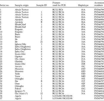
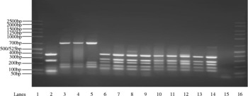
‘BLAST’ searching and phylogenetic analysis of the partial cox1 gene for 37 snails showed that the overwhelming majority (35) were B. truncatus while only two were B. globosus (table 2). The two B. globosus isolates were collected from Ipogun (Ondo state) and Oju Alaro (Lagos state) in the south-western region of Nigeria. All these sequences have been submitted to the EMBL database and their accession numbers are as shown in table 2. PCR-RFLP analysis of the its region from 138 snails confirmed that the dominant species present in those areas surveyed was B. truncatus with only two samples being characterized as B. globosus. A number of its amplicons following restriction with RsaI are displayed in fig. 2. All snails shown in this figure had their species identity confirmed through sequencing but the restriction fragment profiles are sufficiently distinct between the three different species encountered to enable easy identification.
Sample origins of snail species Bulinus truncatus and B. globosus (*).

NA, not applicable.
Gel showing species-specific banding patterns obtained from digestion of the 1.3 kbp PCR product containing the its region using RsaI. Lanes: 1 and 16, size marker (Cambio ladder 50–2500 bp); 2, Bulinus globosus; 3–5, Physa acuta; 6–14, Bulinus truncatus; 15, negative control.

Haplotype network
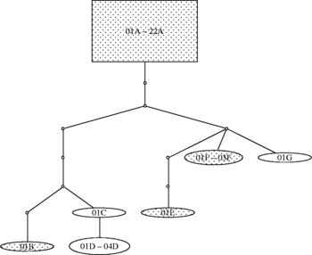
There were seven haplotypes, designated A to G, contained within the network (fig. 3). No clear distinctions were revealed between south-west and north-central Nigeria. Two groupings of haplotypes emerged from the ‘ancestral’ cluster (square box containing 22 sequences). The unlabelled small circles (nodes) correspond to intermediate forms which were inferred by the program but not encountered in the sampling. Each link between the haplotypes represents one mutational difference.
Haplotype network for Nigerian B. truncatus cox1 sequences using the software TCS. Each haplotype is indicated by a letter. Particular samples may be identified through reference to table 2. Haplotypes from south-west Nigeria are indicated in groupings containing spots, while those from north-central Nigeria have no fill. A 95% connection limit was employed in the analysis.

Snail infection status
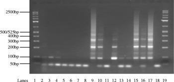
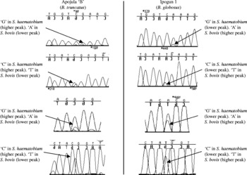
PCR amplification of the schistosome Dra1 repeat (fig. 4) from the genomic DNA of 138 snails showed that 41 (29.7%) from the 23 study sites were positive for schistosome infection. However, certain PCR amplifications using the Dra1 primers resulted in partial, ambiguous profiles, where it was unclear as to whether the snails were only lightly infected or if there had been miracidial penetration of the snail surface but no actual development of the parasite. Lanes 2–4, 13 and 14 demonstrate this situation. The Dra1 repeat is a useful tool to determine whether or not a snail has been exposed to schistosomes; however, it will not inform as to which species of the parasite is responsible. An attempt to establish the species present in these snails was made through the amplification of a part of the schistosome ribosomal its region in a small sample of 11 snails. Amplicons were obtained from four and sequence produced for each. Analysis of the sequence electropherograms for two of the snails showed dual peaks at those locations which are polymorphic between S. haematobium and S. bovis (fig. 5). Further, a snail from Ipogun (lane 10 in fig. 4), typed by sequencing as P. acuta, clearly showed penetration by the parasite and, in order to investigate this feature more fully, schistosome primers for cox1 were used to amplify two weak parasite amplicons from the snail's genomic DNA extract. The fragments were re-amplified using one of the original primers and also different internal primers. Two non-overlapping Schistosoma cox1 fragments were obtained, which, when sequenced, and ‘BLASTed’ typed the infecting DNA as being from S. bovis. However, further work involving partial amplification of the schistosome its showed a similar situation as described in the two B. truncatus isolates mentioned previously, in that dual peaks were observed in the electropherograms at locations polymorphic between S. bovis and S. haematobium, the implication being that Physa, like Bulinus, is either being penetrated by both species of parasite or a hybrid of such. In either circumstance, there may be implications regarding the presence of Physa in zones where S. haematobium or a hybrid of such are transmitted. To complement the apparent penetration of Physa by schistosome miracidia some snails belonging to this genus were collected from Ipogun and examined in the laboratory for shedding cercariae over a period of 21 days. None was found to shed cercariae, but when screened using Dra1-PCR, some gave a positive signal for schistosome DNA.
Agarose gel stained with ethidium bromide showing the infection status of 15 snail samples using the Dra1 repeat. Lanes: 1 and 19, size marker (Cambio ladder 50–2500 bp); 5–8 and 11, uninfected snails; 9, 10, 12, 15 and 16, infected snails; 17, DNA from an adult S. haematobium as a positive control; 18, negative control; 2–4, 13 and 14 showing weak/ambiguous profiles. One of the infected snails was confirmed by sequencing to be B. globosus (lane 9), three were B. truncatus (lanes 12, 15 and 16) and one was Physa acuta (lane 10).

Electropherograms showing additive peaks at positions of nucleotide variation between S. haematobium and S. bovis within the ribosomal its. The profile indicates either a dual exposure of the snails to both species or possibly a hybrid form. The relative peak height between the two isolates is inverted. The reason for this is unknown but, if a dual infection has occurred, it may indicate disproportionate levels of exposure to the two species of parasite. Alternatively, the efficiency of parasite proliferation for either a pure schistosome form or hybrid may depend on the species of molluscan host infected.

Discussion
The majority of Bulinus snails screened belonged to the species B. truncatus with only two being identified as B. globosus, from Ipogun (Ondo state) and Oju Alaro (Lagos state) both from the south-western part of Nigeria. Conchologically the snails can be confused without detailed inspection of the shell microsculpture and collumella regions. Employing a neighbour-joining phylogenetic tree, the B. globosus snails clustered most closely with other West African B. globosus from Niger, Senegal and Burkina Faso. Sampling in the current study was heavily biased towards the south-western region of Nigeria and, in consequence, the small number of B. globosus collected in Ondo and Lagos states might be an aberration of sampling strategy. The PCR-RFLP technique of Stothard et al. (Reference Stothard, Hughes and Rollinson1996) appears to be a rapid and cost-effective method of assessing Bulinus species distribution in Nigeria and would seem to be the way forward for screening snails from other regions of that country.
The use of sequencing for species determination in Nigeria, while very effective, is expensive, slow and should probably only be employed where RFLP banding patterns are atypical or highlight the presence of cryptic species. The restriction profiles for B. truncatus closely match those obtained by Stothard et al. (Reference Stothard, Hughes and Rollinson1996) for the same species, whereas that obtained for B. globosus differs. The difference may be due to either the very wide geographical distribution of this snail and the increased likelihood of encountering variant forms, or the presence of a secondary band in the initial amplification of the particular B. globosus snail shown in fig. 2. The secondary band implied possible parasite its amplification, as this snail was found to be heavily infected, or potential length variation within the tandemly repeated molluscan ribosomal its units.
The sequence-based network in fig. 3 does not show any clear geographical segregation of haplotypes within the sampling areas but reveals that there are two lineages arising from an ancestral pool. Snails from south-western and north-central Nigeria are found in both secondary lineages. A microsatellite study of B. truncatus populations in Niger (Viard et al., Reference Viard, Bremond, Labbo, Justy, Delay and Jarne1996) has shown that this species preferentially reproduces through self-fertilization, and this has also been found to be the case for B. forskalii from southern Cameroon (Gow et al., Reference Gow, Noble, Rollinson, Tchuem Tchuente and Jones2005). The preference for self-fertilization in B. truncatus may explain the relatively few cox1 haplotypes encountered in the study, although it is likely that should larger numbers of samples and other geographical areas be included in the screening process, the network would reveal further haplotypes.
All snails used in this study were field-caught specimens and nearly 30% were shown to be naturally infected, or at least penetrated, with some species of schistosome miracidia, as revealed by the Dra1 amplification. However, 5 of 17 Dra1 amplifications that were difficult to interpret are shown in fig. 4 and it was not clear if some of the snails were truly infected and shedding or whether they were simply exposed to, or penetrated by, miracidia and the snails were carrying a prepatent infection. One of the sites, Oju Alaro, was shown to have two Bulinus species present, namely B. truncatus and B. globosus, and these were both found to be positive for schistosome infection when screened, implying that both species are responsible for the transmission of the disease in this area.
Attempts to identify the species of schistosome present in infected snails through sequencing of the schistosome ribosomal its provided some interesting results. Analysis of the sequence electropherograms for two of the snails showed dual peaks at those locations which are polymorphic between S. haematobium and S. bovis, indicating two possible scenarios (fig. 5). The first is that the snails in question have been penetrated independently by both S. haematobium and S. bovis miracidia or, secondly, that the schistosomes are hybrids between these two species. The second possibility is the more concerning as it indicates that a situation might exist in parts of Nigeria similar to that recently described by Huyse et al. (Reference Huyse, Webster, Geldof, Stothard, Diaw, Polman and Rollinson2009), namely the presence of S. haematobium and S. bovis hybrids in northern Senegalese children. Partial sequencing of the cox1 gene will not resolve this issue as only the maternal mitochondrial molecule will be present and this will simply reflect whichever of the two species fulfilled this role. The dual electropherogram peaks observed in the two isolates described above were found to alternate in their respective heights. The highest peak is the one interpreted by the sequencing software as being the ‘correct’ nucleotide. When ‘BLASTed’, the bases, as called by the software, will often give the closest match in terms of ‘pure’ species identification, i.e. either S. bovis or S. haematobium. However, when the electropherogram is scanned by eye it is clear that there are elements of both species present to a greater or lesser extent (fig. 5). Further screening of eggs and miracidia from infected children and cercariae from snails is recommended to determine whether interactions are occurring between S. haematobium and S. bovis. The observation that Physa can apparently be penetrated either by both S. haematobium and S. bovis miracidia or a hybrid has implications for the presence of this snail species in any area endemic for schistosomiasis. Physa might act as an ‘ecological sponge’, soaking up a proportion of miracidia from either species or their hybrid and, in so doing, ultimately influencing the level of subsequent human infection.
One of the key factors in monitoring the present and future levels and distribution of S. haematobium infection in Nigeria is to improve our understanding about the freshwater snails that play an indispensable role in its transmission. The current study has made a significant contribution to discovering the identity of some snail species, which may also be responsible for the transmission of schistosomiasis in the study areas and has revealed the dominant intermediate host species in these regions, which is B. truncatus. The observations made by Stothard et al. (Reference Stothard, Hughes and Rollinson1996) that restriction fragment analysis of the ribosomal its when applied to Bulinus intermediate host identification could be a cheaper and more rapid method than sequencing is confirmed and, as a consequence, has been employed in the Nigerian Institute of Medical Research for this purpose. The use of its primers to supplement those for Dra1 in an attempt to identify which species of schistosome is infecting the intermediate host snails has raised some interesting questions relating to the possible species status of the parasite and this needs to be addressed in future studies. The current work has concentrated on specific regions of Nigeria but the procedures used show that a larger-scale study across the entire country is feasible and could add significantly to our knowledge of intermediate host distribution and infection status, and hence highlight those populations at risk within the country as a whole.
Acknowledgements
This work was funded by a Systematics Research Fund award by the Linnean Society of London and the Systematics Association, UK to OPA. Additional support was provided by the Nigerian Institute of Medical Research, Yaba, Lagos, Nigeria. The study was carried out at the Molecular Parasitology Laboratory, Public Health Division, Nigerian Institute of Medical Research, Lagos, Nigeria and the Wolfson Wellcome Biomedical Laboratories, Department of Zoology, Natural History Museum, London, UK.