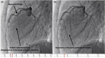
A 12-year-old male was presented with sudden cardiac arrest while playing basketball. He was successfully resuscitated on the field and evaluation revealed asymmetrical left ventricular hypertrophy with a maximum diastolic interventricular septal thickness of 25 mm. Haemodynamics were significant for diastolic dysfunction. There was no gradient across the left ventricular outflow tract at rest or with isoproterenol provocation. There was significant ST elevation with heart rate ~115 bpm after isoproterenol infusion. Selective coronary angiography demonstrated a superficial myocardial bridge (with no significant systolic compression of bridge segment) in the proximal to mid left anterior descending (LAD) artery. Interestingly, there was systolic compression and complete obliteration of flow in the septal branches from LAD (Fig 1, Video 1 in Supplementary material). Troponin was elevated at baseline [0.6–0.9 (ref range 0–0.045) ug/L].
Lateral projection demonstrating selective left coronary artery angiogram showing the myocardial bridge in the left anterior descending coronary artery ( a ). Note the systolic compression ( a ) of the septal perforator branches with almost disappearance of all branches. In diastole ( b ), the septal perforator branches from the left anterior descending coronary artery are visible (Video 1 in supplemental images).

Angiographic finding of myocardial bridge is common in patients with hypertrophic cardiomyopathy (HCM). However, the finding of systolic disappearance of the septal perforators was impressive and explains the ST changes with provocative testing. Normal coronary flow Reference Raphael, Cooper, Parker, Collinson, Vassiliou and Pennell1 is governed by the forward compression wave (generated by ventricular contraction) and the backward expansion wave (due to the decompression of the microcirculation as the ventricle relaxes). There is an opposing backward compression wave (BCW) produced as the intramyocardial vessels are compressed during ventricular systole. Normally, the BCW is small and has a negligible effect on coronary circulation Reference Raphael, Cooper, Parker, Collinson, Vassiliou and Pennell1 . However, Raphael et al have demonstrated a large BCW due to compressive deformation of the intramyocardial coronary arteries in ventricular systole in patients with HCM Reference Raphael, Cooper, Parker, Collinson, Vassiliou and Pennell1 . Coronary flow is, therefore, deranged due to this opposing force to forward flow. We believe that the angiographic appearance of the disappearance of the septal perforator branches in our patient is a sign of this backward compression wave with severe physiological perturbance to the coronary flow. Baseline troponin leak and ST-segment elevation with slight tachycardia show the perfusion deficit precipitated by shortened diastolic time and increased myocardial demand. We considered this angiographic finding in combination with ST changes with tachycardia and baseline troponin leak as an ominous sign and referred our patient for a heart transplant.
Supplementary material
To view supplementary material for this article, please visit https://doi.org/10.1017/S104795112000428X.
Acknowledgements
None.
Financial support
None.
Conflicts of interest
None.
Ethical standards
The authors assert that this report complies with the ethical standards of the Helsinki convention.