I. INTRODUCTION
Fluorescent nanoparticles have been demonstrated in a wide range of applications such as optoelectronic devices, live cell imaging, and/or intracellular dynamics. Reference Burroughes, Bradley, Brown, Marks, Mackay, Friend, Burns and Holmes1 Currently, available fluorescent nanoparticles are mostly semiconductor quantum dots such as CdS, CdSe or CdTe and dye-doped silica/polymer particles. Reference Chan and Nie2–Reference Santra, Wang, Tapec and Tan4 However, cytotoxicity is a critical problem for the heavy metal-containing quantum dots, whereas the dye-doped beads have a number of limitations such as their relatively large size and limited dye-loading content due to self-quenching. Reference Chan and Nie2–Reference Santra, Wang, Tapec and Tan4 A promising highly attractive alternative is the use of fluorescent π-conjugated polymer nanoparticles, so there is growing interest in their synthesis and applications. Reference Bolinger, Traub, Brazard, Adachi, Barbara and Vanden Bout5,Reference Tuncel and Demir6 So far, π-conjugated polymer nanoparticles have been synthesized by the miniemulsion or reprecipitation method. Reference Tuncel and Demir6 In the miniemulsion technique, a polymer is typically dissolved in a water-immiscible organic solvent, and the resulting solution is injected into an aqueous solution including an appropriate surfactant. Reference Landfester7,Reference Sarrazin, Chaussy, Vurth, Stephan and Beneventi8 The mixture is rapidly stirred to form stable miniemulsions containing small polymer droplets, then the organic solvent is evaporated to obtain a stable polymer nanoparticle dispersions. Reference Landfester7 In this method, the size can be changed from several tens to hundreds of nanometers depending on the concentration of the polymer solution. However, the reprecipitation method consists of a hydrophobic conjugated polymer dissolved in a good (organic) solvent that can be poured into a poor solvent (typically water), which is miscible with the good (organic) solvent. Reference Wu, Szymanski and McNeill9 The resulting mixture is stirred vigorously to assist the formation of nanoparticles. After the nanoparticle formation, the organic solvent is sometimes removed to leave behind water dispersible.
For organic nanoparticles composed of small (or low molecular weight) molecules, we have developed a new synthesis route based on “ion-association” that has advantages in simplicity and versatility. Reference Yao and Enseki10,Reference Yao and Funada11 This method uses the formation of water-insoluble ion-pair aggregates in aqueous phases by association of a chromophoric ion with a hydrophobic counterion to fabricate organic nanoarchitectures. Using this approach, we have succeeded in synthesizing highly fluorescent organic nanoparticles. Reference Yao and Ashiba12 In the present study, we contemplate transposing the wet chemical synthesis of organic nanoparticles from small molecular systems to ion-based polymer materials. A novel approach for preparing dispersions of fluorescent π-conjugated polymer nanoparticles is presented on the basis of NanoAgglomeration via Polyion Association (we call this technique “NAPA” method), which includes polyion complex formation and subsequent globulization to fabricate nanoarchitectures in water-based liquid. In considering the utility of π-conjugated polymers, it is evident that control of ionic properties/interactions that would not be possible for typical organic-solvent systems will expand physicochemical parameters such as size and spectroscopic characters. In contrast to the nanoparticle dispersions of organically soluble (hydrophobic) π-conjugated polymers such as nonionic poly(phenylene vinylene) (PPV) derivatives, which can be typically prepared by the simple reprecipitation method, Reference Wu, Szymanski and McNeill9 the present NAPA method is found to prevent further bending or kinking of the polymer backbone upon nanoparticle formation as judged by UV–vis spectroscopy. In addition, size-dependent fluorescence properties are demonstrated.
It is well known that there is a complex relationship between molecular conformation and spectroscopic properties of π-conjugated polymers Reference Grey, Kim, Norris, Miller and Barbara13 because conjugated polymer conformation determines the dipole–dipole interactions between segments of polymer chains. The present study also features on a detailed investigation of how the spectroscopic properties of nanoparticles synthesized by the NAPA approach evolve in the size range between a single (monomolecular) polymer and the bulk material.
II. EXPERIMENTAL DETAILS
A. Materials
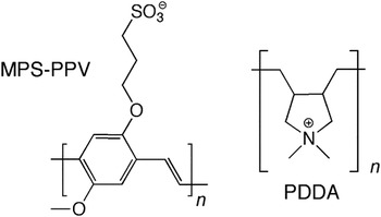
The anionic π-conjugated polymer, poly(2-methoxy-5-propyloxysulfonate phenylene vinylene), potassium salt, (MPS-PPV; 0.25 wt% in water, chemical structure is shown in Fig. 1), one of a larger class of related poly(phenylene vinylene) (PPV) derivatives, was received from Aldrich and used without further purification. The precise molecular weight of MPS-PPV has not been disclosed, but on the basis of the hydrodynamic radius of 20–60 nm for the same product measured by Cosa et al., Reference Ngo, Karam, Fuller, Burger and Cosa14 the molecular weight could be estimated as 1.8–16 × 105. Reference Flory15 Positively charged polycation poly(diallyldimethylammonium), chloride; PDDA; average M w = 1–2 × 10⁵, was chosen as an ion-associable counterion (see also the chemical structure in Fig. 1). 2-Propanol (GR grade) was obtained from Wako Pure Chemicals (Osaka, Japan). Pure water was obtained by a water-distillation supplier (Advantec GS-200, Advantec Japan, Tokyo, Japan).
Chemical structures of MPS-PPV (potassium salt) and PDDA (chloride).

B. Synthesis: MPS-PPV nanoparticles
The MPS-PPV polymer nanoparticles were prepared by means of a newly developed approach based on NAPA. Typically, rapid addition of a mixture containing MPS-PPV (aqueous, 0.1 wt%, 0.5 mL) and PDDA (aqueous, 0.2 wt%, 0.13 mL) into ultrasonicated 2-propanol/water mixture produced almost clear orange suspension. The final volume ratio of 2-propanol/water (denoted as p/w) was set to 1 or 4. In the synthesis, the monomer mole ratio, that is, [PDDA]/[MPS-PPV] in monomer units, was unity. The resulting suspension was then stored overnight at room temperature and examined by absorption/fluorescence spectroscopy and electron microscopy. Note here that spectroscopic properties are predominated by the chromophoric MPS-PPV moieties, so we call these samples “MPS-PPV nanoparticles.”
C. Synthesis: Control experiments
For comparison, we prepared a series of reference samples in the absence of PDDA or 2-propanol. That is, (i) addition of aqueous pristine MPS-PPV solution (with K+ countercations) into the 2-propanol/water-mixed solvent (final mixing volume ratio of 1 or 4) gave the reference samples and (ii) mixing of aqueous MPS-PPV and PDDA (1:1) gave the “MPS-PPV/PDDA” polyion complex in water. The suspensions obtained were also stored overnight at room temperature and then examined by absorption/fluorescence spectroscopy and electron microscopy. In addition, the “MPS-PPV bulk film” was prepared by a drop-cast from an aqueous solution of pristine MPS-PPV onto a glass plate. The “MPS-PPV/PDDA film” was also prepared in a similar manner to that of the MPS-PPV film, but by a drop-cast from the mixture of MPS-PPV/PDDA in water.
D. Instrumentation
Morphology and size of the polymer nanoparticles were examined with a Hitachi S-4800 scanning transmission electron microscope (STEM; Hitachi High-Technologies Corp., Tokyo, Japan). A specimen for STEM observations was prepared by dropping the suspension on an amorphous carbon-coated copper mesh. UV–vis absorption spectra were recorded on a Hitachi U-4100 spectrophotometer. Fluorescence spectra were obtained with a Hitachi F-4500 spectrofluorometer. Note that the obtained spectra are not corrected for the wave length-dependent response of the detector (photomultiplier R-928) system. For the measurements, all samples were excited at 456 nm. Fluorescence quantum yields (Φf) were estimated by comparing the emission spectra of MPS-PPV in water with the quantum yield of Φf = 0.02 reported by Swager and coworkers. Reference McQuade, Pullen and Swager16
E. Computations
We made simple structural models of 1:1 ion-pair complex of trimer cation-anion species, (MPS-PPV)3–PDDA3, to examine how the PDDA countercation in the vicinity of anionic PPV unit influences its geometry. The ground-state geometry optimizations of the related model complexes were carried out with the Gaussian 09 program at the semi-empirical PM6 level. We included solvent (water) effects by applying the conductor polarizable continuum model (C-PCM) present in Gaussian 09. Reference Frisch, Trucks, Schlegel, Scuseria, Robb, Cheeseman, Scalmani, Barone, Mennucci, Petersson, Nakatsuji, Caricato, Li, Hratchian, Izmaylov, Bloino, Zheng, Sonnenberg, Hada, Ehara, Toyota, Fukuda, Hasegawa, Ishida, Nakajima, Honda, Kitao, Nakai, Vreven, Montgomery, Peralta, Ogliaro, Bearpark, Heyd, Brothers, Kudin, Staroverov, Keith, Kobayashi, Normand, Raghavachari, Rendell, Burant, Iyengar, Tomasi, Cossi, Rega, Millam, Klene, Knox, Cross, Bakken, Adamo, Jaramillo, Gomperts, Stratmann, Yazyev, Austin, Cammi, Pomelli, Ochterski, Martin, Morokuma, Zakrzewski, Voth, Salvador, Dannenberg, Dapprich, Daniels, Farkas, Foresman, Ortiz, Cioslowski and Fox17
III. RESULTS AND DISCUSSION
A. Pristine MPS-PPV and MPS-PPV/PDDA polyion complex in water
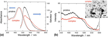
It is known that charged polymers (or polyelectrolytes) can form complexes with oppositely charged amphiphilic polymers, and the polyion complex may have a very different geometrical conformation from the free- (liberated) charged polymer. Reference MacKnight, Ponomarenko and Tirrell18–Reference Kuhn, Levin and Barbosa20 Hence, we first examine that combining the π-conjugated anionic MPS-PPV polymer with oppositely charged PDDA can make well-defined nanoarchitectures (nanoparticles) or not, and demonstrate a change in the geometrical conformation of the polymer, thereby tune its optical properties. The absorption spectrum of pristine MPS-PPV in aqueous solution shows a broad band at 400–600 nm with a peak position of 450 nm, which is due to transitions within the conjugated π-delocalization [Fig. 2(a)]. The emission spectrum exhibits a broad peak at ∼520 nm [Fig. 2(a)]. When PDDA was added into the MPS-PPV solution, a change in the spectroscopic properties was relatively small, unlike the case of surfactant addition. Reference Chen, Xu, McBranch and Whitten21 However, the addition of PDDA caused an overall red shift of the absorption peak position to 456 nm, and in the fluorescence spectra, the appearance of emission around 610 nm was eminent. The excitation spectra monitored at 610 nm exhibited different features between the pristine MPS-PPV and MPS-PPV/PDDA aqueous samples. That is, in the excitation spectra shown Fig. 2(b), intensity at the longer wave length region (≥∼470 nm) of MPS-PPV/PDDA is stronger than that of the pristine MPS-PPV, indicating that red-site emission predominantly comes from transition of chromophores with lower energies. This excitation energy-dependent (or site-selective) fluorescence is a signature of a heterogeneous distribution of fluorophores (corresponding to the effective range of π-conjugation). Hence, the red-site emission of MPS-PPV/PDDA in water can be due to fluorophores with long conjugation-length segments caused by polyion association between anionic MPS-PPV and cationic PDDA, suggesting an extension in the MPS-PPV effective conjugation or reduction in the conformational disorder of polymer backbone. Reference Landfester7,Reference Sarrazin, Chaussy, Vurth, Stephan and Beneventi8 Note that the PPV-unit aggregation in the MPS-PPV/PDDA sample may also be the origin for the red-site emission, but it is less likely because of (i) the observation of overall red shift in the absorption spectrum and (ii) the absence of a noticeable band caused by the strong excitonic interactions, which should appear in the lower energy region compared with that of the single chromophore transitions. The complexation between the cationic/anionic polymers should be driven by a combination of Coulombic attraction and by favorable entropy changes resulting from release of interfacial water molecules, Reference MacKnight, Ponomarenko and Tirrell18 so such interactions would serve to inhibit the folding of the polymer, reducing the conformational disorder in the π-conjugated backbone and thus contributing to a spectral red shift in the absorption of MPS-PPV/PDDA.
(a) Absorption and fluorescence spectra of pristine MPS-PPV (black curves) and MPS-PPV/PDDA polyion complex (red curves) in water. Fluorescence spectra are obtained upon excitation of 456 nm. (b) Excitation spectra of pristine MPS-PPV and MPS-PPV/PDDA complex in water. The fluorescence is monitored at 610 nm. Typical morphology of the MPS-PPV/PDDA complex obtained by STEM observations is shown in the inset.

One should bear in mind that polyion association (in the present case, charge neutral) can lead to hydrophobic nature of the polyion complex. Hence, to examine whether the MPS-PPV/PDDA polyion complex in water consequently produces nanoparticles or not, electron microscopy was conducted. A typical STEM image for the aqueous MPS-PPV/PDDA is shown in the inset of Fig. 2(b). Some aggregated particles with the diameter range of 20–100 nm could be seen; however, a film-like feature was dominant in the image. This means that nanoparticle formation is very partial (or incomplete). In a similar MPS-PPV/PDDA dispersion system in water, nanoaggregate formation has been suggested on the basis of the fluorescence correlation spectroscopic data, Reference Abe and Chen22 that is, the diffusion time from the autocorrelation function obtained was so long that it most likely corresponded to the larger aggregated particles instead of the simple MPS-PPV/PDDA complex. In the present case, formation of the aggregated polymer (= nanoparticles) was very partial in pure aqueous phases, so a more sophisticated method to prepare π-conjugated polymer nanoparticles via polyion association (or polyion complex formation) is to be considered.
B. MPS-PPV polymer nanoparticles
Well-defined MPS-PPV nanoparticles were successfully synthesized by the NAPA approach using PDDA. The dispersions obtained are clear (not turbid), with colors similar to those of the polyion complex. It should be emphasized here that, in contrast to previous nanoparticle systems composed of the hydrophobic and nonionic conjugated polymers, such as MEH-PPV (poly(2-methoxy-5-(2′-ethylhexyloxy)-p-phenylene vinylene)), synthesized by the reprecipitation method, Reference Wu, Szymanski and McNeill9,Reference Szymanski, Wu, Hooper, Salazar, Perdomo, Dukes and McNeill23 which commonly yield blue-shifted absorption spectra as compared with those of the liberated polymer solution, a remarkable feature of our system involves no shift in the absorption bands in comparison to that of the solution-phase polyion complexes. This means that conformational disorder of the polymer backbone does not occur upon formation of the ion-based nanoparticles.
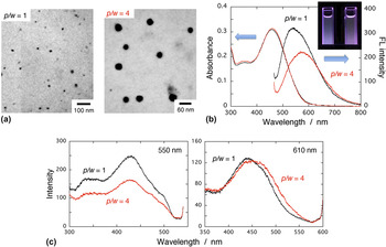
Figure 3(a) displays typical results on STEM images of the MPS-PPV polymer nanoparticles prepared by the NAPA method under the condition of p/w = 1 or 4. The STEM images reveal that roughly spherical nanoparticles have the diameters of 15–30 nm (p/w = 1) or 30–60 nm (p/w = 4). Note that, while keeping the solutions overnight, some large agglomerates might settle down onto the bottom of sample vials. From the images, as the mixing volume ratio p/w increases, the particle size also increases. 24 Interestingly, no coalescence of the soft nanoparticles having an interconnected network was observed, and thus, well-dispersed nanoparticle formation was achieved.
(a) Typical STEM images of the MPS-PPV polymer nanoparticle samples prepared at p/w = 1 and 4. (b) Absorption and fluorescence spectra of MPS-PPV nanoparticles prepared by the NAPA approach using PDDA. Black and red curves stand for the spectra of nanoparticles prepared at p/w = 1 and 4, respectively. Fluorescence spectra are obtained upon excitation of 456 nm. Inset is a photo of the fluorescence from these samples. (c) Excitation spectra of the MPS-PPV nanoparticle samples. The fluorescence is monitored at 550 or 610 nm.

Figure 3(b) shows UV–vis absorption and fluorescence spectra of suspensions of the corresponding MPS-PPV polymer nanoparticles. The absorption peak of the nanoparticle samples was almost identical with that of the MPS-PPV/PDDA complex in water. This means that conformations of the PPV moiety are still retained upon nanoparticle formation, despite the fact that the compact globulization or nanoagglomeration inevitably requires geometrical constraint that may bring about an overall decrease in the conjugation length attributable to the bending or kinking of the polymer backbone. Reference Padmanaban and Ramakrishnan25 The involved PDDA counteractions would then release the conformational constraint, preventing a decrease in its effective conjugation length. This behavior is in sharp contrast to typical observations that hydrophobic π-conjugated polymer nanoparticles prepared by the simple reprecipitation method commonly exhibit blue shift of the absorption peak as compared with those of the liberated polymer solution; for example, blue-shift detection for nanoparticles of polyfluorene derivatives (PFO and PFPV) and MEH-PPV, which suggests an overall decrease in the effective conjugation length. Reference Wu, Szymanski and McNeill9,Reference Szymanski, Wu, Hooper, Salazar, Perdomo, Dukes and McNeill23
Fluorescence spectra of the MPS-PPV nanoparticles are more red shifted as compared with those of the MPS-PPV/PDDA complex in water [Fig. 3(b)], so the fluorescence color changes to reddish gradually (see the inset). Particular emphasis is placed on the facts that (i) fluorescence quantum yield increased upon nanoparticle formation as compared with that of the MPS-PPV/PDDA complex in water; (ii) fluorescence peak position was strongly dependent on the p/w value (i.e., size dependent). A dilute solution of pristine MPS-PPV in water was used as a standard for quantum yield determination (Φf = 0.02), Reference McQuade, Pullen and Swager16 and the estimated Φf was 0.024, 0.085, or 0.064, for samples of MPS-PPV/PDDA complex in water, the polymer nanoparticles prepared at p/w = 1 or 4, respectively. The increase in the fluorescence efficiency is explained by loss of intrachain and/or interchain quenching (i.e., by reduction of nonradiative decay channels in the main chain such as internal conversion and/or that of exciton energy transfer between (within) chains to quenching sites), Reference Padmanaban and Ramakrishnan25,Reference Montano, Dattelbaum, Wang and Shreve26 which can be caused by interstitial incorporation of PDDA between the MPS-PPV polymer backbones. However, the origin for the size-dependent fluorescence properties can be considered on the basis of their fluorescence excitation spectra. Figure 3(c) displays fluorescence excitation spectra of the MPS-PPV nanoparticle samples (p/w = 1 and 4) monitored at 550 or 610 nm. Monitoring at 550 nm, the peak positions of excitation spectra were different from the absorption maximum and exhibited shorter wave length, whereas those monitored at 610 nm were almost identical with that of the absorption. Moreover, the excitation spectra of samples with p/w = 1 and 4 monitored at the same wave length were moderately similar to each other. This means that emission in the low-/high-energy region (red-/blue-site emission) mainly stems from the polymer segments with relatively long/short effective conjugation length, respectively. In other words, in the MPS-PPV nanoparticle samples, a major contribution to the red-site emission (at ∼610 nm) is the almost sole excitation of the chromophoric segments having a long effective conjugation length. Note that the absorption spectral shapes including the peak position were almost identical among all MPS-PDDA-related samples (i.e.,, two polymer nanoparticle samples with different particle sizes and the polyion complex in water), so the reduction in the blue-site emission intensity (∼500 nm) observed in the large-sized nanoparticle sample (prepared at p/w = 4) can probably be due to self-quenching caused by chain–chain interactions. Reference Szymanski, Wu, Hooper, Salazar, Perdomo, Dukes and McNeill23,Reference Padmanaban and Ramakrishnan25,Reference Montano, Dattelbaum, Wang and Shreve26 From a viewpoint of surface chemistry, a small (large) particle has a large (small) surface-to-volume ratio, so the blue-site (red-site) fluorescence is strongly associated with the surface site (inner site) of the nanoparticles, respectively. This also means that the surface (or interface) of the polymer nanoparticles may have a considerably mobile region for the chromophoric segments.
C. Bulk films
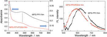
The absorption and fluorescence spectra of the MPS-PPV nanoparticle suspensions are compared with those of the pristine MPS-PPV or MPS-PPV/PDDA thin solid film prepared by a drop cast of the corresponding purely aqueous solution. The spectra for these films are shown in Fig. 4. Note that these films on glass substrates were not transparent [see the additional background scattering in Fig. 4(a)], so it was hard to determine the precise absorption peak positions of the films; however, the positions were very close to each other (470–480 nm). The emission peak of MPS-PPV/PDDA film was ∼595 nm, more red shifted as compared with those of the nanoparticle samples prepared at both p/w = 1 and 4 (λem = 545 and 575 nm, respectively). However, the solid-state pristine MPS-PPV film exhibited more red-shifted emission (∼620 nm) as compared with that of the MPS-PPV/PDDA film possessing similar absorption properties. It is expected that PDDA polyion can prevent chain–chain interactions in (or between) the MPS-PPV polymer(s) through its incorporation or spacing between the backbones, Reference Lin, Jiang, Xu, Lin and Tsukruk27,Reference Tang, Li, Wang, Wang, Sun and Yang28 so it may be possible that the more red-shifted emission from the pristine MPS-PPV film (620 nm) is due to nonradiative excitation energy transfer originated from the chromophore arrangements in a closely packed structure, Reference Wu, Szymanski and McNeill9,Reference Lin, Jiang, Xu, Lin and Tsukruk27 which brings about the energy lowering in the emission peak toward the more red sites. It is worth noting here that the emission behavior of the present π-conjugated polymer nanoparticles lies in between the solution phase (polyion-complex state) and the bulk solid phase (dense-packed state), suggesting that MPS-PPV polymers (or backbones) are distributed in a relatively loose packing manner inside the ion-based nanoparticles compared with that in the solid film. In any case, π-conjugated polymer nanoparticles synthesized via the NAPA approach can tune the fluorescence properties by controlling the nanoscopic size of the nanoparticles.
Absorption (left) and fluorescence spectra (right) of MPS-PPV and MPS-PPV/PDDA films on a glass substrate. Fluorescence spectra, which are normalized to the peak intensity, are obtained upon excitation of 456 nm.

D. Application of the simple reprecipitation method: Pristine MPS-PPV in 2-propanol/water mixtures
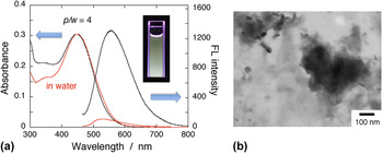
If the solubility of MPS-PPV (potassium salt) in 2-propanol is very low, the polymer nanoparticles may be produced by just adding the pristine aqueous MPS-PPV solution into 2-propanol (or 2-propanol/water mixtures) without using PDDA, which corresponds to a simple reprecipitation method. Hence, we examined what is perceived when the reprecipitation approach is applied for preparing the polymer nanoparticles. Figure 5(a) shows absorption and fluorescence spectra (black curves) of the sample prepared by mixing the pristine MPS-PPV solution with 2-propanol at the mixing volume ratio of p/w = 4. The red curves represent absorption (normalized) and fluorescence spectra of the pristine MPS-PPV in water (absorption peak; ∼450 nm, fluorescence peak; ∼530 nm). For the spectra of MPS-PPV in the 2-propanol/water mixture, we found that (i) absorption peak was unchanged; (ii) the low-energy region of absorption (≥∼460 nm) was substantially reduced; (iii) fluorescence intensity became much stronger with a slight red shift to ∼555 nm (Φf ∼0.29 estimated; see also Fig. 2). Broad absorption spectrum with a red (low energy) tail is frequently observed for solid-state nanoparticles of conjugated polymers (or aggregates) such as MEH-PPV, Reference Szymanski, Wu, Hooper, Salazar, Perdomo, Dukes and McNeill23 and indicates the presence of considerable chromophore (here the PPV unit) interactions. Reference Nguyen, Martini, Liu and Schwartz29 Hence, the disappearance of such red tail as well as the increase in the fluorescence intensity strongly suggest that pristine MPS-PPV (potassium salt) is more or less soluble in 2-propanol/water mixture (p/w = 4).
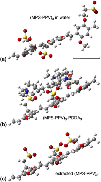
(a) Absorption and fluorescence spectra of pristine MPS-PPV in 2-propanol/water mixture (p/w = 4) in the absence of PDDA. For comparison, absorption and fluorescence spectra of pristine MPS-PPV in water are also shown (red curves). Absorption spectra are normalized at the peak position. The inset is the fluorescence image of the sample. (b) Typical STEM image for the MPS-PPV in 2-propanol/water mixture (p/w = 4).

Indeed, the choice of solvent can have a significant effect on the (main and/or side) chain structures of conjugated polymers dissolved solution, particularly on their fluorescence performance. It is well established that conjugated polymer chains (or backbones) in dilute solution are isolated, so the fluorescence of such isolated chains mainly comes from intrachain excitons. Reference Nguyen, Martini, Liu and Schwartz29 In contrast, the polymer chains in contact with each other can form weakly emissive interchain species. Reference Nguyen, Martini, Liu and Schwartz29 MPS-PPV has hydrophobic phenylene vinylene backbone with hydrophilic sulfonate side chains. Generally, in aqueous solution, polar side chains adopt a conformation where the hydrophilic units are exposed to water, and the hydrophobic units are stacked inside to form apparent segregation owing to hydrophobic and/or π–π interactions of aromatic rings. Reference Gu, Bao, Zhang, Wang and Shen30 Using this argument, it is reasonably considered that MPS-PPV in aqueous solution can exist in a slightly aggregated state. At the same time, the polar sulfonate groups can extend into water, resembling a micelle where a hydrophobic core is protected by a hydrophilic shell. Under the condition, when 2-propanol is added into the aqueous phase containing the coiled-up polymers, the organic solvent gradually takes the place of water, and the polymer chain may be less aggregated, resulting in the enhancement of fluorescence efficiency of MPS-PPV that is linked to the highly emissive intrachain excitons. Reference Gu, Bao, Zhang, Wang and Shen30 Similar phenomena have been observed upon addition of DMSO (dimethylsulfoxide) into the aqueous MPS-PPV solution. Reference Ng, Yu, Gopal, Rome, Monbouquette and Tolbert31 When the MPS-PPV polymer is dissolved in DMSO/water mixtures, the chains have more open and isolated structures, yielding the enhanced fluorescence intensity, in good agreement with the present case of the 2-propanol/water mixture. Reference Ng, Yu, Gopal, Rome, Monbouquette and Tolbert31
To confirm whether the pristine MPS-PPV is (partly) dissolved in 2-propanol/water mixture (p/w = 4) or not, electron microscopic measurements were carried out for the morphology observation. Figure 5(b) shows a typical STEM image of the sample. Some particulate species or flocculate of the polymer could be seen in the image, but film-like morphologies were predominantly observed, implying that the reprecipitation method using 2-propanol was inappropriate for preparing the MPS-PPV nanoparticles.
E. Qualitative interpretation by geometry computations
To investigate how the PDDA countercation in the vicinity of the anionic PPV influences its polymer backbone geometry, we made (and calculated) simple structural models of ion-pair complexes of the MPS-PPV/PDDA trimer. We optimized the ground-state geometry of (MPS-PPV)3 or (MPS-PPV)3–PDDA3 with the Gaussian 09 program at the semi-empirical PM6 level. Reference Yao and Ashiba12,Reference Frisch, Trucks, Schlegel, Scuseria, Robb, Cheeseman, Scalmani, Barone, Mennucci, Petersson, Nakatsuji, Caricato, Li, Hratchian, Izmaylov, Bloino, Zheng, Sonnenberg, Hada, Ehara, Toyota, Fukuda, Hasegawa, Ishida, Nakajima, Honda, Kitao, Nakai, Vreven, Montgomery, Peralta, Ogliaro, Bearpark, Heyd, Brothers, Kudin, Staroverov, Keith, Kobayashi, Normand, Raghavachari, Rendell, Burant, Iyengar, Tomasi, Cossi, Rega, Millam, Klene, Knox, Cross, Bakken, Adamo, Jaramillo, Gomperts, Stratmann, Yazyev, Austin, Cammi, Pomelli, Ochterski, Martin, Morokuma, Zakrzewski, Voth, Salvador, Dannenberg, Dapprich, Daniels, Farkas, Foresman, Ortiz, Cioslowski and Fox17,Reference Stewart32 The solvent (water) effect was also incorporated using the dielectric continuum model provided by the conductor polarizable continuum model (C-PCM). Figures 6(a) or 6(b) displays a typical optimized structure of (MPS-PPV)3 or (MPS-PPV)3–PDDA3, respectively. Figure 6(c) shows the side view of the MPS-PPV trimer moiety extracted from Fig. 6(b). Interestingly, our simple model can be a good indication for evaluating MPS-PPV polymer chain conformations in the presence/absence of PDDA countercation. The calculations suggest that the geometry of liberated MPS-PPV (even trimer) with a longer conjugation length is less stable compared with that of the MPS-PPV/PDDA complex [see Figs. 6(a) and 6(c); in Fig. 6(a), the terminal phenyl ring is rotated out of the main molecular plane], that is, PDDA makes the effective conjugation of MPS-PPV longer as compared with that of the free polymer. This is in qualitatively agreement with our spectroscopic observations as described before. Due to the presence of PDDA chains and Coulombic interactions between these MPS-PPV and PDDA polyions, defect or kink formation in the MPS-PPV polymer backbone may be reduced even upon the nanoparticle formation, and consequently, the π-conjugated polymer nanoparticles synthesized via the NAPA method did not deteriorate their spectroscopic properties. We believe successful synthesis of such fluorescent conjugated polymer nanoparticles in desired properties possessing biocompatibility and nontoxicity will make these materials highly attractive for applications in various areas such as optoelectronics, bioimaging, and nanomedicine in the future.
(a and b) Optimized structures of MPS-PPV (trimer) and its polyion (trimer–trimer) complex of (MPS-PPV)3–(PDDA)3 calculated at the PM6 level. (c) Sole MPS-PPV geometry extracted from the complex shown in (b).

IV. CONCLUSIONS
In summary, a facile method was developed to synthesize fluorescent π-conjugated polymer nanoparticles. The polymer we chose was anionic poly(2-methoxy-5-propyloxysulfonate phenylene vinylene); MPS-PPV. The synthesis was based on NanoAgglomeration via Polyion Association (called as NAPA method) that includes polyion complex formation between MPS-PPV and cationic polyammonium and subsequent globulization to fabricate nanoarchitectures in aqueous-based suspension. In contrast to a common observation that hydrophobic-conjugated polymer nanoparticles made by a simple reprecipitation method show a blue shift in their UV–vis absorption peak, the NAPA-based nanoparticles exhibited no spectral shift during the nanoparticle formation, suggesting the absence of further bending or kinking of the polymer backbone in the polyion complexes. The fluorescence peak position was particle size dependent, which is dominantly due to the increased contribution of sole excitation of the chromophoric segments that have a long effective conjugation length in large-sized nanoparticles. A small (large) particle has a large (small) surface-to-volume ratio, so the blue-site (red-site) fluorescence should be reasonably associated with the surface site (inner site) of the polymer nanoparticles, respectively. We believe that such nanoengineering methodology for functional polymers will play a fundamental role in an entire new class of fluorescent organic nanoparticles.
Supplementary Material
To view supplementary material for this article, please visit http://dx.doi.org/jmr.2014.244.