Implications
The study of molecules involved in early embryo–uterine interactions in single ovulating species remains a challenge due to difficulties encountered to isolate the endometrium adjacent to the pre-implantation embryo when present in the uterus. However, knowledge of the embryo–maternal interactome at the time of in vitro embryo development can lead to improving assisted reproduction techniques (ART). Such improvements will contribute to counteract the reduced pregnancy rates and higher miscarriage associated with ART, also leading to increased proportions of healthy individuals born. Ultimately, the increased efficiency or ART will reduce costs of breeding and it may be translated to human ART.
Introduction
Embryos that develop in vivo are superior in quality to in vitro-produced embryos, survive cryopreservation better and are less prone to developmental abnormalities (Rizos et al., Reference Rizos, Ward, Duffy, Boland and Lonergan2002; Farin et al., Reference Farin, Piedrahita and Farin2006 and Reference Farin, Farmer and Farin2010; Havlicek et al., Reference Havlicek, Kuzmany, Cseh, Brem and Besenfelder2010). Therefore, improved knowledge of the very early uterine environment (i.e. day 5 to day 8) can lead to improved embryo culture conditions in vitro. However, the small size of embryos during pre-hatching development (i.e. very early stages of pre-implantation) makes it difficult to identify and isolate the endometrium adjacent to the embryo, complicating the direct analysis of zona-enclosed (ZE) embryo–endometrial interactions. As a consequence, an in vivo model with ZE embryos is lacking, and it is, therefore, unknown whether in vitro studies can reliably reflect in vivo embryo–maternal interactions. Using a multiple embryo transfer (ET) model, we found several differentially expressed proteins (DEP) in the uterine fluid (UF) on day 8 (Muñoz et al., Reference Muñoz, Corrales, Caamaño, Díez, Trigal, Mora, Martín, Carrocera and Gómez2012; Gómez et al., Reference Gómez, Caamaño, Corrales, Díez, Correia-Álvarez, Martín, Trigal, Carrocera, Mora, Pello-Palma, Moreno and Muñoz2013); some of these UF proteins are submitted to different bilateral regulations between horns (Trigal et al., Reference Trigal, Díez, Muñoz, Caamaño, Goyache, Correia-Alvarez, Corrales, Mora, Carrocera, Martin and Gómez2014). The UF interface is representative of interactions between embryos, endometrium and immune cells in utero, which can also affect conceptus and endometrial growth by increasing the concentration of cytokines (Leung et al., Reference Leung, Derecka, Mann, Flint and Wathes2000; Oliveira and Hansen, Reference Oliveira and Hansen2008; Ideta et al., Reference Ideta, Hayama, Nakamura, Sakurai, Tsuchiya, Tanaka, Yamaguchi, Fujiwara, Imakawa and Aoyagi2010; Muñoz et al., Reference Muñoz, Corrales, Caamaño, Díez, Trigal, Mora, Martín, Carrocera and Gómez2012). The UF contains not only live cells (immune, detached endometrial) but also secreted products and cell debris, such as epithelial cells that are in constant renewal and extrusion into the lumen (Welsh Reference Welsh1993; Fahey et al., Reference Fahey, Schaefer and Wira2006). As early embryos normally live in this environment, the analysis of UF samples recovered from live animals with multiple embryos developing in uterus increases the likelihood of detecting embryo-induced changes (Muñoz et al., Reference Muñoz, Corrales, Caamaño, Díez, Trigal, Mora, Martín, Carrocera and Gómez2012; Gómez et al., Reference Gómez, Caamaño, Corrales, Díez, Correia-Álvarez, Martín, Trigal, Carrocera, Mora, Pello-Palma, Moreno and Muñoz2013; Trigal et al., Reference Trigal, Díez, Muñoz, Caamaño, Goyache, Correia-Alvarez, Corrales, Mora, Carrocera, Martin and Gómez2014).There is no evidence demonstrating that the development of an individual ZE embryo is compromised by the simultaneous presence of other ZE embryos in the uterus. However, further empirical demonstration that the presence of multiple embryos in the uterus does induce physiological reactions is necessary. In this study, we show evidence that the metabolic and functional UF, and blood plasma profiles obtained with multiple ET, are consistent with those observed when a single embryo develops. Our results support the multiple ET model as an efficient system to study very early embryo–maternal interactions in the cow.
Material and methods
All experimental procedures involving animals were performed according to the European Community Directive 2010/63/EU (Spanish Real Decreto 53/2013), and were sanctioned by the Animal Research Ethics Committee of SERIDA. All reagents were purchased from Sigma (Madrid, Spain), unless otherwise stated.
Animals and ET
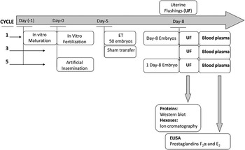
Procedures involving animal feeding and management, oestrus synchronization, ET, embryo recovery by uterine flushings, blood sampling and analysis have been described elsewhere (Muñoz et al., Reference Muñoz, Corrales, Caamaño, Díez, Trigal, Mora, Martín, Carrocera and Gómez2012; Gómez et al., Reference Gómez, Caamaño, Corrales, Díez, Correia-Álvarez, Martín, Trigal, Carrocera, Mora, Pello-Palma, Moreno and Muñoz2013; Trigal et al., Reference Trigal, Díez, Muñoz, Caamaño, Goyache, Correia-Alvarez, Corrales, Mora, Carrocera, Martin and Gómez2014). Figure 1 shows a general outline of the experiments performed. Synchronized recipients were allocated to the following experimental groups: (1) sham transfer (ST) (i.e. intra-uterine deposition of holding medium on Day 5); (2) ET performed on Day 5 with day 5 morulae (n=50) produced in vitro with unsorted (Muñoz et al., Reference Muñoz, Corrales, Caamaño, Díez, Trigal, Mora, Martín, Carrocera and Gómez2012) and sex-sorted sperm (Gómez et al., Reference Gómez, Caamaño, Corrales, Díez, Correia-Álvarez, Martín, Trigal, Carrocera, Mora, Pello-Palma, Moreno and Muñoz2013); and (3) recipients subjected to artificial insemination (AI). Recovery of the UF and embryos was performed on day 8 in all groups. In brief, the ipsilateral horns of recipients were first flushed with 45 ml of phosphate buffered saline (PBS)+10 µl/ml protease inhibitor (Protease Arrest; GE Healthcare, Madrid, Spain). Recovery of the diluted UF was performed by aspirating with syringe only while a steady flow could be achieved. Those recipients transferred with embryos were then extensively flushed with PBS+1 mg/ml poly-vinyl-pyrrolidone. The embryos were identified using a stereomicroscope and were rapidly separated from the UF, which was centrifuged (2000×g) at 4°C, aliquoted and stored at −145°C. Embryos were counted and classified. With this flushing system, the cathether was positioned to collect contents from the craneal and middle horn thirds (Muñoz et al., Reference Muñoz, Corrales, Caamaño, Díez, Trigal, Mora, Martín, Carrocera and Gómez2012), which may exclude collection of more caudal embryos (Sartori et al., Reference Sartori, Suárez-Fernández, Monson, Guenther, Rosa and Wiltbank2003). Nonetheless, rates of viable embryos remain as 60% out of the total collected (Muñoz et al., Reference Muñoz, Corrales, Caamaño, Díez, Trigal, Mora, Martín, Carrocera and Gómez2012). Once UF paired samples (i.e. UF recovered from an embryo-transferred recipient – ET-UF, and UF recovered from the same recipient after a sham transfer – ST-UF) were obtained, recipients were subjected to AI at oestrus; only when one live embryo was recovered, the Day 8 UF was processed. Animals were treated at 2- to 3-months intervals throughout non-consecutive oestrus cycles. Recovered UFs were categorized as follows: ET-UF: with at least n=5 viable embryos recovered (n=9.8 on average); AI-UF: recovered when a single embryo was obtained after AI, once stated that one morphologically normal embryo was recovered and it was able to develop in vitro for 24 h in single culture; and ST-UF: without embryos transferred. Blood plasma samples were collected on day 8 by coccygeal vein puncture and collected in ethylenediaminetetraacetic acid -evacuated tubes. Blood tubes were immediately refrigerated at 4°C, and centrifuged at 2000×g within 30 min after recovery. Supernatant plasma was aliquoted and stored at −80°C until prostaglandin (PG) analysis.
Typical pattern of treatments applied to synchronized recipients. On day 5, in the first cycle, IVP embryos are transferred to the uterus and recovered 3 days later (day 8) together with diluted uterine fluid (UF). The following cycle, second oestrus cycle, is left as inoperative. Subsequently, the animal is sham transferred on day 5 and UF is recovered on day 8. Treatment application was balanced, n=4 animals followed the above schedule, whereas the other n=4 animals, conversely, were sham transferred in the first cycle and received embryos in the third cycle. During the fourth cycle, no procedures were applied to recipients. Subsequently, in the fifth cycle, the animal is artificially inseminated at oestrus; only when one live embryo is recovered, the UF is processed for validation purposes (artificial insemination was repeated once again when no live embryo was obtained). The recovered UFs are processed for western blot and ion chromatography. Day 8 blood plasma is processed for prostaglandin analysis. ET=embryo transfer.

Protein analysis
For validation purposes, targeted proteins, previously shown to be differentially expressed between ET-UF and ST-UF, were quantified using western blotting procedure, as previously described (Muñoz et al. Reference Muñoz, Corrales, Caamaño, Díez, Trigal, Mora, Martín, Carrocera and Gómez2012). Antibodies used were raised against bovine proteins (β-actin, NFkB) or proteins from other species sharing high sequence identity with the bovine proteins (clusterin, immunoproteosome 20S β5i subunit–i20S). Antisera specific to (1) NFK β – 2 µg/ml (ab72555; Abcam, Cambridge, UK); (2) Clusterin – 2 µg/ml (Sc-5289; Santa Cruz Biotechnologies, Santa Cruz, CA, USA); (3) β-Actin – 0.3 µg/ml (A2228; Sigma-Aldrich, Madrid, Spain); and (4) i20S –1 : 10 000 (BML-PW8355; Enzo Life Sciences, Axxora, UK) were used at the stated dilutions.
The intra- and inter-assay CVs for protein analysis were as follows: β-Actin – 3.9% and 8.3%; clusterin – 6.4% and 14.5%; i20S – 7.9% and 16.2% and NFK β – 9.8% and 12.4%, respectively.
Hexose analysis
Concentrations of glucose and fructose in UFs were analysed using an ion chromatography method as previously described (Gómez et al., Reference Gómez, Caamaño, Corrales, Díez, Correia-Álvarez, Martín, Trigal, Carrocera, Mora, Pello-Palma, Moreno and Muñoz2013).
The intra- and inter-assay CVs for hexose analysis were as follows: glucose – 5.1% and 5.8% and fructose – 9.0% and 13.4%, respectively
PG analysis
PGF2 α and PGE2 in blood plasma from the same groups of cows were analysed by ELISA method following supplier recommendations (Prostaglandin F2 α EIA Kit, 516011; Prostaglandin E2 EIA Kit, 514010; Cayman, Ann Arbor, Michigan, USA). In brief, 50 µl of plasma was added to each experimental well, followed by 50 µl of PGF2 α or PGE2 Ache tracer and 50 µl of PGF2 α or PGE2 EIA antiserum. Subsequently, ELISA plates were incubated for 18 h at 4°C. Plates were washed five times with the provided wash buffer, and 200 µl of Ellman’s reagent was added to each well. Finally, the plates were covered with a plastic film and allowed to develop in the dark. The absorbance was measured at 450 nm in a microplate reader (model 680; Sigma). Positive and negative sera controls were added to each microtitre plate for normalization. Controls and sera samples were analysed in duplicate.
The intra- and inter-assay CVs for PG analysis were as follows: PGF2 α – 9.1% and 9.2% and PGE2 – 3.7% and 9.7%, respectively.
Subsequently, based on the hexose and protein UF profiles or PG plasma profiles obtained with multiple ET, AI and ST samples, we defined the endometrial (i.e. UF) and plasma response models (RM), shown in Table 1.
Proposed classification of responses to embryos observable in blood plasma or uterine fluid of recipients after ET, Sham-ET and AI

ET=embryo transfer; Sham-ET=sham transfer; AI=artificial insemination; RM=response model.
Responses that did not fit with the criteria shown in Table 1 were considered as non-validated (i.e. non-physiological; as an example: AI>ET>ST).
Statistical analysis
Sources of variation affecting abundance of DEP, hexose and PG concentrations were identified fitting linear models using the Proc GLM of SAS/STAT (Version 9.2; SAS Institute Inc., Cary, NC, USA). The models included, where appropriate, the following fixed effects: UF type (ET, AI and ST), recipient and replicate. Least square means were estimated for each level of fixed effects with a significant F value. R.s.d. was calculated for each variable analysed. Ryan–Einot–Gabriel–Welsch Q-test compared raw means between levels from fixed effects. Proteins were expressed as a twofold change, whereas hexoses and PGs were measured in µg/ml and pg/ml, respectively.
Results
Samples of ET-UF (n=12) were recovered from n=10 cows. The average total recovery rate for ET embryos was 32%, being 20% (n=9.8 embryos/50 transferred) of the recovered embryos classified as viable. The compact, late morula+early blastocysts (i.e. difficult to distinguish provided that flushing medium did not contain protein and the embryos collapsed frequently) accounted for 28% of the viable embryos; blastocysts, expanded blastocysts and hatching blastocysts accounted for 17%, 54% and 3%, respectively; no embryo was fully hatched.
Samples of AI-UF (n=9) were recovered from n=7 cows. The embronic stages recovered were two blastocysts and seven late early blastocysts/morulae. In order to confirm whether these embryos were alive, all AI embryos used were cultured in vitro for 24 h and all (n=9) of them developed to the expanded blastocyst stage.
Protein profiles in UF
As shown in Table 2, actin, clusterin, i20S and NFkB did not show significant differences between ET, AI and ST-UFs. Therefore, the expression patterns of these proteins were consistent with a RM type 3, which is explained as a lack of embryonic response.
Relative abundance of proteins analysed by western blotting on Day 8, diluted uterine fluid recovered by flushing from cows that, on Day 5, were embryo transferred, were artificially inseminated or were transferred with embryo culture medium alone (i.e. sham transfer)

n=numbers of cows per treatment or uterine fluids samples; LSM=least square means.
Data are expressed as LSM and r.s.d.
1 Clusterin: total values resulting from pro-form and mature forms.
Hexose contents in UF
Hexose content in the uterus was examined in ET-UF, ST-UF and AI-UF (Table 3). The profile obtained for glucose and fructose concentrations and their ratio fits with a RM type 3, as no significant differences were obtained between ST, ET and AI-UFs. Numerically, glucose values show a continuous variation RM type 4, ET>AI>ST, which is consistent with a significant, positive correlation observed between glucose concentration and viable embryos present in the UFs (r=0.42; P<0.01).
Glucose and fructose concentrations in Day 8 diluted uterine fluid recovered by flushing from cows that, on Day 5, were embryo transferred, were artificially inseminated or were transferred with embryo culture medium alone (i.e. sham transfer)

n=numbers of cows per treatment or uterine fluids samples; LSM=least square means.
Effects: cow, treatment and replicate analysis.
Data are expressed as LSM and r.s.d.
1 Glucose and fructose concentrations normalized to total protein.
PG in blood
PGE2 and PGF2 α were analysed in blood plasma. Table 4 shows equivalent PG profiles between AI-UF and ET-UF, although both AI and ET profiles differed from ST-UF in PGF2 α but not in PGE2. These values are consistent with RM type 1 and RM type 3 for PGF2 α and PGE2, respectively.
Prostaglandin concentration (pg/ml) in Day 8 blood plasma of cows that, on Day 5, were embryo transferred, were artificially inseminated or were transferred with embryo culture medium alone (i.e. sham transfer)

n=numbers of cows per treatment; PGF2 α=prostaglandin F2 α; PGE2=prostaglandin E2; LSM=least square means.
Data are expressed as LSM and r.s.d.
a,bValues within a row with different superscripts differ significantly at the indicated P value.
Discussion
Our study demonstrates that some plasma molecules (i.e. PGs) may reflect the presence of embryos in the uterus at very early stages. This suggests systemic changes that would exceed the uterine compartment. Our results are consistent with other studies in utero, as the total amount of PGs were over three times higher in Day 13 pregnant after multiple ET than in cyclic heifers (Spencer et al., Reference Spencer, Forde, Dorniak, Hansen, Romero and Lonergan2013). In particular, concentrations of PGF2 α normally exceed several times that of PGE2 (Bartol et al., Reference Bartol, Thatcher, Bazer, Kimball, Chenault, Wilcox and Roberts1981; Lewis et al., Reference Lewis, Thatcher, Bazer and Curl1982; Shelton et al., Reference Shelton, Parkinson, Hunter, Kelly and Lamming1990; Ulbrich et al., Reference Ulbrich, Frohlich, Schulke, Englberger, Waldschmitt, Arnold, Reichenbach, Reichenbach, Wolf, Meyer and Bauersachs2009). At such stages, PGs activate a number of IFNT-stimulated genes, and it seems that PGs could be important recognition signalling molecules in cattle (Spencer et al., Reference Spencer, Forde, Dorniak, Hansen, Romero and Lonergan2013; Lonergan and Forde, Reference Lonergan and Forde2014). In contrast with these observations later in development, our results in blood on Day 8 showed that ZE embryos did not affect PGE2 concentrations. However, the increase in PGF2 α concentration triggered by early ZE embryos is consistent with the above uterine data. Interestingly, Ulbrich et al. (Reference Ulbrich, Frohlich, Schulke, Englberger, Waldschmitt, Arnold, Reichenbach, Reichenbach, Wolf, Meyer and Bauersachs2009) found that the stable metabolites 15-keto PGF2 α (PGFM) and 15-keto PGE2 (PGEM), which accurately represent changes in their respective hormones, increase on Day 15 in pregnant animals. Similarly, pregnant cows on days 5 and 6 show concentrations four to five times higher for PGFM than for PGEM (Richardson et al., Reference Richardson, McNiven, Petit and Duynisveld2013), consistent with our results where PGF2 α and PGE2 concentrations were analysed on Day 8.
The endometrium is not the only source of UF PGs, as bovine blastocysts produce substantial amounts of PGF2 α with higher abundance of PGF2 α than PGE2 (Shemesh et al., Reference Shemesh, Milaguir, Ayalon and Hansel1979; Lewis et al., Reference Lewis, Thatcher, Bazer and Curl1982). The reported increase in UF PGs, at least close to the implantation time, has been suggested to come from the embryonic side (Ulbrich et al., Reference Ulbrich, Frohlich, Schulke, Englberger, Waldschmitt, Arnold, Reichenbach, Reichenbach, Wolf, Meyer and Bauersachs2009; Spencer et al., Reference Spencer, Forde, Dorniak, Hansen, Romero and Lonergan2013). In contrast, our results suggest that on Day 8 such an increase is an endometrial response, provided that multiple embryos (ET) v. a single (AI) did not differ. The trophoblast also expresses the PGF2 α receptor PTGFR (Ulbrich et al., Reference Ulbrich, Frohlich, Schulke, Englberger, Waldschmitt, Arnold, Reichenbach, Reichenbach, Wolf, Meyer and Bauersachs2009). PGF2 α may, thus, act in a paracrine or autocrine manner on the embryo. The embryo, therefore, seems sensitive towards PGF2 α, and the physiological benefit remains undetermined thus far.
The proteins NFkB, clusterin, i20S and β-actin analysed in our study were successfully validated, and the UF reflected no changes in the presence of one or more embryos – that is, RM type 3. NFkB is a nuclear transcription factor central in inflammatory response regulation. Clusterin, a protein associated with receptivity in mice (Brown et al., Reference Brown, Moulton, Witte, Swertfeger and Harmony1996), participates in hexose metabolism, antioxidative activity and protease activity. Immunoproteasome subunit 20S (β5i) (i20S) distinguishes proteasome activity from immunoproteasome activity (Angeles et al., Reference Angeles, Fung and Luo2012). The activation of proteasome/immunoproteasome subunits and the association of such a complex with factors linked to interferon‐γ (IFNG; a close analogue to the ruminant IFNT) suggest that embryonic recognition at these early stages does not involve antigen processing and likely participation of interferons, contrary to the differences we previously observed between embryos of different sex (Gómez et al., Reference Gómez, Caamaño, Corrales, Díez, Correia-Álvarez, Martín, Trigal, Carrocera, Mora, Pello-Palma, Moreno and Muñoz2013). In our previous work, we could also demonstrate that bovine serum albumin (BSA), purine nucleoside phosphorylase (PNP) and heat shock 70 kDa protein 5 (HSPA5) showed Type 1, Type 3/Type 4 and Type 3 validation profile, respectively. The cow endometrium predominantly transcribes PNP (Forde et al., Reference Forde, McGettigan, Mehta, O’Hara, Mamo, Bazer, Spencer and Lonergan2014). Unaltered PNP and HSP70 levels indicate no changes in purine metabolism and no stress due to cytotoxic damage.
Changes in both glucose and fructose UF concentrations are consistent with Type 3 validation, as no differences were found between ET, AI and ST hexoses profiles. In a previous study with male and female embryos in the uterus, we found glucose-phosphate-isomerase upregulated in UF that contained male embryos v. female UF. Upon subsequent measurement of hexoses in UF, UF glucose was found unchanged with embryonic sex, but fructose increased in female UF (Gómez et al., Reference Gómez, Caamaño, Corrales, Díez, Correia-Álvarez, Martín, Trigal, Carrocera, Mora, Pello-Palma, Moreno and Muñoz2013). In the present study, in contrast and as expected, we did not observe changes related to embryonic sex (consistent with the presumed 1 : 1 male to female ratio within embryos produced with non-sex-sorted sperm, and/or equilibrated numbers of transfers of male and female embryos produced with male- and female-sorted sperm).
In this study, we provide various independent data sets supporting the normalcy of embryo-triggered responses dependent on multiple ETs (i.e. four proteins validated and hexose contents in the UF, and blood PGs). These findings are consistent with validation results shown in a previous work, involving total protein, embryotrophic effects of ET-UF in vitro and proteins (i.e. PNP, BSA and HSP70). However, all comparisons were performed on Day 8 of development, and it is unknown whether the specific embryonic stages found in AI and ET flushes may influence our results. These studies may lead to valuable knowledge to improve in vitro embryo development technology, not only in cattle but also in species like humans where such approaches are ethically not allowed.
Acknowledgements
The authors thank C. Ponsart for information supply and valuable comments, and F. Goyache and P. Lonergan for their valuable discussion and criticisms. Project support was provided by the Spanish Ministry of Science and Innovation – MICINN, project AGL2012-37772: Principado de Asturias, Plan de Ciencia, Tecnología e Innovación 2013-2017 (GRUPIN 14-114) and FEDER, and by the European Union Seventh Framework Programme (FP7/2007-2013) under the grant agreement no. 311776. M. M. was supported by MICINN-RYC08-03454. The authors are members of the COST Action FA1201 Epiconcept: Epigenetics and Periconception Environment.