Introduction
The genus Psoroma Michx. is defined by its generitype P. hypnorum (Vahl) S. F. Gray, which is a tripartite species, brown from melanins and without any TLC-detectable compounds. It is primarily terrestrial, and most often grows on a bryophyte cover, but can sometimes grow on trunks, particularly on the decomposing bases or fallen logs. Its asci have a characteristic apical ascus structure stained blue by IKI and referred to as a manubrium, and its ellipsoid proper spores are surrounded by strongly verrucose perispores (see illustrations in Jørgensen Reference Jørgensen1978). Its amyloid structure has later been referred to as an ‘apical ring-structure’ (Jørgensen Reference Jørgensen2001). About 15 primarily terricolous species share most of these characteristics and belong to the P. hypnorum group. There is no monograph on these species and some related corticolous species have uncertain generic affiliations. All the species are austral, but in addition to P. hypnorum the following three species have also reached the Northern Hemisphere: P. tenue Henssen (Jørgensen Reference Jørgensen2004), P. cinnamomeum Malme (Jørgensen Reference Jørgensen2001), and P. paleaceum (Fr.) Timdal & Tønsberg (Timdal & Tønsberg Reference Timdal and Tønsberg2006).
Until a decade ago, Psoroma was a collective name for all tripartite Pannariaceae species with thalline excipuli. However, most foliose species have now been placed in Pannaria (Ekman & Jørgensen Reference Ekman and Jørgensen2002; Elvebakk & Galloway Reference Elvebakk and Galloway2003; Passo & Calvelo Reference Passo and Calvelo2006; Passo et al. Reference Passo, Stenroos and Calvelo2008). Among the remaining squamulose and primarily corticolous species, Elvebakk & Bjerke (Reference Elvebakk and Bjerke2005) transferred Psoroma isabellinum Vain. to Pannaria, although they believed it merits a position within an as yet undescribed genus. Its position in Pannaria was maintained by Passo et al. (Reference Passo, Stenroos and Calvelo2008), who also transferred Psoroma implexum Stirt. to Pannaria. They also included Pannaria hispidula (Nyl.), a species mostly treated as a Psoroma, in Pannaria.
Ekman & Jørgensen (Reference Ekman and Jørgensen2002) showed two distinct Pannaria and Psoroma clades in their phylogenetic trees. This was confirmed by Passo et al. (Reference Passo, Stenroos and Calvelo2008) with a much better selection of taxa. They treated the two usnic acid species, Psoroma pholidotoides (Nyl.) Trev. and P. soccatum R. Br. ex Crombie, in a separate clade, and included three corticolous species (P. pholidotum (Mont.) Müll. Arg., P. fuegiense (Zahlbr.) Henssen & P.M. Jørg., and P. aphthosum Vain.) in their Psoroma clade.
At present the Psoroma clade is monophyletic, based on the analysis of a limited number of sequences. However, it is an assemblage of species with no clear set of shared characters. Its apical amyloid ascus structure separates it clearly from neighbouring Pannaria species, but is rather similar to structures in other species in the Pannariaceae (see illustrations by Keuck Reference Keuck1977) with visual patterns also depending on the IKI concentrations applied. Its photobiont pattern is now shared with many species within Pannaria s. lat., and the Psoroma clade also includes the cyanospecies Santessoniella polychidioides (Zahlbr.) Henssen (Ekman & Jørgensen Reference Ekman and Jørgensen2002; Passo et al. Reference Passo, Stenroos and Calvelo2008). The genus Psoroma is still heterogeneous, with a large variation in thallus morphology including paleotropical foliose species, with subgroups which are primarily either terricolous or corticolous and with considerable variation in chemistry, whereas important characters such as perispore and amyloid ascus structures have not yet been sufficiently studied. In view of this situation, we have attempted to find monophyletic entities within this group, which can be characterized by better defined synapomorphies than those valid for Psoroma as defined by its present members.
This objective is in close agreement with views expressed by Jørgensen (Reference Jørgensen2003), who listed 38 accepted Psoroma species in his review of Pannariaceae, although seven of these have later been transferred to other genera. He accepted only “c. 20” species within the Psoroma hypnorum group (an earlier estimate of “≈15” was indicated by Ekman & Jørgensen Reference Ekman and Jørgensen2002) as part of the genus, whereas the remaining ones “must be placed in appropriate genera eventually”.
In addition to their uncertain generic affiliations, many Psoroma species also remain poorly understood, as critical characters such as chemistry, perispore structure and internal ascus structures normally were not included in the original descriptions. As a contribution to an increased understanding of this group of lichens, the present study addresses species' definitions and the generic positions of two complexes at present retained within Psoroma. Among the corticolous and squamulose Psoroma species, P. pholidotum (Mont.) Müll. Arg. has the earliest basionym (Montagne Reference Montagne1835, as Parmelia pholidota Mont). It was described from the Chilean Juan Fernández Islands, but has been interpreted from various other areas. When Psoroma microlepideum Malme from southern Chile was described, it was considered to be a closely related species (Malme Reference Malme1925). The poorly known species P. fuegiense (Zahlbr.) Henssen & P. M. Jørg. also needs to be evaluated in this context. It has not been studied in modern times, except for being transferred from Pannaria to Psoroma by Jørgensen (Reference Jørgensen1999), and positioned in phylogenetic trees by Ekman & Jørgensen (Reference Ekman and Jørgensen2002) and Passo et al. (Reference Passo, Stenroos and Calvelo2008).
During our own field studies in the Juan Fernández Islands, Psoroma pholidotum proved to be a greenish species, and both dry and up to two year old herbarium specimens look superficially quite similar to Psoroma soccatum, except for the soredia in the latter. Therefore, this study also includes this last species and also the other usnic acid-containing species, until now treated mainly as P. pholidotoides. They have been considered to be rather well-known species with a distinct chemistry. The aim of the present study is therefore to describe the accepted species of these two groups in relation to other species of Psoroma, and to define their genetic positions supported by a phylogenetic analysis based on ITS and LSU rDNA.
Materials and Methods
Specimens and DNA extraction
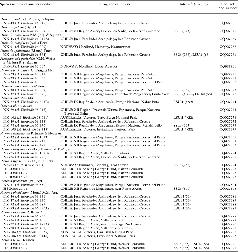
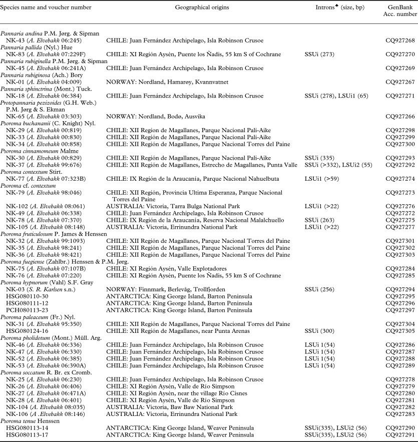
Thirty-nine specimens representing five Pannaria species and ten Psoroma species were analyzed in this study (Table 1). Voucher specimens of all samples are deposited at TROM, and Antarctic specimens also at Korea Polar Research Institute. Collection information is included in Table 1. Collection information on the additional GenBank samples added in Fig. 2 has recently been presented by Passo et al. (Reference Passo, Stenroos and Calvelo2008) and is not included here. Protopannaria pezizoides was included to root the tree.
Details of Pannaria and Psoroma specimens and GenBank accession numbers of the ITS and LSU rDNA sequences generated for this study
The lichen materials were ground using a freeze-crusher (SK200, Tokken, Japan) after freezing in liquid nitrogen, and genomic DNAs were extracted using Plant DNA mini-kit (Qiagen, Germany) according to the manufacturer's guide.
PCR amplification and sequencing
ITS1-5.8S-ITS2 and partial LSU rDNA (1-948 nucleotide positions, S. cerevisiae numbering) were amplified using the primers ITS1F (5′-CTT GGT CAT TTA GAG GAA GTA A-3′) (Gardes & Bruns Reference Gardes and Bruns1993) and LR5 (5′-ATC CTG AGG GAA ACT TC-3′) (Vilgalys & Hester Reference Vilgalys and Hester1990). Touch-down PCR amplifications were performed in a T-gradient thermocycler (Biometra, Germany) with the following cycling parameters: 5 min. initial denaturation at 94°C, 15 touch-down cycles of 30 s. denaturation at 94°C, 1 min annealing at 62–55°C at the ramp of 0.5° per cycle and 2 min extension at 72°, followed by 30 cycles of 30 s. denaturation at 94°C, 1 min annealing at 55°, and 1 min extension at 72°, and 10 min. final extension at 72°. Sequences of ITS and LSU rDNA regions were determined with primers, ITS1F, ITS4 (TCC TCC GCT TAT TGA TAT GC) (White et al., Reference White, Bruns, Lee, Taylor, Innis, Gelfand, Sninsky and White1990), LR0R (5′-GTA CCC GCT GAA CTT AAG C-3′) (Rehner & Samuels Reference Rehner and Samuels1994), and LR5 using ABI 3730XL automated sequencer (Applied Biosystems, USA). The sequences were proof read, edited and aligned using the jPHYDIT program (Jeon et al., Reference Jeon, Chung, Park, Hur, Lee and Chun2005).
Phylogenetic analysis
Sequence alignment of ITS1, 5.8S, ITS2 and partial LSU rDNA were conducted by the program ClustalX ver. 1.83 (Thompson et al. Reference Thompson, Gibson, Plewniak, Jeanmougin and Higgins1997) and manually adjusted. Ambiguously aligned sites were excluded from the phylogenetic analyses. Phylogenetic trees were inferred from the data sets by neighbor-joining (NJ), maximum parsimony (MP), and maximum likelihood (ML) analyses. NJ tree was reconstructed using NJ algorithm of PAUP 4.0b10 (Swofford Reference Swofford2002) under the Kimura's 2-parameter model (Kimura Reference Kimura1980). Maximum parsimony analysis was conducted by heuristic search option of PAUP 4.0b10 with 1000 replicates of random sequence additions, tree bisection reconnection (TBR) branch swapping algorithm, and MulTrees options in effect. All gaps were treated as missing data. Weighted MP analysis (wtMP) was conducted after weighting characters by rescaled consistency index (RC) values with the same search parameters as MP analysis. ML tree was searched by PhyML ver 3.0 (Guindon & Gascuel, Reference Guindon and Gascuel2003) with the HKY evolutionary model (Hasegawa et al. Reference Hasegawa, Kishino and Yano1985) and the search options of best tree topology finding by branch swapping of NNIs and SPRs, initial tree constructed by BioNJ method, and parameter estimation for proportion of invariant and transition/transversion ratio. Supports for internal branches were tested by the bootstrap analyses of 1000 and 100 replications for MP and ML searches, respectively.
Morphological and anatomical analysis
Type material of most of the species studied, in addition to our own collections and selected collections from other herbaria, were subjected to morphological, anatomical and chemical studies and compared with related species from our own and other collections available. Chemistry was studied using standard TLC methods based on Culberson (Reference Culberson1972) and Orange et al. (Reference Orange, James and White2001), involving solvents A and C. Iodine+ reactions were studied by adding IKI to mounts pretreated with KOH (Orange et al. Reference Orange, James and White2001), but perispore structures were studied on material mounted in water only. Nomenclature of ascospore structures follows Nordin (Reference Nordin1997). Because of the challenges in separating the species, anatomical comparisons were particularly thorough with copies of detailed drawings of 6–15 spores and IKI+ ascus structures of all cited specimens with mature apothecia enclosed with the herbarium specimens.
Results
PCR and sequencing
Approximately 1·5 kb DNA fragments were amplified from most of the specimens with the primers ITS1F and LR5, but size variation was observed in some of the specimens with larger DNA fragments ranging between 1·6 kb and 1·85 kb (data not shown). Sequence alignment with reference sequences revealed that size variation was mostly caused by the presence/absence of intron sequences after 1770 nt position (SSUi; S. cerevisiae numbering) of SSU rDNA and/or after 915 and 917 nt positions (LSUi1 and LSUi2; S. cerevisiae numbering) of LSU rDNA (Table 1).
Phylogenetic analysis
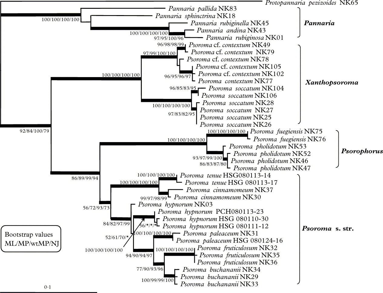
Combined sequences of ITS1-5.8S-ITS2-partial LSU rDNA sequences excluding intron sequences and ambiguously aligned nucleotide sites constituted a total of 1344 characters. Among them 1102 sites were constant, 56 sites were variable but parsimony-uninformative, and 186 sites were parsimony-informative characters. The ML tree is presented with bootstrap support values by ML, MP, wtMP, and NJ searches in Fig. 1. Strict consensus of six and two equally parsimonious trees by MP and weighted MP analyses and NJ tree were very similar to the tree by ML search. Branches conserved in four independent phylogenetic analyses were presented as thick lines. Most of the branches that were strongly supported by high bootstrap values were maintained in trees by different phylogeny methods. Disagreement among trees was found in relationships among samples of Psoroma contextum and among samples of P. hypnorum from the Antarctic. The difference was also found in the position of Pannaria sphinctrina NK18 and Pannaria pallida NK83. Phylogenetic trees reconstructed using the ITS region and LSU rDNA separately, maintained the relationships of main lineages which were conserved in combined sequence analyses (data not shown).
Maximum likelihood tree based on combined sequences of ITS1-5.8S-ITS2-partial LSU rRNA gene. Branches that were conserved in ML, MP,wtMP and NJ are represented as thick lines. Bootstrap values are indicated when values were above 50%.
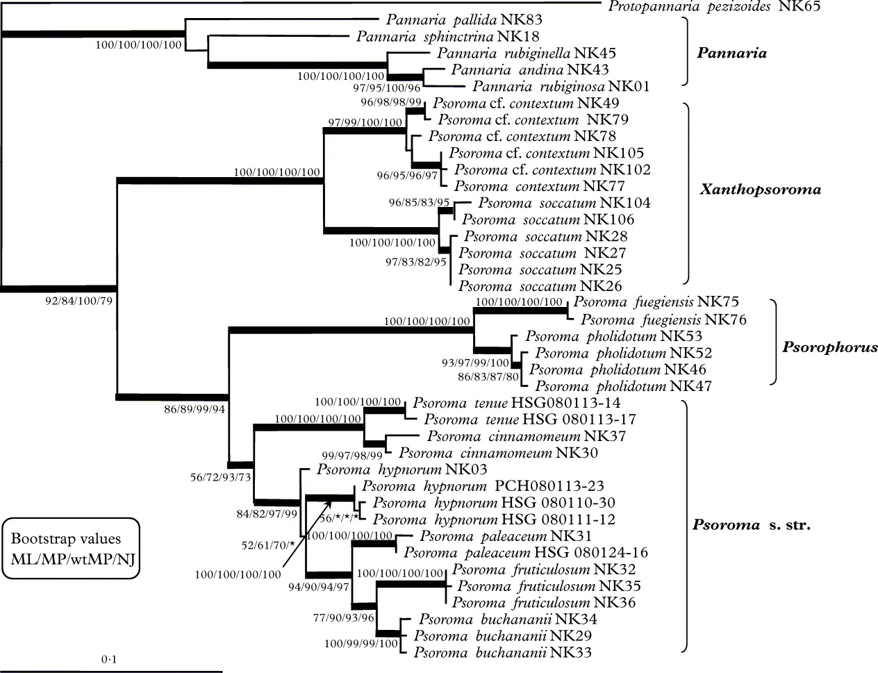
In the phylogenetic trees by ML, MP, wtMP and NJ analyses, Pannaria species and Psoroma s. lat. species were clearly distinguished. Monophyly of each lineage was strongly supported by high bootstrap values. Within the Psoroma s. lat. lineage, corticolous species were separated from the terricolous species. The Xanthopsoroma lineage was composed of the corticolous species, Psoroma soccatum and P. contextum, and monophyly of the lineage was supported by 100% bootstrap values. The two species P. soccatum and P. contextum s. lat. were also clearly separated. The Psorophorus lineage was composed of Psoroma pholidotum and P. fuegiense, also corticolous species, and monophyly of the lineage was supported by 100% bootstrap value. The species were also very well separated. The Psoroma s. str. lineage was composed of terricolous species from Chile, Norway and the Antarctic. Monophyly of the lineage was maintained in ML, MP, wtMP and NJ trees and bootstrap support was moderate (ML, MP and NJ) or high (wtMP). Psoroma cinnamomeum and P. tenue formed two clades well isolated from other species within Psoroma s. str. The samples of P. buchananii, P. fruticulosum and P. paleaceum formed strongly supported monophyletic species clades, whereas the only European sample of P. hypnorum included, did not associate well with the Antarctic samples. Psoroma buchananii is reported as new to Chile here.
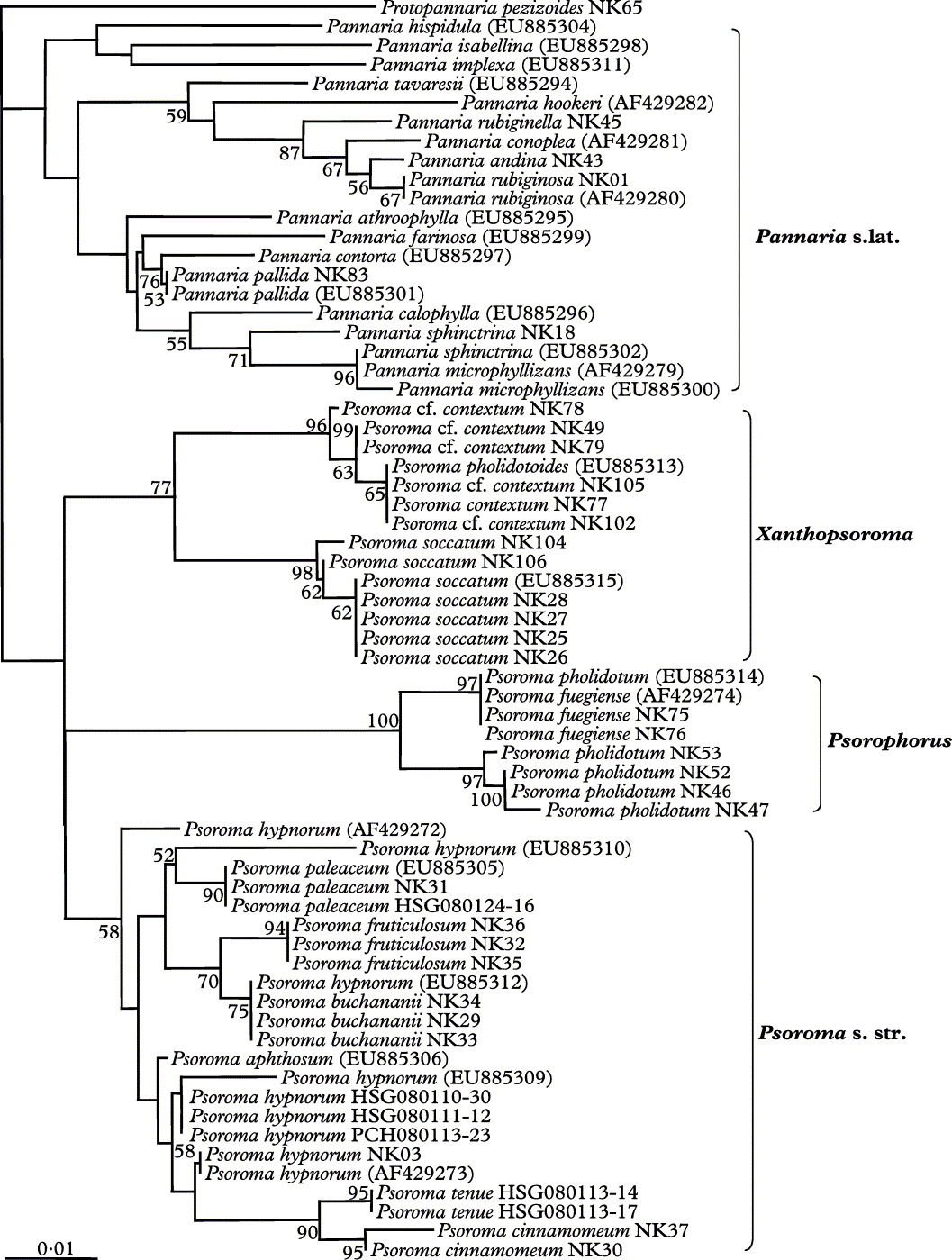
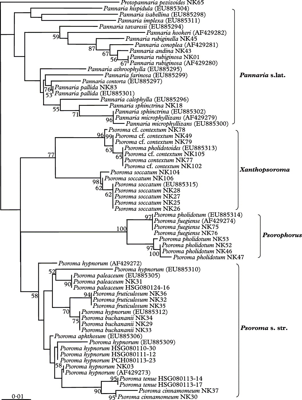
ITS sequences from this study and others retrieved from GenBank database were aligned together and phylogeny was reconstructed by the NJ method (Fig. 2). In this analysis, the four main lineages defined by combined sequences of ITS and LSU rDNA (Fig. 1) were maintained. Supports for the monophyly of Xanthopsoroma and Psorophorous clades were relatively high (77% and 100%), but supports for the monophyly of Pannaria s. lat. and Psoroma s. str. were low (<50% and 58% respectively). Within the Pannaria clade, the five samples included in Fig. 1 grouped nicely with samples from the same or closely related species in the corresponding clade of Fig. 2, now numbering 20 samples. The two GenBank sequences added to the Xanthopsoroma clade were almost identical to sequences included in Fig. 1. The same was the case in the Psorophorus clade, although a sample previously analyzed phylogenetically as Psoroma pholidotum (Passo et al. Reference Passo, Stenroos and Calvelo2008), grouped with fuegiense instead of pholidotum. The GenBank sequence of P. paleaceum grouped nicely with the two sequences already included in Fig. 1, whereas P. hypnorum is more heterogeneous after the addition of another three GenBank sequences, in addition to one more sequence (EU885312) appearing in Fig. 2 as a possibly misidentified specimen of P. buchananii.
Neighbor-joining tree based on the ITS1-5.8S-ITS2 rRNA sequences. Accession numbers are included for the sequences retrieved from GenBank. Bootstrap values are indicated when values were above 50%.
The Genera and the Species
Psorophorus Elvebakk & Hong gen. nov
Psoromati sensu stricto (ut Psoroma hypnorum id definit) similis, sed corticola plerumque, hypothallo vel prothallo nigro distincto, squamulis et cephalodiis tenuibus et valde ad substratum adpressis, structura ascorum amyloidea interna tubuliformi instructus, et melaninis destitutus.
Generitypus: Psorophorus pholidotus (Mont.) Elvebakk (2010).
Thallus squamulose, 2–15 cm wide, corticolous. Chlorobiont squamules 70–100 μm thick, densely appressed to the substratum, up to 1 mm wide but mostly 0·1–0·5 mm, circular to irregularly incised, discrete, becoming confluent centrally, strongly salad green when wet, distinctly pale greenish when dry, a colour maintained in specimens several years old but which turn olive-brown after decades of storage. Hypothallus 10–15 μm thick, very distinct and black, protruding into a 2–5 mm wide prothallus, also present below squamules. Upper surface glabrous and glossy. Epicortex 15–20 μm thick, of thin-walled (c.1 μm thick ) cells, 3–5 μm in size. Chlorobiont of cf. Myrmecia cells, irregularly globose to ellipsoid, 6–12 × 7–13 μm in size, chlorobiont layer 50–70 μm thick. Medulla 10–15 μm thick. Hypocortex lacking. Cephalodia grey, very abundant, located directly on the prothallus/hypothallus, very rarely on squamules, 0·6–1·2 mm wide, when young more or less placodioid, later developing small erect lobules. Cyanobiont Nostoc, cells greyish green, irregularly subglobose or ellipsoid, 3–6 × 4–7 μm in size, in 20–40 μm large, indistinct glomeruli, without visible chain structures.
Apothecia sessile to subsessile, laminal, 0·8–1·5 mm wide. Discs at first dark chestnut brown or almost blackish, gradually becoming pale brown with an orange to reddish tinge, and flat or convex when old. Thalline excipulum 40–120 μm wide on mature apothecia when seen from above, crenulate-striate, with anatomy as in the squamules, irregular and present or evanescent on older apothecia, basal parts mostly hirsute. Epithecium light brown, c. 10 μm thick. Hymenium colourless, but strongly IKI + blue, c. 80 μm thick. Asci clavate, 15 × 70–80 μm in size, with 8 ascospores and a tube-like, IKI+ blue, apical structure, sometimes with one or two additional cap-like structures. Proper ascospores hyaline, non-septate, subglobose to ellipsoid, 12–20 × 6·5–12 μm. Perispores of the same shape, 12–22 × 7–13 μm, distinctly verrucose, commonly with thick, nodulose apical extensions, 2·5–4 μm in size. Paraphyses 4–5-septate, simple to sparingly branched, apices 2–4·5 μm wide, distinctly swollen with short articulated segments. Hypothecium light brown, c. 70 μm thick, IKI negative.
Chemistry. No compounds detected by TLC, and no visible melanins/pigments except in the apothecia.
Etymology. The name refers to the very distinct and black hypothallus/prothallus ‘carrying’ discrete squamules and cephalodia.
Key to the species of Psorophorus
1 Cephalodia pale grey, rosettiform to umbilicate when young; mature apothecia strongly convex with evanescent margins, long-ellipsoid, often rather irregular proper ascospores, and perispores commonly with nodulose apical extensions ... ... P. pholidotus
Cephalodia dark grey, irregularly cushion-formed when young; mature apothecia weakly convex without evanescent margins, subglobose to short-ellipsoid proper ascospores, and perispores without nodulose apical extensions ... P. fuegiense
Psorophorus pholidotus (Mont.) Elvebakk comb. nov
Basionym: Parmelia pholidota Mont., Annls. Sci. Nat. Bot. Ser. 2, 4:18 (1835); type: Ins. Juan Fernández, in cortice Drymis (canelo), April 1830, Bertero 1626 (PC-MONT, ‘holotype’ (Jørgensen Reference Jørgensen2003), here selected as neotype). —Pannaria pholidota (Mont.) Nyl., Annls. Sci. Nat. Bot. Sér.4, 3: 182 (1855).—Psoroma pholidotum (Mont.) Müll. Arg., Flora 71: 45 (1888).
Pannaria reticulata Hue, Nouv. Arch. Mus. Nat. Hist. Paris Sér. 4, 8: 261 (1906), nom. superf., see Jørgensen (Reference Jørgensen2003: 71).—Psoroma reticulatum (Hue) Zahlbr., Catal. Univ. Lich. III: 294 (1925), nom. superf., see Jørgensen (Reference Jørgensen2003: 71).
Notes on typification. Jørgensen (Reference Jørgensen2003) cited the type as “Juan Fernández, ad corticem Drymidis, April 1830, Bertero 1626 (PC-MONT, holotype !)”. However, Montagne (Reference Montagne1835) did not mention Bertero 1626, but only cited Bertero 1623, “crescit ad corticem Drymis chilensis, vulgò Canelo”. Searches in PC only revealed an unnumbered Bertero collection ‘ex Psidium’, and Bertero 1626. The latter carries the information “In cortice Drymis (canelo), Ins. Juan Fernandez, 1830 april”. According to D. J. Galloway (pers. comm.) this has been written in Bertero's handwriting, but there is no evidence of Montagne's handwriting. There is a possibility that Montagne (Reference Montagne1835) erroneously wrote ‘1623’ instead of ‘1626’. Gay (Reference Gay1852: 147) in his survey of Chilean lichens cited both Bertero 1623 and 1626 as collections of this species. The same is the case with Zahlbruckner (Reference Zahlbruckner and Skottsberg1924). However, the Bertero 1623 collection was not cited by Hue (Reference Hue1908) who studied P. pholidotus extensively. It probably does not exist, not in H-NYL either. In conclusion, Bertero 1626 is defined as a neotype here. Both PC collections are in good agreement with the collections of the species made recently in the Juan Fernández Islands.
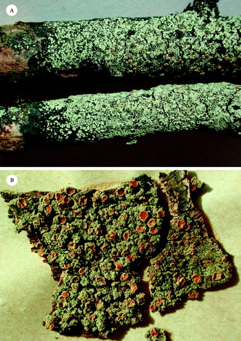
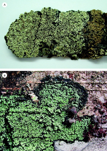
Psorophorus species. A, P. pholidotus photographed of an almost 3-year old herbarium specimen (A. Elvebakk 06:336); B, P. fuegiensis (holotype).

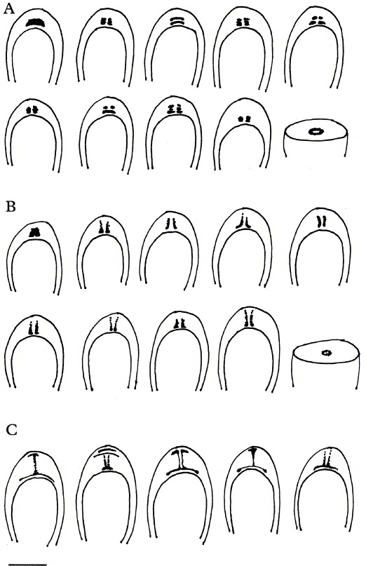
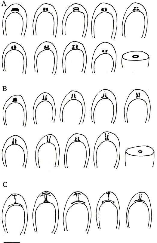
IKI+ ascus apical tube structures. A, Psorophorus pholidotus (the first one studied with high IKI concentration); B, P. fuegiensis (the first one studied with high IKI concentration); C, Psoroma hypnorum. Scale = 10 μm.
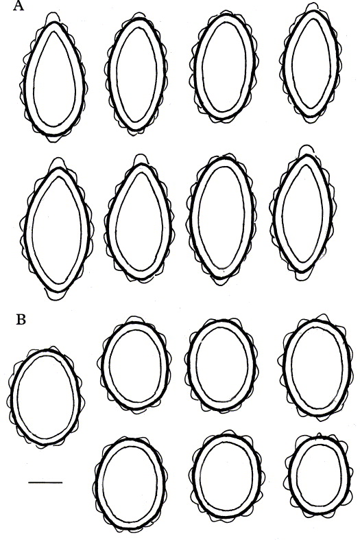
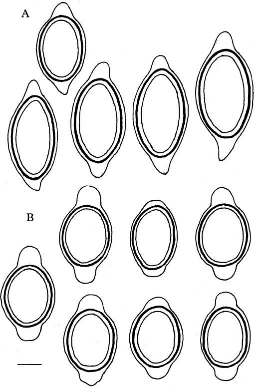
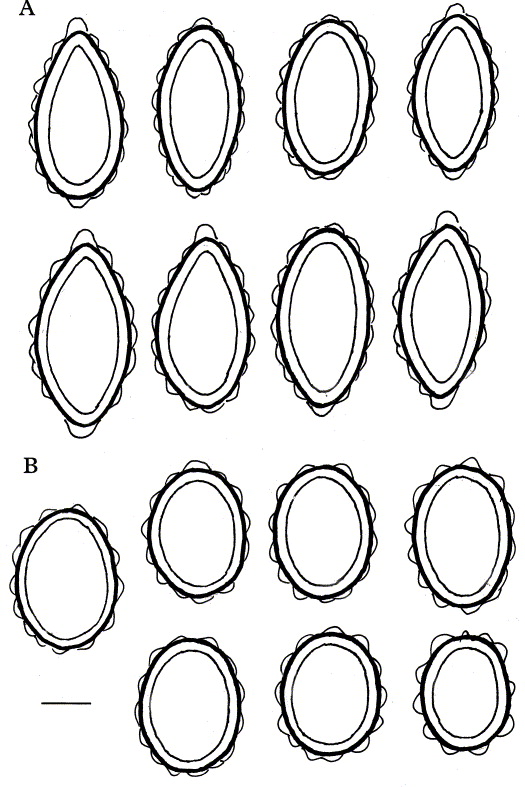
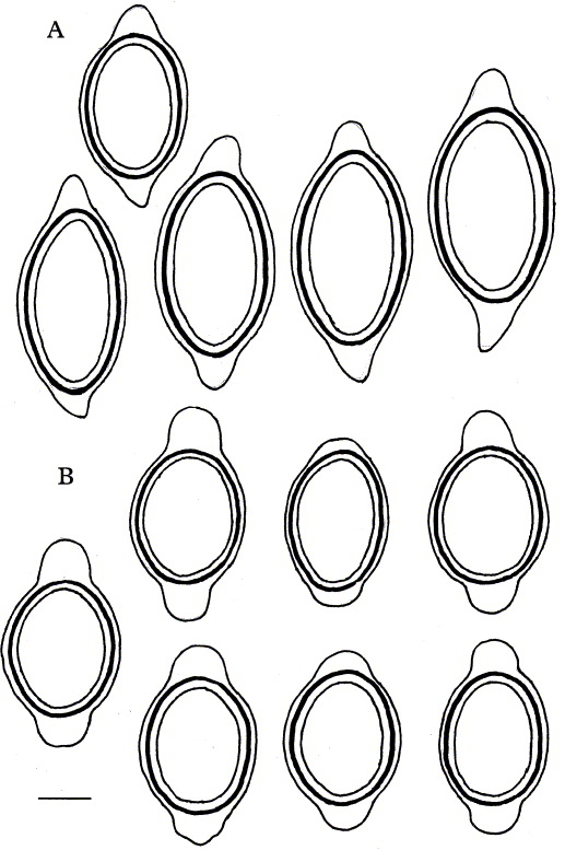
Psorophorus, ascospores. A, P. pholidotus; B, P. fuegiensis. Scale = 5 μm.
Characters as stated for the genus, except for the following:
Cephalodia pale grey, when young regularly rosettiform, placodioid to weakly umbilicate, later developing small erect lobules. Apothecium discs developing a strong convexity when old. Thalline excipulum mostly evanescent on older apothecia. Asci with a short and broad, tube-like, IKI+ blue, apical structure. Proper ascospores long-ellipsoid, often weakly lacrymiform or weakly acuminate, 14–20 × 6·5–10 μm; perispores of the same shape, 16–22 × 7–12 μm, distinctly verrucose, commonly with thick, nodulose 2·5–4 μm apical extensions.
Additional specimens studied. Chile: Arquipiélago de Juan Fernández: Isla Robinson Crusoe, Miradór de Selkirk and 0·5 km E of Miradór de Selkirk, 33°38′S, 78°51′W, 2006, A. Elvebakk 06:311 (SGO); 06:315; 06:319A; 06:330; 06:336; 06:337 (TROM); 1·5 km W of San Juan Bautista, 33° 38′S, 78° 50′W, 2006, A. Elvebakk 06:257 (TROM); Plazoleta del Yunque, 33°38′S, 78° 50′W, 2006, A. Elvebakk 06:385; 06:386; 06:390A; 06:393; 06:394 (TROM); 06:397 (UPS); Cordón Pesea de los Viejos, 1975, W. Quilhot s.n. (UV). XIV Región de los Ríos: Lago Riñihue, Riñihue, Cerro Tralcan, 1940, R. Santesson 3805 (S). X Región de los Lagos: Chiloé, Cucao, Parque Nacional de Chiloé, near Guardería, at Tepual path, 42° 37′S, 74° 06′W, 2000, A. Elvebakk 00:521 (TROM). XI Región Aysén: 14 km E of Tortel, 47° 47′S, 73° 23′ W, 2007, A. Elvebakk 07:246 (TROM).
Psorophorus fuegiensis (Zahlbr.) Elvebakk & Hong comb. nov
Basionym: Pannaria fuegiensis Zahlbr., Kungl. Sv. Vetensk.-Akad. Handl. 57(6): 13 (1917); type: (Chile): Feuerland, im Walde inweit der Mündung des Rio Azopardo, auf Baumrinden (W— holotype!).—Psoroma fuegiense (Zahlbr.) Henssen & P. M. Jørg. (as “fuegense”), New Zeal. Journ. Bot. 37: 261 (1999).
Psoroma microlepideum Malme, Ark. Bot. 20; 6: 17 (Reference Zahlbruckner1925);—type: 'Patagonia occident., Isla Desolacion, Puerto Angosto (Chile), Dusén Fueg. # 215 (S—holotype!), syn. nov.
Psoroma microlepideum var. pallidum Räsänen, Ann. Bot. Soc. Zool.-Bot. Fenn. ‘Vanamo’ 2, I-VI: 45 (1932); —type: Chile, Fuegia occ., Fiordo Finlandia, Bahia Kairamoi, in silva paludosa Nothofagi antarcticae ad corticem arboretum, 3 March 1929, H. Roivainen (H—holotype).
Identical or broadly overlapping with characters of P. pholidotus, except in the following:
Cephalodia dark grey, starting as irregular, placodioid, weakly convex cushions, gradually developing small erect lobules and becoming similar to those of P. pholidotus. Apothecia remaining rather flat or only weakly convex when mature. Thalline excipulum rarely evanescent on old apothecia. Asci with a small and narrow, tube-like IKI + blue apical structure. Proper ascospores subglobose to short-ellipsoid 10–17 × 9–12 μm, never lacrymiform nor apiculate; perispores 12–19 × 10–13 μm, rarely with any apical extensions.
Additional specimens studied. Chile: IX Región de la Araucanía: Parque Nacional Conguillío, 1986, W. Quilhot s.n. (UV); Road from Melipeuco to Lago Conguillío, road to Administration Centre, 38° 49′ S, 71° 40′ W, 1986, B. J. Coppins, D. J. Galloway, G. Guzmán & P .W. James 5647 (BM). X Región de los Lagos: Provincia de Llanquihue, Parque Nacional Vicente Pérez Rosales, near Petrohué, 41°09′ S, 72°23′W, 2001, J. W. Bjerke 517/01 (TROM); 2000, A. Elvebakk 00:570 (TROM). XIV Región de los Ríos: Lago Riñihue, Enco, 1940, R. Santesson 7616a (S); Lago Panguipulli, Volcán Choshuenco, 1940, R. Santesson 3938B (S); 11·4 km SW of Choshuenco near Río Enco, 39° 53′ S, 72° 81′ W, 1986, B. J. Coppins, D. J. Galloway, G. Guzmán & P. W. James 4143 (BM). XI Región Aysén: Laguna San Rafael National Park, on slope N of Río Salton, 46° 38′ N – 73° 51′W, 1997, M. Wedin 6104, 6108 (BM); 38 km along the road N of Bahía Murta, 5 km W of Laguna Cofré, 46°11′S, 72°45′W, 2007, A. Elvebakk 07:312B (TROM); Valle Explotadores, Cabañas Alacaluf, 45 km along the road west of Puerto Tranquilo, 46°30′ S, 73°04′W, 2007, A. Elvebakk 07:098; 07:107B (TROM); 55 km SW along the road of Cochrane, 3·5 km N of Puente Los Ñadis, 47°35′ S, 72°52′W, 2007, A. Elvebakk 07:220 (SGO, TROM, UPS); 7 km N of the road junction Tortel and Puerto Yungai, 47°44′ S, 73°14′W, 2007, A. Elvebakk 07:267B (TROM); 0·5 km E of Tortel, 47° 47′ S, 73°31′W, 2007, A. Elvebakk 07:233B (TROM); 4 km E of Tortel, 47° 48 S, 73° 29′ W, 2007, A. Elvebakk 07:236A (TROM).
Discussion of Psorophorus species
All the names in this group have been widely misunderstood, particularly in terms of their photobiont associations. Zahlbruckner (Reference Zahlbruckner1917) interpreted his Pannaria fuegiensis as a bipartite cyanospecies, whereas Malme (Reference Malme1925) described the now synonymous Psoroma microlepideum as a green algal species without any cephalodia observed. Nylander (Reference Nylander1855a, Reference Nylander1859, Reference Nylander1863) evidently considered Psorophorus pholidotus to be a bipartite cyanospecies, as he separated the genera Psoroma and Pannaria principally on their choice of photobionts, and treated this species as Pannaria pholidota, a combination overlooked in recent treatments. This probably explains why he also reported the species from Mexico (Nylander Reference Nylander1859), an area with many bipartite species, but lacking tripartites. Hue (Reference Hue1892, Reference Hue1902, Reference Hue1908) continued this interpretation, but with Pannaria now in a broader sense, and cited it from the island Sao Tomé off West Africa, Bolivia and Sri Lanka (‘Ceylon’), and provided a very thorough description, including cephalodia. One Sri Lankan collection (Thwaites 52, S) has been seen by us, and corresponds to the Palaeotropical ‘Psoroma sphinctrinum’ complex, which is planned to be accommodated in a new genus by A. Elvebakk & P. M. Jørgensen (unpublished).
The species has also been confused with Xanthopsoroma. Nylander (Reference Nylander1855b, Reference Nylander1863) reported a collection from the mainland Chilean cordillera as P. pholidota (Lechler 853, not studied by us). Hue (Reference Hue1902) indicated that this collection, together with a Gay collection from southern Chile, represented a new species, named Pannaria reticulata Hue, however, invalidly, because no description was given at the time. When the description was provided later (Hue Reference Hue1906), he included the name Pannaria pholidota (Mont.) Nyl. as a synonym, and his new species, apparently meant for what we treat here as Xanthopsoroma contextum, therefore became a synonym and a nom. superf. for P. pholidotus, a conclusion already stated by Jørgensen (Reference Jørgensen2003).
Our studies have shown Pannaria fuegiensis and Psoroma microlepideus to be conspecific, with the former name taking priority. Also original spore measurements published by Zahlbruckner (Reference Zahlbruckner1917) and Malme (Reference Malme1925) are very similar. The holotype of Pannaria fuegiensis has a somewhat broader and more robust thalline excipulum than the holotype of Psoroma microlepideum and most other specimens initially determined accordingly, with the exception of Quilhot s.n. However, their anatomy and other characters are identical and the thalline excipulum character is not considered sufficient to merit their recognition as separate species.
The type of Parmelia pholidota is very similar in habit to the types referred to above. Except for their type collections, these species have previously been interpreted so variably that no records are reliable. Malme (Reference Malme1925) wrote, even on the label of the holotype specimen of Psoroma microlepideum, that it might be conspecific with Psoroma pholidotum. A second specimen of Psoroma fuegiense was recorded from Ushuaia by Malme (Reference Malme1925). It was, however, redetermined as Fuscopannaria minor (Darb.) P. M. Jørg. by Jørgensen (Reference Jørgensen1999). The report from the Juan Fernandez Islands by Zahlbruckner (Reference Zahlbruckner and Skottsberg1924) is probably also erroneous. Räsänen (Reference Räsänen1932) described P. microlepideum var. pallida, characterized by a whitish pale thallus, but it has already been included in P. microlepideum by Jørgensen (Reference Jørgensen2003). Follmann (Reference Follman1965) accepted P. microlepideum as a separate rare species, known only from the Region of Magallanes, whereas Psoroma pholidotum was also considered to be uncommon, but with a wide distribution in mainland Chile in addition to its occurrence in the Juan Fernández Islands. Psoroma pholidotum has been accepted for Argentina by Grassi (Reference Grassi1950), Calvelo & Liberatore (Reference Calvelo and Liberatore2002) and Passo et al. (Reference Passo, Stenroos and Calvelo2008), but these records need to be confirmed.
In the present study Psorophorus pholidotus and Psorophorus fuegiensis are genetically very distinct. Morphologically, the characters cited are quite good, but not completely reliable. An a posteriori determination test on the 37 collections studied showed that about 85 % of them could be determined on the external apothecium and/or cephalodium characters alone, including several collections from the Juan Fernández Islands where mature apothecia were lacking or poorly developed. It is now shown here that they are very distinct anatomically. Short-ellipsoid vs. long-ellipsoid spores is a very reliable character. The size ranges quoted represent 98% of the measurements. Whereas P. fuegiensis spores are always very regularly elliptic, those of P. pholidotus are frequently either weakly apiculate or lacrymiform.
Surprisingly, we also found clear differences between the two species in the IKI+ ascus apical structures. Cross- and longitudinal sections show a distinct tube structure in both species, which is broad in P. pholidotus, sometimes with additional sheet structures, and narrower in P. fuegiensis. This is a character which has only been previously used at generic or sectional level in Pannariaceae, but is now found to be taxonomically very important at the species level. The IKI+ difference may be considered as the strongest visible manifestation that these two species are very distinct in spite of their morphological similarity. Interestingly, both recent collections used in the phylograms by Ekman & Jørgensen (Reference Ekman and Jørgensen2002) and Passo et al. (Reference Passo, Stenroos and Calvelo2008) can, quite unambiguously, be confirmed by our phylogenetic reanalysis to represent Psorophorus fuegiensis (Passo 126, published as Psoroma pholidotum, and Wedin 6108, published as P. fuegiense).
Psorophorus pholidotus is very common on Isla Robinson Crusoe, but it is not a Juan Fernández endemic. Three well-defined specimens have been studied from the Chilean mainland (from Regions XIV, X and XI), but P. fuegiensis is a far more common species there; it is listed from Regions IX–XII and the recently established Region XIV (between IX and X). It is probably widespread also in the austral, wettest forests of Argentina, but no specimens have been seen by us. The genus Psorophorus is a southern South American endemic.
Xanthopsoroma Elvebakk & Hong gen. nov
Psoromati sensu stricto (ut Psoroma hypnorum id definit) simile, sed corticola plerumque, hypothallo vel prothallo nigro distincto, squamulis valde ad substratum adpressis, perisporis planis et magnis extensionibus apicalibus nodulosis sive truncatis praeditis, structura ascorum interna tubiformi amyloidea valde distincta et tholo generatim IKI+ circumdata instructum, acidum usneicum et nonnulla terpenoida incognita continens.
Generitype: Xanthopsoroma contextum (Stirt.) Elvebakk (2010).
Thallus squamulose, 2–15 cm wide, corticolous, occasionally saxicolous. Chlorobiont squamules 120–180 μm thick, appressed to the substratum, in peripheral parts up to 2 mm wide, in central parts strongly imbricate, yellowish green, turning more yellow to ochraceous after decades of storage. Hypothallus very distinct and black, protruding into a 2–5 mm wide prothallus. Upper surface glabrous and glossy. Epicortex 40–60 μm thick, paraplectenchymatous, cell walls 3–4 μm thick, lumens 5–10 μm, the lowermost elongated and directed towards the surface, the uppermost strongly sclerenchymatic. Chlorobiont of cf. Myrmecia cells, irregularly globose to ellipsoid, 6–15 × 7–15 μm, chlorobiont layer 30–60 μm thick. Medulla 40–60 μm thick. Hypocortex lacking. Cephalodia brown, common, located directly on the prothallus/hypothallus or on the the squamules, 0·3–1·5 mm wide, globose to irregularly pulvinate. Cyanobiont Nostoc, cells bluish green, irregularly subglobose or ellipsoid, 3·5–7 × 4–8 μm, in 15–30 μm large, indistinct glomeruli, without visible chain structures.
Apothecia sessile to subsessile, laminal, 0·5–2·5 mm wide. Discs reddish brown to chestnut-brown, mostly flat. Thalline excipulum 0·1–0·3 mm wide on mature apothecia when seen from above, regularly crenulate-striate, with anatomy as in the squamules. Epithecium light brown, c. 10 μm thick. Hymenium colourless, but strongly IKI + blue, c. 70–80 μm thick. Asci clavate, c. 20 × 70–80 μm, with 8 ascospores and a long and narrow, sometimes apically widening tube-like structure, IKI + blue, sometimes laminated, in addition to a more diffuse large apical IKI+ structure, most often also a small IKI+ external apical cap. Proper ascospores hyaline, non-septate, short-ellipsoid, 12–20 × 8–12 μm; perispores 18–29 × 9–13 μm, ellipsoid, even, and with two broad apical extensions. Paraphyses weakly septate, mostly unbranched, apices 2–3 μm wide, weakly swollen. Hypothecium light brown, c. 70 μm thick, IKI−.
Chemistry. Usnic acid (major/minor) and up to 13 unidentified terpenoids, at present not known from any other lichens and with one to four of them always present in major quantities.
Etymology. Named after its yellow colour due to usnic acid.
Key to the Xanthopsoroma species
1 Soredia present; apothecia not uncommon, but less than 1 mm wide and mostly with a thalline central plug, proper ascospores 15–20 μm long and perispores with nodulose apical extensions. Contains ‘X. soccatum terpenoids’, one in major quantities ... . X. soccatum
Esorediate; apothecia very common, up to 2 mm wide and without thalline central plugs. Proper spore/perispore shapes and chemistry variable ... 2
2(1) Apothecia with thick margins, short-ellipsoid proper ascospores (12–16 μm) and perispores mostly with large, truncate apical extensions. Contains ‘X. contextum terpenoids’, normally three in major quantities ... X. contextum
Deviations from one or more of these four characters ... X. cf. contextum
Xanthopsoroma contextum (Stirt.) Elvebakk comb. nov
Basionym: Psoroma contextum Stirt., Proc. Phil. Soc. Glasgow 10: 294 (1877); type: New Zealand, near Wellington, 1974, J. Buchanan 46/74 (BM— lectotype!; WELT— isolectotype!).
Xanthopsoroma species. A, X. contextum (WELT— isolectotype); B, X. soccatum.

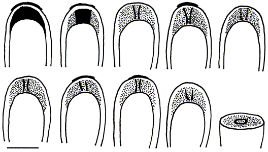
Xanthopsoroma, IKI+ ascus apical structures (two upper left samples with high IKI concentrations). Scale = 10 μm.
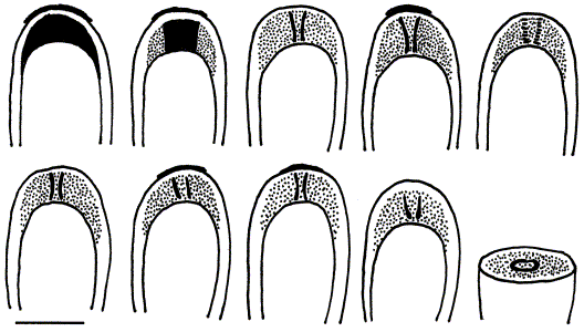
Xanthopsoroma, ascospores. A, X. soccatum; B, X. contextum. Scale = 5 μm.
Characters as in the description of the genus, except for the following:
Chlorobiont squamules 150–180 μm thick, appressed to the substratum, in peripheral parts up to 2 mm wide, in central parts strongly imbricate, yellowish green, turning more yellow after decades of storage. Epicortex 40–60 μm thick. Chlorobiont 7–15 × 6–12 μm, chlorobiont layer 50–60 μm thick. Medulla 50–60 μm thick.
Apothecia common, subsessile, laminal, 0·5–2·5 mm wide. Thalline excipulum 0·2–0·3 mm wide on mature apothecia when seen from above. Asci long and narrow, sometimes apically widening, IKI + blue, tube-like structure. Proper ascospores short-ellipsoid, 12–16 × 8–10·5 μm; perispores 18–25 × 9–11·5 μm, with two 3–4 μm broad apical extensions, either short (c. 1 μm) or enlarged to regularly truncate extensions, 4–5 μm long, occasionally deviating, even vesicular.
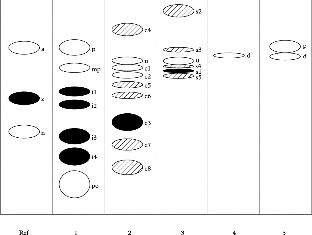
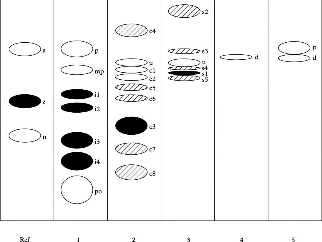
Chemistry. Usnic acid (minor) and three to eight unidentified terpenoids (Fig. 9), at present not known from any other lichen. ‘X. contextum terpenoids 1–3’ are constantly present, mostly in major quantities, ‘X. contextum terpenoid 4’ is present in only 20–30% of the specimens sampled, but in major quantity, ‘X. contextum terpenoids 5–8’ appear mostly in trace quantities, and their presence appears to be depending mainly on the concentrations of the TLC samples.
Chromatogram showing TLC-detectable compounds in solvent C in 1) Pannaria isidiosa Elvebakk & Elix, 2) Xanthopsoroma contextum, 3) X. soccatum, 4) Phyllopsora sp., and 5) Pannaria pallida (Nyl.) Hue. Only major compounds are included among the latter, p = pannarin, mp = methylplacodiolic acid, i1–i4 = ‘Pannaria isidiosa terpenoids 1–4’, po = porphyrilic acid, c1–c8 = ‘Xanthopsoroma contextum terpenoids 1–8’, u = usnic acid, s1–s5 = ‘Xanthopsoroma soccatum terpenoids 1–5’, d = dechloropannarin. References: a = atranorin, z = zeorin, n = norstictc acid. Terpenoids occurring in major quantities are shown in black.
Additional selected specimens examined. Argentina: Tierra del Fuego: Lago Fagnano, E end, Cabezera Lago, 1940, R. Santesson 7927 (S).—Chile: Archipiélago de Juan Fernández: Isla Robinson Crusoe, Miradór de Selkirk, 33° 38′ S, 78° 51′ W, 2006, A. Elvebakk 06:338 (TROM). IX Región de la Araucanía: Parque Nacional Nahuelbuta, Pehuenco, 37° 36′ S, 72° 48′ W, 2008, A. Elvebakk 08: 323B (TROM). XI Región Aysén: 3 km N of the village Río Cisnes, 44° 41′ S, 72° 15′ W, 2006, A. Elvebakk 06:469 (TROM); Valle Explotadores, 10 km W of Punta Río Tranquilo, 46° 37′ S, 72° 51′ W, 2007, A. Elvebakk 07:051 (TROM); 38 km N of Bahía Murta along the road, W of Laguna Cofré, 46° 11′ S. 72° 45′ W, 2007, A. Elvebakk 07:314 (TROM); Puerto Aysén, 1940, R. Santesson 4347 (S). XII Región de Magallanes: Laguna el Parrillar, 40 km SW of Punta Arenas, 53° 22′ S, 71° 07′ W, 1999, A. Elvebakk 99:739 (TROM); Punta Arenas, Tres Puentes, 1940, R. Santesson 8078 (S); Isla Riesco, Mina Elena, 1940, R. Santesson 2051, 7818 (S).—Australia: Tasmania: Pelion Plains, c. 1 km SW of Pelion Hut, saxicolous, 41° 50′ S, 146° 02′ E, 1992, G. Kantvilas 215/92 (HO); Grass Tree Hill, 42° 47′ S, 147° 22′ E, 1981, G. Kantvilas 706/81 (HO); Mt. Sprent, 42° 48′ S, 145° 58′ E, 1987, G. Kantvilas s. n. (HO 12450); Lake Skinner Track, 42° 56′ S, 146° 42′ E, 1980, G. Kantvilas 236/80 (HO); South Sister, near summit, 41° 32′ S, 148° 10′E, 2004, J. A. Elix 28583 & G. Kantvilas (CANB).—New Zealand: South Island: Golden Bay Co., Cobb, Fenella Hut, 41° 03′ S, 172° 31′ E, 1975–1985, J. K. Bartlett s. n. (AK 178956); Buller Co., Mount Arthur, 1975–1985, J. K. Bartlett s.n. (AK 197994).
Xanthopsoroma soccatum (R. Br. ex Crombie) Elvebakk comb. nov
Basionym: Psoroma soccatum R. Br. ex Cromb., Journ. Linn. Soc., Bot. 17: 398 (1879); type: Tasmania, Table Mt. [= Mt. Wellington], R. Brown, Iter austr. 502 (BM— lectotype).
Characters as in the description of the genus, except for the following:
Chlorobiont squamules 120–40 μm thick, appressed to the substratum, in peripheral parts up to 1 mm wide, in central parts strongly imbricate, yellowish green, turning ochraceous after decades of storage. Soralia common, labriform to irregular and laminal, of soredia c. 0·05 mm. Epicortex 40–50 μm thick. Chlorobiont 7–13 × 6–12 μm, chlorobiont layer 30–40 μm thick. Medulla 40–50 μm thick.
Apothecia uncommon, sessile to subsessile, laminal, 0·5–1·0 mm wide, often with a central, thalline plug. Thalline excipulum 0·1–0·2 mm wide on mature apothecia when seen from above. Asci with a long and narrow, tube-like IKI+ blue structure, only occasionally apically widening. Proper ascospores 15–20 × 8–12 μm; perispores 22–29 × 9–13 μm, with two large, irregular and mostly nodulose apical extensions, 2–4 × 2–6 μm.
Chemistry. Usnic acid (major), one to five unidentified ‘X. soccatum terpenoids’, 1 in minor to major quantities, the others mostly as traces (Fig. 9). The terpenoids are presently not known from any other species.
Additional selected specimens examined. Argentina: Prov. Neuquén: Nahuel Huapi National Park, between Puerto Blest and Laguna los Cantaros, 41° 01′ S, 71° 50′ W, 1989, M. Wedin 1831 (UPS).—Chile: Archipiélago de Juan Fernández: Isla Robinson Crusoe, 1·5 km W of San Juan Bautista along the path to Miradór de Selkirk, 33° 38′ S, 78° 51′ W, 2006, A. Elvebakk 06:230; 06:273; 06:284A (TROM). XIV Región de los Ríos: Lago Panguipulli, Volcán Choshuenco, 1940, R. Santesson 3938A (S); Lago Riñihue, Enco, 1940, R. Santesson 7617 (S). XI Región Aysén: Puerto Aysén, 1940, R. Santesson 4350 (S); 15 km S of Lago Torres NE of Puerto Aysén, 44° 57′ S, 72° 10′ W, 2006, A. Elvebakk 06:429 (TROM). XII Región de Magallanes: Península Brunswick, 10–15 km S of Fuerte Bulnes, near Río San Pedro, 53° 36′ S, 70° 58′ W, 1998, A. Elvebakk 98:270B, J. W. Bjerke & E. Domínguez (TROM).—Australia: Victoria: Errinundra National Park, Rainforest Walk, 300 m S of Cobb Hill, 37° 19′ S, 148° 50′ E, 2008, A. Elvebakk 08:122; 08:130; 08:146 (TROM); Bemm River Scenic Reserve, 45 km E of Orborst, 37° 38′ S, 148° 53′ W, 2008, A. Elvebakk 08:085B (TROM). Tasmania. Arve Valley, west of Geeveston, Keough's Creek Forest Walk, 43° 09′ S, 146° 48′ E, 2008, A. Elvebakk 08:219B (TROM); Cradle Mountain-Lake St. Clair National Park, S end of Lake Clair, Cynthia Bay, 42° 07′ S, 140° 10′ E, 2008, A. Elvebakk 08:171 (TROM).—New Zealand: North Island: Wellington, Central Volcanic Plateau, N side of Mt. Ruapehu, 1·5 km S of Whakapapa Village, 39° 12′ S, 175° 32′ E, 2002, A. Elvebakk 02:402 (TROM). South Island: Canterbury, c. 10 km N of Lake Wanaka, 1 km N of Makarora, near Pipson Creek, 44° 14′ S, 169° 15′ E, 2002, A. Elvebakk 02:434 (TROM).
Discussion of Xanthopsoroma species
In both phylograms, X. contextum and X. soccatum appear as distinct clades. The latter species is very homogeneous in all its characters studied. Apart from its soredia it differs from X. contextum in having smaller and thinner squamules, smaller and fewer apothecia mostly with thalline central plugs, narrower thalline excipuli, long-ellipsoid spores, narrower and often nodulose apical perispore extensions, and different terpenoids. There are no significant differences between specimens from South America and New Zealand/Australia. The species are so different that X. soccatum should not be considered a sorediate counterpart of X. contextum as stated, for example by Kantvilas & Jarman (Reference Kantvilas and Jarman1999), who provide good illustrations of both species. Here, and in all other recent literature, X. contextum has been interpreted as Psoroma pholidotoides (Nyl.) Trev. The holotype of the latter, H-NYL 30773!, however, defines a very different species with pannarin as its major secondary compound. This analysis was unambiguous, and in accordance with a previous and unpublished analysis by J. A. Elix.
The chemistry of these species has also been misunderstood. Jørgensen & Galloway (Reference Jørgensen and Galloway1992) cited X. soccatum to contain dechloropannarin and usnic acid, a report repeated by later studies. Dechloropannarin was first reported for X. contextum (as Psoroma reticulatum) by Quilhot et al. (Reference Quilhot, Piovano, Arancibia, Garbarino and Gambaro1989), who reported three chemical strains, with usnic acid and various combinations of three other substances, probably a result of mixed samples. This is the source of the frequently cited chemosyndrome of ‘vicanicin, ± pannarin, ±leprolomin and ±usnic acid’ for the species previously interpreted as P. pholidotoides. Dechloropannarin was described by Elix et al. (Reference Elix, Lajide and Galloway1982) from the lichen Psoroma caesium Müll. Arg., now Phyllopsora sp. (Jørgensen Reference Jørgensen2003). A sample of ‘Psoroma caesium’ was obtained from J. A. Elix. Dechloropannarin is instead a compound closely associated with pannarin, in TLC overlapping with pannarin in solvent A, but slightly below pannarin in solvent C and becoming brownish after storage, compared with pannarin. It appears to have been correctly reported from Pannaria pallida by Quilhot et al. (Reference Quilhot, Piovano, Arancibia, Garbarino and Gambaro1989), but reported as norpannarin from the same species by Passo & Calvelo (Reference Passo and Calvelo2004).
The unidentified terpenoids reported here for the two species are unknown from Pannariaceae, and not known to the present authors to be identical to terpenoids from any other lichen species. They are all different from those of Pannaria isidiosa Elvebakk & Elix, the only other Pannariaceae species, except Pannaria durietzii (P. James & Henssen) Elvebakk & D. J. Galloway, with terpenoids present as major compounds (Fig. 9). They are also different from zeorin, reported from the latter species, but probably erroneously reported from Pannariaceae, as stated by Elvebakk & Elix (Reference Elvebakk and Elix2006).
The chemosyndromes of X. soccatum and X. contextum are very different. The former was found to be very homogeneous in its chemistry as well as in other characters. Xanthopsoroma contextum, on the other hand, was found to be much more complex. A number of specimens are in close accordance with the type, and are referred to in the description and included as cited specimens. They are also widely distributed from southern South America, to New Zealand and Australia, where also X. soccatum is a common species. Among the deviating specimens in the X. contextum complex, several primarily fertile specimens, including NK 78 and 79 in the phylograms, had X. soccatum chemistry so that we searched for a primarily fertile counterpart species of the latter. However, characters derived from a limited number of specimens did not form any clear pattern. Five specimens among these deviating types are represented in the phylograms, where they were clustered with contextum and not with soccatum. These deviating samples are at present referred to as X. cf. contextum, and further studies based on a larger selection of samples are needed.
The genus Xanthopsoroma is very distinct in several characters, and its primarily fertile species can be expected to be evolutionarily old, allowing for some variation which is not yet understood.
General Discussion
The genera
The trees presented in Figs 1 & 2 confirm the previously published pattern of a major subdivision into a Pannaria s. lat. and a Psoroma s. lat. clade. In the Pannaria clade, the bipartite species were separated from the tripartite ones, as in the study by Passo et al. (Reference Passo, Stenroos and Calvelo2008). In Fig. 2, the squamulose and foliose tripartite species are in different positions. However, this tree is based on ITS only, the number of added sequences is low, and relationships within the Pannaria s. lat. clade are outside the scope of the present study.
Psorophorus is morphologically a distinct genus, with its small and very thin, corticolous squamules. They maintain a distinct green colour much longer than other similar species such as the pannarin-containing species such as P. implexum, which soon become more uniformly grey after storage. Psorophorus has a very massive chlorobiont layer, a poorly specialized epicortex, with very thin-walled cells. Together with the lack of secondary compounds this probably accounts for the maintenance of the green algal colour in the lichen. Thus the colour of the genus is surprisingly similar to the usnic acid genus Xanthopsoroma. It should also be noted that there is no abrupt change of colour in these lichens when water is added to dead specimens as in Xanthopsoroma and tripartite Pannaria species with a TLC-detectable chemistry (see Elvebakk Reference Elvebakk2007). However, there is a slow colour change to olive-brown after decades of storage.
The cephalodia are very characteristic, often larger than the chlorobiont squamules, and always develop directly on the hypothallus/prothallus, although this is less evident in central parts of the thallus where the squamules are confluent. In Psorophorus pholidotus in particular, cephalodia are very conspicuous and often represent 20–40 % of total cover of squamules on the hypothallus. The lack of secondary cortical compounds and specialized cortical cells probably represents an adaptation to humid areas. The unusual high biomass of cephalodia might be an adaptation to low light intensities. The habitat conditions for both species known to us are in old-growth forests of high rainfall areas in Chilean Patagonia and in Isla Robinson Crusoe. In the latter area, P. pholidotus was collected in a protected, moist area (Plazoleta del Yunque, at 250–300 m altitude, with a strong preference for trunks of Drimys confertifolia) and on a light-exposed, but much fog-affected pass (Miradór de Selkirk, 540–570 m altitude, on thin twigs of shrubs), which would be in agreement with the species' obvious requirement for humidity on the mainland.
Chemically, the genus is TLC negative, similar to most species in Psoroma s. str., and different from most other small-squamulose, tripartite Pannariaceae species. It differs from the former in many characters, such as its strictly corticolous habitat, thin and strongly adpressed squamules, shape of cephalodia, lack of melanins, and microscopically by its small tube-like IKI+ ascus apical structure. However, Psorophorus is a sister group to Psoroma in the phylogram, and the closeness of these two groups illustrates the taxonomic importance of chemistry in psoromoid lichens.
Xanthopsoroma is now shown to be a very distinct genus. Its general appearance differs from Psoroma s. str. in being corticolous with adpressed squamules on a very distinct hypothallus, and from Psorophorus by having much thicker squamules and a differentiated cortical cell structure. Its unique chemistry and perispore structure separate it from all other genera in Pannariaceae. Except for its usnic acid colour it looks quite similar to other corticolous squamulose species, such as Psoroma implexum Stirt., now transferred to Pannaria by Passo et al. (Reference Passo, Stenroos and Calvelo2008).
Apical amyloid structures have long been considered an important character in Pannariaceae (Keuck Reference Keuck1977; Jørgensen Reference Jørgensen1978), but have rarely been studied in detail, although Keuck (Reference Keuck1977) included an illustration (fig. 180, of ‘Psoroma reticulatum’), most probably referring to X. contextum. The present study shows that both Psorophorus and Xanthopsoroma have long, IKI+ apical tubes without the basal and apical internal sheets mostly seen in the asci of Psoroma hypnorum. In addition, Xanthopsoroma also has a general, but less IKI+ active tholus, which can hide the tube structure when studied in high IKI concentrations. Most asci of Xanthopsoroma also have an external apical IKI+ sheath.
These findings confirm several previous statements that chemistry and ascus IKI structures are important synapomorphies in the phylogeny of Pannariaceae lichens. Our phylogenetic analyses have also indicated that a stable classification is obtained only after a reasonable number of samples are represented in each clade. This is valid also for single species defined as study targets, and is an aspect mostly lacking from previous phylogenetic studies involving Pannariaceae.
Psoroma s. str. species
Psoroma s. str. was introduced primarily as a reference to the phylogenetic study of the species now positioned in Psorophorus and Xanthopsoroma. The selection of material included several collections of each of six terricolous/muscicolous Psoroma s. str. species. The genus is monophyletic in both our trees. For comparison, Psoroma hypnorum itself has been studied, and found to differ from the two new genera in its terricolous/ muscicolous habitat, inconspicuous hypothallus, thick squamules not densely adpressed to the substratum, presence of brownish melanins, perispores with more robust verrucae, and large, verruciform apical extensions. The IKI+ apical ascus structure in P. hypnorum was called a ‘manubrium’ by Jørgensen (Reference Jørgensen1978), a properly chosen name meaning ‘handle’. More detailed illustrations here confirm this structure which is clearly different from those in the two new genera.
Most of these characters are valid also for the remaining Psoroma s. str. species included in this study, although detailed studies on IKI + apical structures have not yet been carried out, and perispore structures also need to be more carefully compared. The Psoroma s. str. clade also shows a dichotomy, with the four sequences of P. tenue and P. cinnamomeum forming a very well-supported subclade. This was unexpected, as there are no evident synapomorphies characterizing these two clades separately, as opposed to characters of each of the species involved. This phylogenetic analysis reveals that these groups obviously have had a long-lasting separate evolutionary history, and we believe this is the primary reason for the rather low bootstrap support of the present Psoroma s. str. clade. This will be explored more thoroughly in a future study involving many more planned sequences.
Although nested within the Psoroma s. str. clade in Fig. 2, Psoroma aphthosum is a deviant species. It is corticolous, has pannarin instead of melanins and has a different squamule structure. Further detailed studies are needed before its taxonomic position can be confirmed.
After the present treatment above, 26 of the species listed within Psoroma by Jørgensen (Reference Jørgensen2003) still remain. A final definition of Psoroma s. str. after the segregation of Psorophorus and Xanthopsoroma also depends on future studies of additional primarily corticolous species, particularly from New Zealand and south-eastern Australia, and a group of foliose species centred in tropical South East Asia. Psoroma also needs to be defined vs. the poorly understood genus Santessoniella. With its biatorine apothecium and amyloid apical ascus ring/tube, it was originally thought to be most closely related to the bipartite cyanolichen genus Parmeliella (Henssen 1997). However, the tube structure could instead reveal a relationship with Psoroma, and Santessoniella species. Some of them should be studied further to investigate any potential homologies of their tube structures with Psoroma cephalodia, a hypothesis first put forward by Ekman & Jørgensen (Reference Ekman and Jørgensen2002).
The study by Passo et al. (Reference Passo, Stenroos and Calvelo2008) did not resolve the relationship between the studied species in the Psoroma hypnorum group. In the present study P. fruticulosum forms a well-defined group of three closely related samples. Psoroma buchananii also forms a well-defined clade of closely related samples. In Fig. 2 the Passo 39 sample of P. hypnorum is found to be genetically almost identical to one of the present samples of P. buchananii and it is believed to belong to the latter species. Psoroma paleaceum also forms a well-defined clade, including the sample Passo 22 shown in Fig. 2.
Two Chilean samples of Psoroma cinnamomeum and two Antarctic samples of Psoroma tenue formed a very well-defined subclade within the Psoroma s. str. group. This is a phylogenetic position in need of further studies, which is also the case with P. hypnorum itself, which appears heterogeneous in the present phylograms.
The authors are indebted to the curators of AK, BM, CANB, H, HO, PC-MONT, TROM, S, UV, W, and WELT for loans and for the opportunity to study at their herbaria, to CONAF for collection permits in Chile, to the Dept. of Sustainability and Environment in Victoria and the Dept. of Primary Industries, Water and Environment, Tasmania, for collection permits in Australia, and to J. A. Elix, Australian National University, for a reference sample. The first author also appreciated Jack Elix's hospitality and ‘5 o'clock beer discussions’ when in Australia, and nice company in the field together with him, P. McCarthy and G. Kantvilas, as well as hospitality shown by D. J. Galloway, Landcare Research, and W. Quilhot and C. Rubio, Universidad de Valparaíso, during visits to New Zealand and Chile, respectively. Per P. Aspaas, Tromsø University Library, kindly translated the diagnoses into Latin. This research was partly supported by the Korea Polar Research Institute (Grants PE08050 and PE09130).