GBS (Group B Streptococcus/-cocci) is a leading agent of severe, invasive bacterial infection in human newborns. Neonatal infection with this opportunistic pathogen can present as early-onset or late-onset disease. In early-onset cases, bacteria are transferred from the mother to the infant in utero, following ascending infection of the placental membranes, or during passage through the birth canal, by aspiration of infected vaginal fluids. Early-onset neonatal infection manifests within the first few hours or days of life, often presenting as pneumonia and respiratory failure, which can quickly progress to bacteraemia and septic shock. By contrast, late-onset GBS disease can occur in infants up to several months old, and is distinguished by bloodstream infection with a high rate (40–60%) of progression to meningitis (Ref. Reference Johri1). Infants that survive GBS meningitis can suffer serious long-term neurological consequences, such as seizures, hearing loss and cognitive impairment. Serious GBS infections are increasingly recognised in adult populations, particularly in the elderly and individuals compromised by underlying medical conditions. More than 40% of all invasive GBS cases in the USA occur past infancy (Ref. Reference Edwards and Baker2).
The development of GBS disease reflects successful bacterial colonisation of the vaginal epithelium, penetration of placental or epithelial barriers, resistance to immune clearance allowing bloodstream survival and, in cases of meningitis, the ability to breach the endothelial blood–brain barrier (BBB). In overcoming these obstacles, GBS expresses a diverse array of surface-associated and secreted virulence factors that mediate specific host-cell interactions and interfere with innate immune clearance mechanisms. The present review explores knowledge of GBS virulence mechanisms at each key step of disease progression, with particular emphasis on the most recent molecular insights gained from studies of isogenic bacterial mutants using in vitro and in vivo models of GBS infection.
Adherence to host epithelial surfaces
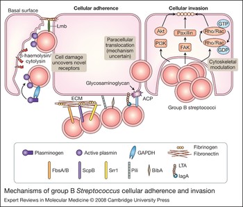
The pathogenesis of GBS disease can first be traced to asymptomatic mucosal colonisation, particularly of the maternal urogenital tract. Approximately 25% of healthy adults carry GBS, and the majority of babies born to a mother who harbours the bacteria will also become colonised (Ref. Reference Campbell3). GBS bind avidly to human vaginal epithelial cells under the low pH conditions characteristic of vaginal mucosa, through the low avidity interactions of cell-wall-associated lipoteichoic acid (LTA) and via higher-affinity interactions mediated by hydrophobic GBS surface proteins. Many of these GBS–host-cell interactions involve attachment of the bacterium to extracellular matrix (ECM) molecules such as fibronectin, fibrinogen and laminin, which in turn bind host-cell-surface proteins such as integrins (Fig. 1).
Mechanisms of group B Streptococcus cellular adherence and invasion. Surface-expressed proteins FbsA/B, ScpB, Srr1, pili, BibA, LTA and ACP mediate group B Streptococcus (GBS) binding to host cells and ECM components, such as fibrinogen and fibronectin. Secreted β-haemolysin/cytolysin promotes GBS invasion, possibly by breaking down host barriers to reveal novel receptors on the basement membrane, such as laminin. GBS also use GAPDH to activate host plasminogen and degrade the ECM. Intracellular GBS invasion is enhanced by bacterial-dependent cytoskeletal rearrangements triggered by host PI3K/AKT- and FAK-signalling pathways and the Rho family of GTPases. Alternatively, GBS can also use an unknown mechanism to cross host epithelial barrier by a paracellular route. Several GBS adhesins, including FbsB, ScpB, pili, LTA and ACP, also contribute to cellular invasion. Abbreviations: ACP, alpha C protein; BibA, GBS immunogenic bacterial adhesin; ECM, extracellular matrix; FAK, focal adhesion kinase; FbsA/B, fibrinogen-binding proteins A and B; GAPDH, glyceraldehyde 3-phosphate dehydrogenase; GDP, guanosine diphosphate; GTP, guanosine triphosphate; Lmb, laminin-binding protein, LTA, lipoteichoic acid; PI3K, phosphoinositide 3-kinase; ScpB, C5a peptidase; Srr1, serine-rich repeat domain protein 1.

ScpB, a GBS cell-surface protein previously characterised for its ability to cleave the complement-derived chemoattractant C5a, was identified in a phage-display screen for fibronectin binding (Ref. Reference Beckmann4). The dual functionality of ScpB was confirmed by decreased fibronectin binding of isogenic GBS ScpB deletion mutants and the direct interaction of recombinant ScpB with solid-phase fibronectin (Ref. Reference Cheng5). ScpB contains five distinct domains, including an N-terminal protease domain and three fibronectin type III domains (Fn1–Fn3) at the C-terminus. RGD motifs in the protease domain and between Fn1 and Fn2 bind to integrins, which may promote both cellular adherence and complement proteolysis by stabilising ScpB to allow C5a binding (Ref. Reference Brown6). Naturally occurring ScpB variants with a deletion that destroys peptidase function retain the capacity to bind fibronectin (Refs Reference Cleary7, Reference Tamura8). Further targeted-mutagenesis studies demonstrate that GBS adherence to laminin involves the adhesin Lmb (Ref. Reference Spellerberg9); attachment to fibrinogen is mediated by repetitive motifs within the surface-anchored protein FbsA (Ref. Reference Schubert10); and the serine-rich repeat domain protein Srr-1 binds human keratin 4 (Ref. Reference Samen11). In each case, these receptor–ligand interactions promote GBS adherence to epithelial cells. A recombinant form of GBS surface protein LrrG, containing the leucine-rich-repeat (LRR) motifs found in many bacterial invasins, binds to epithelial cells in a dose-dependent manner, suggesting that it may also serve an adhesin function during GBS infection (Ref. Reference Seepersaud12).
GBS were recently revealed to express pili (Ref. Reference Lauer13), filamentous cell-surface appendages better studied in Gram-negative bacteria, where they are known to facilitate host-cell attachment and colonisation (Ref. Reference Sauer14). Among eight sequenced GBS genomes, two genetic loci encoding pili were identified, the second existing in one of two variants, although not all genomes contain both loci (Ref. Reference Rosini15). ‘GBS pilus island 2’ includes the genes encoding PilB, an LP(x)TG-motif-containing protein that polymerises to form a pilus backbone, and accessory pilus proteins PilA and PilC (Refs Reference Dramsi16, Reference Maisey17). Epithelial cell adherence was reduced in isogenic GBS mutants lacking PilA or PilC, but not those lacking PilB (Ref. Reference Dramsi16). The crystal structure of the PilC homologue in GBS pilus island 1 reveals two IgG-like fold domains (N1 and N2), the latter of which is required for epithelial cell binding (Ref. Reference Krishnan18).
Invasion across host epithelial barriers
Following cellular adherence and colonisation, GBS can use secreted toxins or employ cell-surface virulence factors, known as invasins, to promote bacterial entry and survival within host cells (Fig. 1). Some of these factors promote invasion by exploiting the ECM and/or host cellular signal transduction pathways – mechanisms that are just beginning to be understood in the context of GBS infection. Ultimately, entry into epithelial cells provides GBS with an intracellular niche for survival, but can also result in breakdown of host tissue integrity and inflammatory activation, both of which may contribute to disease pathology. Importantly, infection of placental cells can promote ascending in utero infection, whereas invasion of pulmonary epithelium and endothelium promote systemic dissemination.
Migration of GBS through freshly isolated chorioamniotic membranes has been documented by electron microscopy (Ref. Reference Galask19). GBS invade primary chorion cells efficiently in vitro, and are capable of transcytosing through intact chorion cell monolayers without disruption of intracellular junctions (Ref. Reference Winram20). GBS also secrete hyaluronate lyase, which is capable of degrading an important ECM component that is abundant in placental tissues (Ref. Reference Lin21). Intracellular invasion of both alveolar epithelial and pulmonary endothelial cells by GBS was first noted in newborn macaques following intra-amniotic challenge (Ref. Reference Rubens22), and later confirmed in human tissue culture lines (Refs Reference Rubens23, Reference Gibson24). Electron microscopy studies demonstrate that host cytoskeletal changes are triggered by GBS, which lead to endocytotic uptake of the bacterium within a membrane-bound vacuole (Refs Reference Valentin-Weigand25, Reference Nizet26).
FbsB, the surface-anchored GBS–epithelial-cell adhesin binds fibrinogen via its N-terminal domain (Ref. Reference Schubert10), Lmb mediates ECM adherence (Refs Reference Spellerberg9, Reference Tenenbaum27) and ScpB interacts with fibronectin (Ref. Reference Cheng5). Each of these plays a demonstrable role in promoting efficient epithelial or endothelial cell invasion. Another GBS surface protein, Spb1, was identified by subtractive hybridisation to play a specific role in serotype III GBS invasion of epithelial cells (Ref. Reference Adderson28). In addition, the surface-anchored alpha C protein (ACP) is known to mediate GBS invasion of human cervical epithelial cells, and ACP deletion renders GBS less virulent in a neonatal mouse model of infection (Refs Reference Bolduc29, Reference Li30). ACP specifically interacts with host cell glycosaminoglycan (GAG) on the epithelial cell surface to promote bacterial internalisation (Ref. Reference Baron31). A GBS strain expressing an ACP variant with a charge-neutralising mutation in the GAG-binding residue cluster was deficient in invasion of cervical epithelial cells (Ref. Reference Baron32). In a second possible mechanism, one of two N-terminal ACP domains promotes GBS invasion by binding α1β1-integrins on the epithelial cell surface (Ref. Reference Bolduc and Madoff33).
The intracellular uptake of GBS involves activation of cytoskeletal rearrangements in the target cell. Rho family GTPases, which are small, ubiquitous signalling molecules found in the eukaryotic cytosol, are known to be manipulated by pathogenic bacteria at the cell surface to trigger downstream regulation of actin polymerisation and cytoskeletal rearrangement (Ref. Reference Dumenil and Nassif34). GBS infection of epithelial cells increases activated levels of Rho family members RhoA, Rac1 and Cdc42, and GBS invasion can be inhibited by dominant-negative expression of these proteins and by Rho family GTPase inhibitors (Ref. Reference Burnham, Shokoples and Tyrrell35). Furthermore, Rac1 and integrin-β1 are also involved in macrophage phagocytosis of GBS and subsequent phagosome maturation (Ref. Reference Wang36). GBS invasion mediated by the ACP surface protein proceeds in a Rho-GTPase-dependent manner (Ref. Reference Baron31). Another host signal transduction pathway involved in GBS uptake involves phosphoinositide-3 kinase (PI3K)/Akt. PI3K is a lipid kinase that catalyses the recruitment, phosphorylation and activation of the intracellular effector Akt, which in turn triggers downstream signalling to modulate cytoskeletal activities. Akt phosphorylation is demonstrated in the epithelial cell response to GBS infection, and chemical inhibition of PI3K or Akt and genetic inactivation of PI3K results in reduced GBS invasion (Ref. Reference Burnham, Shokoples and Tyrrell37).
Although cellular invasion may play a principal role in bloodstream penetration in late-onset GBS infection, extensive lung epithelial and endothelial destruction may be evident in severe early-onset cases. Cellular damage results largely from the actions of the GBS β-haemolysin/cytolysin, a pore-forming toxin that lyses lung epithelial and endothelial cells and compromises their barrier function (Refs Reference Nizet38, Reference Gibson, Nizet and Rubens39). Even at subcytolytic doses, GBS β-haemolysin/cytolysin promotes lung epithelial cell invasion and triggers release of interleukin-8 (IL-8), a principal neutrophil chemoattractant (Ref. Reference Doran40). GBS mutants lacking β-haemolysin/cytolysin expression are less able to penetrate pulmonary barriers and produce systemic infection than wild-type strains in a rabbit model of GBS pneumonia (Ref. Reference Hensler41). The cytolytic, proinvasive and proinflammatory effects of the GBS β-haemolysin/cytolysin are all neutralised by dipalmotyl phosphatidylcholine (DPPC), the major phospholipid constituent of lung surfactant (Ref. Reference Nizet38). This finding may help to explain the increased risk of premature, surfactant-deficient neonates to develop severe lung injury and invasive disease upon GBS infection.
The glycolytic enzyme glyceraldehyde-3-phosphate dehydrogenase (GAPDH) has been implicated in virulence of a number of bacterial pathogens including group A Streptococcus (GAS), through unanticipated dual functionalities that include binding and activation of host plasminogen (Ref. Reference Terao42). GAS acquisition of surface plasmin activity promotes host invasion and systemic spread (Ref. Reference Cole43). GBS GAPDH shares homology with GAS GAPDH and is expressed on the cell surface. GBS can bind lysine residues of host plasminogen via GAPDH, activate the bound proenzyme to plasmin, and thereby gain the ability to degrade host matrix proteins such as fibronectin (Ref. Reference Seifert44). Pretreatment of GBS with plasminogen and exogenous tissue plasminogen activator enhances virulence in a mouse model of infection, possibly because of a plasmin-mediated increase in bacterial invasiveness in host tissues (Ref. Reference Magalhaes45).
Finally, in addition to penetration of host cell barriers by intracellular invasion or direct damage to cells and extracellular matrix, new evidence indicates that GBS can cross cell monolayers via a paracellular route. GBS have been shown to associate with junctional protein complexes in electron microscopic studies. In a TranswellTM model of epithelial cell barrier function, GBS transcytosis proceeded with active and transient junction opening without altering transepithelial electrical resistance (Ref. Reference Soriani46). The GBS strain expressing a GAG-binding-deficient ACP variant could not invade cervical epithelial cells, but could still accomplish transcytosis, indicating that the two processes can occur independently (Ref. Reference Baron32).
Resistance to innate immune clearance
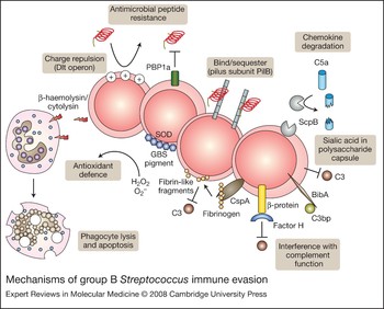
Once GBS penetrates cellular barriers to reach the bloodstream or deep tissues, a broader immunological response is activated to clear the infection, in which host phagocytic cells including neutrophils and macrophages play a critical role. Effective uptake and clearance of GBS by these cells depends upon opsonisation by specific antibodies or serum complement, factors that can be quantitatively and qualitatively deficient in newborns, especially those born prematurely. The propensity of GBS to produce invasive infections further reflects many virulence factors that allow the bacteria to resist opsonophagocytosis or neutralise the bactericidal activities of neutrophils and macrophages (Fig. 2).
Mechanisms of group B Streptococcus immune evasion. Group B Streptococcus (GBS) express several surface-expressed or secreted factors to evade host immune defences and promote survival. The Dlt operon is responsible for increasing incorporation of D-alanine residues in cell-wall teichoic acids, thereby reducing electronegativity and affinity for cationic antimicrobial peptides. PBP1a and the pilB subunit of GBS pili also contribute to antimicrobial peptide resistance. ScpB, the sialic acid capsule, BibA, β protein and CspA all inhibit host clearance of GBS by interfering with complement components C5a, C3 and C3bp. SOD properties of the orange carotenoid pigment shield GBS from killing by phagocyte-generated reactive oxygen species. Alternatively, β-haemolysin/cytolysin can boost GBS survival by cytolytic or proapoptotic injury to host phagocytes. Abbreviations: BibA, GBS immunogenic bacterial adhesin; CspA, cell-surface protease A; PBP1a, penicillin-binding protein 1a; ScpB, C5a peptidase; SOD, superoxide dismutase.

Upon penetration of GBS into the lung tissue or bloodstream of the newborn infant, an immunological response is recruited to clear the microorganism. Central to this response are host phagocytic cells, including neutrophils and macrophages. Effective uptake and killing by neutrophils requires opsonisation of the bacterium by specific antibodies in the presence of complement. However, complement deposition does not affect GBS survival or uptake by macrophages, probably because GBS protect themselves by binding factor H, a host counter-regulator of complement (Ref. Reference Maruvada, Blom and Prasadarao47). Neonates are particularly prone to invasive disease because of their quantitative or qualitative deficiencies in phagocytic cell function, specific antibody, or the classic and alternative complement pathways. In addition to these newborn host susceptibilities, GBS possess a number of virulence determinants that seek to thwart each of the key components of effective opsonophagocytic killing. The sialylated GBS capsular polysaccharide (CPS) represents one such defence factors.
Complement is a system of enzymatic reactions used by the innate immune system to recognise microbes and coat their surfaces with host proteins, making them more easily detected and engulfed by phagocytic cells bearing complement receptors, while simultaneously amplifying other aspects of the inflammatory response. The thick CPS is critical for limiting the effectiveness of host complement defence. The serotype-specific epitopes of ten known GBS CPSs (Ia, Ib, II–VIII and more recently IX) are created by different arrangements of four monosaccharides (glucose, galactose, N-acetylglucosamine and sialic acid) into unique repeating units, but unfailingly these structures contain a terminal sialic acid bound to galactose in an α2 → 3 linkage (Refs Reference Jennings48, Reference Wessels49, Reference Wessels50, Reference Jennings51, Reference Kogan52, Reference Kogan53, Reference Slotved54). This sialic acid molecule provides antiphagocytic protection by impairing surface deposition of opsonically active complement C3 on the bacterial surface. GBS subjected to sialidase treatment, or isogenic GBS mutants lacking capsular sialylation, are more susceptible to neutrophil killing and are less virulent in animal models of infection (Refs Reference Campbell, Baker and Edwards55, Reference Marques56). However, since others have shown that encapsulated and unencapsulated GBS are equally susceptible to macrophage uptake, the role of CPS in resisting phagocytosis per se versus other aspects of immune cell killing remains unclear (Ref. Reference Segura, Cleroux and Gottschalk57).
Sialic-acid-dependent reduction in C3 deposition is correlated with diminished production of C5a, an important complement-derived chemoattractant (Ref. Reference Takahashi58), which works synergistically with ScpB-mediated proteolytic inactivation of C5a to reduce host neutrophil mobilisation. Additionally, a new cell-surface GBS immunogenic bacterial adhesin (BibA) was recently determined to mediate inhibition of other complement components. BibA binds human C3bp, a component of the classical complement pathway, promotes resistance to phagocytic killing, mediates adherence to epithelial cells and contributes to virulence in a mouse model of infection (Ref. Reference Santi59). GBS β-protein was shown to prevent opsonophagocytosis by binding short consensus repeats found in the middle region of factor H, enabling the unbound active region to block C3b deposition on the bacterial cell surface (Ref. Reference Jarva60). The beta antigen of C protein binds human IgA antibody (Ref. Reference Jerlstrom, Chhatwal and Timmis61), and IgA deposited nonspecifically on the bacterial surface probably inhibits interactions with complement. Finally, a cell-surface protease, CspA, targets host fibrinogen, producing adherent fibrin-like cleavage products that coat the bacterial surface and interfere with complement-mediated opsonophagocytic clearance (Ref. Reference Harris62).
Once engulfed and contained in the phagosome, GBS face the rapid release of toxic reactive oxygen species (ROS) produced in the oxidative burst. Unlike Staphylococcus aureus, GBS do not produce catalase, yet are still able to resist killing by ROS and survive inside macrophage phagolysosomes (Refs Reference Cornacchione63, Reference Teixeira64, Reference Wilson and Weaver65). GBS possess an endogenous source of the oxygen-metabolite scavenger glutathione (Ref. Reference Wilson and Weaver65), and the GBS SodA enzyme can neutralise superoxide anions (Ref. Reference Poyart66). GBS also produce an orange carotenoid pigment, a property unique among haemolytic streptococci, that is genetically linked to the cyl operon encoding the β-haemolysin/cytolysin cytotoxin (Ref. Reference Spellerberg67). The free-radical scavenging properties of this carotenoid neutralise hydrogen peroxide, superoxide, hypochlorite and singlet oxygen, and thereby provide a shield against several elements of phagocyte ROS killing (Ref. Reference Liu68).
Antimicrobial peptides (AMPs) that exhibit broad-spectrum activities, such as cathelicidins and defensins, are produced by many immune and epithelial cell types. The small, cationic nature of most AMPs supports the assumption that their mechanism of killing involves their electrostatic attraction to negatively charged microbial cell surfaces, followed by their assembly to create membrane pores or otherwise disrupt membrane integrity (Ref. Reference Gallo and Nizet69). GBS increase their intrinsic resistance to AMPs by incorporation of positively charged D-alanine residues into their cell-wall teichoic acids, thereby reducing surface electronegativity and affinity for the cationic peptides (Ref. Reference Poyart70). A surface-anchored penicillin-binding protein, PBP1a, enhances GBS resistance to cathelicidins and defensins, thereby reducing GBS susceptibility to killing by alveolar macrophages and neutrophils (Ref. Reference Hamilton71), and promoting bacterial survival in a neonatal rat model of aerosolised lung infection (Ref. Reference Jones72). Similarly, expression of the pilus backbone protein PilB renders GBS more resistant to killing by cathelicidin AMPs, and is associated with enhanced phagocyte resistance and systemic virulence (Ref. Reference Maisey73).
Induction of phagocyte apoptosis, or programmed cell death, represents an alternative bacterial defence mechanism to avoid phagocytic clearance. Apoptosis is a carefully regulated signal cascade involving a group of cysteine proteases known as caspases and several pro- and anti-apoptotic regulators belonging to the Bcl-2 family. In contrast to some cell-death ligands, macrophage apoptosis triggered by GBS requires caspase-3 activation and utilises unique changes in regulation and localisation of Bcl-2 family members (Ref. Reference Ulett74). GBS-induced macrophage apoptosis can also progress independently of caspases. Here, calpains, which belong to a different class of cytosolic cysteine proteases, are recruited to cleave and activate Bcl-2 family members and relay the death signal; the dual pathways for phagocyte destruction increase the chances that host defences will be circumvented (Ref. Reference Fettucciari75). The complete role of GBS β-haemolysin/cytolysin in the induction of apoptosis and/or necrotic macrophage cell death remains unclear. Production of this cytolysin was shown to enhance GBS survival in mouse and human blood and this pro-survival phenotype was linked to its ability to induce cytolysis and apoptosis of phagocytes (Ref. Reference Liu68). Furthermore, growth of GBS in high glucose concentrations, which minimises β-haemolysin/cytolysin production, also reduces macrophage apoptosis (Ref. Reference Ulett76). However, it was also demonstrated that in vitro macrophage infection with either wild-type GBS or a GBS mutant lacking β-haemolysin/cytolysin resulted in similar levels of viability, indicating that GBS-induced macrophage apoptosis can also occur by a β-haemolysin/cytolysin-independent mechanism regulated, at least in part, by glucose (Ref. Reference Ulett76).
Finally, a new understanding of GBS immune avoidance by molecular mimicry is emerging. The conserved GBS terminal α2 → 3 linked sialic acid capsular component is identical to a sugar epitope widely displayed on the surface of all mammalian cells. Compared with wild-type strains, capsule-deficient GBS mutants elicit greater degrees of proinflammatory cytokine release from human cells. Like human sialic acids, GBS capsular sialic acids have been demonstrated to engage sialic-acid-recognising immunoglobulin superfamily lectins (Siglecs) on human leukocytes, a family of cell-surface receptors with intracellular domains that send negative signals to limit host cell activation. This interaction suggests that bacterial surface sialylation may have evolved to mimic host ‘self’ antigens, allowing GBS to disguise themselves from immune detection, manipulate phagocyte function and dampen the immune response to GBS infection (Ref. Reference Carlin77).
Inflammatory activation and the sepsis syndrome
When failures in epithelial barrier function and immunological clearance allow GBS to establish bacteraemia in the neonate, development of septicaemia may ensue. Animal models in which GBS are infused intravenously demonstrate a biphasic host inflammatory response (Ref. Reference Rojas78). The acute phase (<1 hour) is manifested by increased pulmonary artery pressure and decreased arterial oxygenation, and is associated with a rise in serum levels of thromboxanes. Pulmonary hypertension and hypoxaemia persist through the late phase (2–4 hours), in which a progressive pattern of systemic hypotension, decreased cardiac output and metabolic acidosis develops together with haematological abnormalities, organ system dysfunction and an increase in inflammatory markers, such as thromboxanes, prostacyclins, tumor necrosis factor-alpha (TNF-α), IL-1 and IL-6.
IL-1, a known stimulator of cyclo-oxygenase and lipo-oxygenase pathways, appears to occupy a proximal position in the deleterious cytokine cascade of septic shock. Treatment with an IL-1 receptor antagonist improves cardiac output and mean arterial pressure and increases survival in piglets receiving a continuous infusion of GBS (Ref. Reference Vallette79). Conversely, the cytokine IL-12, which is elevated 12–72 hours after challenge in animal models, has an important role in regulating the systemic response to GBS infection. Pretreatment with a monoclonal antibody against IL-12 results in greater mortality and intensity of bacteraemia, whereas therapeutic administration of IL-12 is associated with lower mortality and bloodstream replication of the organism (Ref. Reference Mancuso80).
Since the release of TNF-α, IL-l and IL-6 are stimulated by soluble GBS cell-wall antigens (Ref. Reference Vallejo, Baker and Edwards81), studies have sought to identify the specific GBS component(s) of the cell wall that trigger the host cytokine cascade. GBS peptidoglycan is more effective than lipoteichoic acid or capsular polysaccharide as a stimulator of cytokine release from monocytes (Ref. Reference Vallejo, Baker and Edwards82). Knockout studies in mice indicate that cell wall peptidoglycan-induced activation of p38 and NF-κB depends upon the cytoplasmic Toll-like receptor (TLR) adaptor protein MyD88, but does not proceed via the pattern recognition receptors TLR2 or TLR4 (Ref. Reference Mancuso83). GBS activation of TLR2 was shown to depend on surface expression of lipoproteins, which also play a significant role in the development of GBS sepsis (Ref. Reference Henneke84). Structural differences in the linkage, anchoring and backbone of GBS lipoteichoic acid compared with those present in other Gram-positive bacteria may account for its diminished immune activation properties (Ref. Reference Henneke85).
Inhibitor studies have revealed that the mitogen-activated protein kinase (MAPK)/c-Jun N-terminal kinase (JNK) signalling pathway is essential for the NF-κB-dependent inflammatory response of phagocytes to GBS. Since phagocytosis and oxidative killing of GBS were not affected by inhibition of this pathway, JNK may represent a viable therapeutic target for GBS sepsis (Ref. Reference Kenzel86). The nitric oxide (NO) pathway has also been implicated in the overproduction of proinflammatory cytokines, such as IL-6, and initiation of cellular injury during GBS infection of lung tissue (Ref. Reference Raykova87). The GBS cell wall and β-haemolysin/cytolysin act synergistically to upregulate inducible nitric oxide synthase (iNOS) in murine macrophages (Ref. Reference Ring88). The inducible cyclo-oxygenase COX2 is also activated upon GBS infection in human monocytes, probably through MAPK pathway signalling (Ref. Reference Maloney89). GBS infection was also shown to stimulate COX2 and prostaglandin E2 (PGE2) expression in lung tissue in vitro and in vivo. GBS-induced COX2/PGE2 inflammatory response was reduced by treatment with an iNOS inhibitor and restored by addition of a NO donor, indicating that it is at least partially regulated by the NO pathway (Ref. Reference Natarajan90).
The role of complement in GBS-mediated inflammation remains controversial. In one study, whole blood derived from C3 or complement receptor 3 (CR3/CD11b/CD18) knockout mice infected with GBS revealed a tempered TNF-α response (Ref. Reference Levy91). NO was also shown to depend on CR3 expression in macrophages exposed to GBS (Ref. Reference Goodrum, McCormick and Schneider92). However, others have shown that macrophages lacking CR3 demonstrate a normal cytokine response to GBS infection (Ref. Reference Henneke93).
The proinflammatory effects of the GBS β-haemolysin/cytolysin also contribute to sepsis pathophysiology. The toxin acts to stimulate iNOS and NO release in macrophages (Ref. Reference Ring88). In a mouse model of bacteraemia and arthritis, β-haemolysin/cytolysin expression is associated with higher mortality, increased bacterial loads, greater degrees of joint injury and intra-articular and systemic release of the proinflammatory cytokines IL-1 and IL-6 (Ref. Reference Puliti94). Challenge of rabbits with isogenic GBS mutants showed that β-haemolysin/cytolysin production was associated with significantly higher degrees of hypotension, increased mortality and evidence of liver necrosis with hepatocyte apoptosis (Ref. Reference Ring95). Partially purified GBS β-haemolysin/cytolysin preparations produce significant hypotensive actions when infused in rats and rabbits, including death due to shock (Ref. Reference Griffiths and Rhee96). The β-haemolysin/cytolysin toxin contributes directly to cardiomyocyte dysfunction and apoptosis, which may augment its role in the pathophysiological abnormalities of GBS sepsis (Ref. Reference Hensler, Miyamoto and Nizet97).
GBS blood–brain barrier penetration and meningitis
The pathophysiology of GBS meningitis varies according to age of onset. In early-onset disease, autopsy studies demonstrate little or no evidence of leptomeningeal inflammation, despite the presence of abundant bacteria, vascular thrombosis and parenchymal haemorrhage (Ref. Reference Quirante, Ceballos and Cassady98). By contrast, infants with late-onset disease usually have diffuse purulent arachnoiditis with prominent involvement of the base of the brain (Ref. Reference Berman and Banker99). Similar age-related differences in central nervous system (CNS) pathology are evident in the infant rat model of invasive disease (Ref. Reference Ferrieri, Burke and Nelson100). These histopathological differences reflect underdevelopment of the host immunological response in the immediate neonatal period, with a higher proportion of deaths resulting from overwhelming septicaemia.
To produce meningitis, GBS must penetrate the BBB, a specialised structural and functional barrier that maintains homeostasis of the CNS. The BBB consists largely of specialised brain microvascular endothelial cells (BMECs), which guard the brain from circulating toxins and microbes by maintaining tight intercellular junctions and prohibiting pinocytosis. Intracellular invasion and transcytosis of human BMEC tissue culture monolayers has been shown in vitro, and this model has been used to probe the potential role(s) of individual GBS virulence determinants in the initial pathogenesis of GBS CNS infection.
When a GBS transposon mutant library was screened for reduced BMEC invasion, a particularly hypoinvasive mutant was found to harbour a disruption of a gene (iagA) that encodes an enzyme for biosynthesis of diglucosyldiacylglycerol, a membrane glycolipid that functions as an anchor for lipoteichoic acid. Deletion of iagA yielded a GBS mutant that sheds lipoteichoic acid into the medium, exhibits decreased BMEC invasion in vitro and is attenuated in a murine model of meningitis (Ref. Reference Doran101). In separate avenues of research, GBS mutants lacking the GBS fibrinogen receptor FbsA, laminin-binding protein Lmb, or pilus backbone subunit protein PilB also demonstrated reduced adherence or invasion of BMECs in vitro (Refs Reference Maisey17, Reference Tenenbaum27, Reference Tenenbaum102). At high bacterial densities, human BMEC invasion by GBS is accompanied by evidence of β-haemolysin/cytolysin-induced cellular injury (Ref. Reference Nizet26). Correspondingly, β-hemolysin/cytolysin-knockout mutants show decreased BBB penetration and decreased lethality from meningitis in vivo (Ref. Reference Doran, Liu and Nizet103).
GBS invasion of human BMECs can be blocked by inhibition of actin polymerisation, suggesting that GBS trigger rearrangement of the host cytoskeleton and induce their own uptake (Ref. Reference Nizet26). This process may be accomplished, at least in part, by tyrosine phosphorylation of focal adhesion kinase (FAK), which occurs upon GBS infection. Phosphorylation of FAK induces its association with PI3K and paxillin, an actin filament adaptor protein (Ref. Reference Shin104), and is required for efficient GBS BMEC invasion. GBS-infected BMECs also exhibit increased levels of activated Rho family members RhoA and Rac1. Rho family GTPase inhibitors and dominant-negative expression of RhoA and Rac1 are effective in blocking GBS BMEC invasion (Ref. Reference Shin and Kim105).
The host inflammatory response to GBS contributes significantly to the pathogenesis of meningitis and CNS injury. The initiation of the inflammatory response is triggered through the sentinel function of the BBB endothelium, which activates a specific pattern of gene transcription for neutrophil recruitment, including production of chemokines (e.g. IL-8, Groα), endothelial receptors (intracellular cell-adhesion molecule 1, ICAM-1) and neutrophil activators (GM-CSF) (Ref. Reference Doran, Liu and Nizet103). A vascular distribution of cortical lesions in neonatal rats with GBS meningitis indicates that disturbances of cerebral blood flow contribute to neuronal damage (Ref. Reference Kim106). Inflammation of individual brain vessels can lead to focal lesions, whereas diffuse alterations of cerebral blood flow could cause generalised hypoxic/ischaemic injury and cerebral oedema (Refs Reference Kim106, Reference Wahl107). In porcine BMECs, iNOS production is upregulated in response to GBS infection in a NF-κB-dependent manner (Ref. Reference Glibetic108). Further arteriolar dysfunction is associated with the presence of oxygen free radicals thought to be a byproduct of infiltrating neutrophils (Ref. Reference McKnight109). GBS β-haemolysin/cytolysin induces IL-8 and the neutrophil receptor ICAM-1, thereby promoting neutrophil migration across polar BMEC monolayers, suggesting that the toxin is crucial to this particular manifestation of GBS CNS disease (Ref. Reference Doran, Liu and Nizet103).
In the neonatal rat model of meningitis, TNF-α production by astrocytes, microglia and infiltrating leukocytes appears to contribute to apoptosis of hippocampal neurons (Ref. Reference Bogdan110) and further increases in BBB permeability (Ref. Reference Kim, Wass and Cross111). GBS signal through TLR2 to activate and stimulate NO production by microglia cells, resulting in neuronal destruction (Ref. Reference Lehnardt112). Microglial apoptosis is triggered by GBS cell death via the cysteine protease caspase-8, and is hypothesised to represent a self-dampening mechanism that prevents over-stimulation of CNS inflammation (Ref. Reference Lehnardt113). Intraventricular inoculation of newborn piglets with GBS results in an early sharp rise in cerebrospinal fluid TNF-α levels, followed shortly by prostaglandin release and subarachnoid inflammation (Ref. Reference Ling114). In the neonatal rat, simultaneous intracisternal administration of dexamethasone with GBS challenge markedly reduces the magnitude of subarachnoid inflammation, vasculopathy, and neuronal injury (Ref. Reference Kim106).
Clinical applications for vaccine development
An effective vaccine against GBS would represent a major public health advance for newborn infants and other high-risk populations. Purified GBS capsular polysaccharide antigens modelled on the effective campaigns for Haemophilus influenzae type B (HiB) and pneumococcal vaccination in childhood have been coupled to an immunogenic protein carrier. Such glycoconjugate vaccines against serotypes Ia, Ib and II–VIII GBS have been synthesised and found to be immunogenic in preclinical trails in mice, rabbits and/or baboons. Several of these have advanced to Phase I and Phase II clinical trails in healthy adults with an excellent safety profile (Refs Reference Paoletti and Kasper115, Reference Paoletti and Kasper116, Reference Baker and Edwards117). Immunised humans develop serotype-specific anti-CPS antibodies that function well to promote GBS killing during in vitro opsonophagocytic assays. The recent discovery of frequent O-acetyl modifications of the immunodominant terminal α2 → α3-linked sialic acid moiety on several GBS serotypes may provide important insight for optimising CPS purification to retain native structure and maximum immunogenicity (Ref. Reference Lewis, Nizet and Varki118). One challenge faced by the glycoconjugate approach is to develop combination products that would provide appropriately broad-spectrum antigenic coverage for the diverse GBS serotypes associated with disease in any particular demographic group or geographic area.
The investigation of candidate surface-expressed protein antigens distributed more broadly (or ideally universally) among strains of different GBS serotypes has intensified in recent years (Ref. Reference Johri1). The C5a peptidase ScpB is universally expressed by GBS strains capable of eliciting protective IgG antibodies, and may be deliverable in recombinant form within a biodegradable polymer (Refs Reference Cleary7, Reference Santillan, Andracki and Hunter119). Similarly, surface proteins LrrB and Sip are highly conserved across GBS strains of diverse serotypes and each induces protective immunity in mice (Refs Reference Seepersaud12, Reference Brodeur120). The component proteins of newly discovered GBS pili may also represent candidates for a universal vaccine antigen and have been explored as classical antigens and recombinantly expressed in Lactococcus lactis as a live-attenuated vaccine (Refs Reference Lauer13, Reference Buccato121). Although universal protein vaccine antigens may overcome some limitations associated with capsule-based vaccines, introduction of any vaccine during pregnancy (one model for GBS prevention) will meet a concerned and apprehensive target population and will no doubt demand a variety of intensive risk assessment and educational interventions (Ref. Reference Patten122).
Research in progress and outstanding research questions
Each new year of GBS research heralds the discovery of novel virulence determinants or new functions for previously identified surface proteins or secreted factors. Advances can be attributed to the application of new methodologies, such as the use of reverse vaccinology to screen genomes for immunogenic surface antigens, which led to the identification multifunctional immunogenic adhesin BibA and unveiled the surface pili that had been overlooked for more than 50 years of GBS research (Refs Reference Lauer13, Reference Santi59). Functional screening and careful re-examination of previously identified surface structures have also revealed secondary functions, such as a role for the GBS C5a peptidase ScpB in epithelial adherence or capsular sialic acid in engagement of host Siglec receptors. Furthermore, it is likely that GBS will remain a useful model organism for Gram-positive bacterial pathogenesis and for probing the developmental regulation of newborn immune function.
Rising incidences of invasive disease in adults and emerging patterns of antibiotic resistance (Ref. Reference Chohan123) indicate that further attention must be paid to elucidate GBS virulence factors and the mechanisms by which they interact with host cells and our immune system. Indeed, the emergence of GBS strains with decreased susceptibility to β-lactam antibiotics has now been reported in both the USA and Japan (Refs Reference Dahesh124, Reference Kimura125). These GBS isolates harbour mutations in penicillin-binding protein 2x (PBP2x), which is similar to the first-step mutations on the pathway to full β-lactam resistance seen in pneumococcal isolates a few decades ago. Enhanced understanding of the molecular basis of GBS pathogenesis may pinpoint novel bacterial and host molecules that can represent novel therapeutic or immunoprophylactic targets against disease caused by this foremost of neonatal pathogens.
Acknowledgements
The authors appreciate the contribution of anonymous peer reviewers to the completeness of the manuscript. The authors' own research on GBS molecular pathogenesis has been supported by the National Institutes of Health (V.N., K.S.D.), an American Heart Association Established Investigator Award (V.N.), a Burroughs-Wellcome Fund Career Award in the Biomedical Sciences (K.S.D.), and a Graduate Research Fellowship from the National Science Foundation (H.C.M.).