Left aortic arch with right descending aorta is a rare anomaly. In the presence of a right ductal ligament, it constitutes a vascular ring.Reference Ergin, Jayaram and LaCorte 1 , Reference Dominiguez, Sang-Oh and Dorst 2 We report a patient with left arch with severe aortic coarctation beyond the left subclavian artery and right descending aorta with a left patent arterial duct causing left main bronchus compression. The left bronchus was “clamped” between the right pulmonary artery anteriorly and the descending aorta posteriorly. After an accurate anatomical delineation by three-dimensional reconstruction, surgical descending aortic translocation relieved the airway compression and the aortic coarctation.
Case report
A 2-month-old infant presented with a heart murmur. An initial echocardiogram showed a left aortic arch with mild coarctation distal to the left subclavian artery, and a small restrictive ventricular septal defect. In view of clinical stability, he was reviewed 3 months later. During the second clinic visit, he presented with respiratory distress, diminished breath sounds in the left chest, and differential cyanosis. Chest X-ray showed widespread right lung infiltrates with reduced vascularity and hyperinflation of the left lungs. His respiratory distress worsened, quickly warranting mechanical ventilation. A bronchoscopy was performed in view of X-ray findings and showed a slit-like compression of the left bronchus.
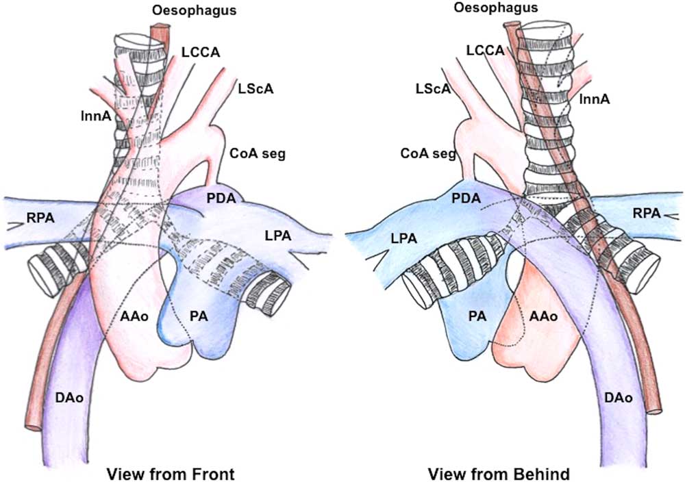
A detailed echocardiography after clinical stabilisation showed intact ventricular septum, left ventricular hypertrophy with good systolic function, confluent pulmonary artery branches with poor flow into a good-sized left pulmonary artery, a left aortic arch with normal arch branching, anatomically severe aortic coarctation with systolic gradient of 16 mmHg, a large left-sided ductus shunting right to left in systole and maintaining the descending aortic flows, and a right-sided descending aorta.Reference Singh, Greenberg and Balsara 3 CT confirmed left arch, normal branching pattern, with a right descending aorta and severe aortic coarctation distal to the left subclavian artery, and left-sided patent arterial duct inserting beyond the coarctation maintaining lower limb perfusion (Supplementary files 1 and 2). The descending aorta coursed in front of the vertebral column from the left to the right, distal to the ductal insertion below the level of carina, to continue as the right descending aorta. Left main bronchus was compressed between the descending aorta posteriorly and a hypertensive right pulmonary artery anteriorly. The oesophagus was pushed to the right by the right descending aorta (Fig 1). The lack of left pulmonary arterial blood flow on echocardiography was caused by left lung hypoventilation owing to left bronchial obstruction.
The anatomy of the vascular clamp is shown from front ( a ) and back ( b ): the left bronchus is compressed between the anterior right pulmonary artery (RPA) and the prevertebral descending aorta (DAo). The aortic arch is severely narrowed after the last left subclavian (LScA) branch and the left-sided duct (PDA) maintains descending aortic perfusion. AAo=ascending aorta; CoA seg=coarctation segment; InnA=innominate artery; LCCA=left carotid artery; LPA=left pulmonary artery; PA=pulmonary artery.

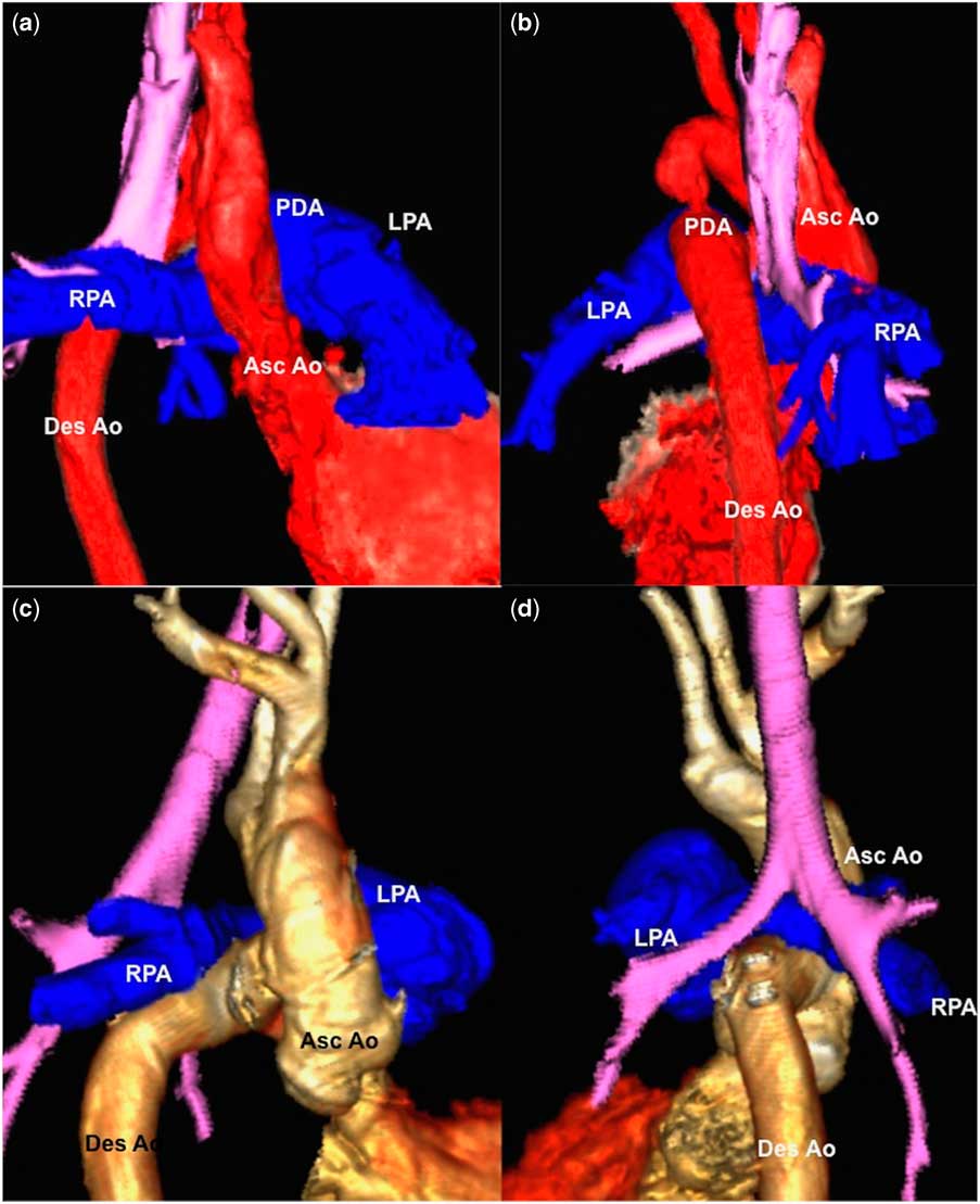
Surgical translocation of the right descending aorta to the posterior wall of the ascending aorta in the transverse sinus below the right bronchus on cardiopulmonary bypass resulted in complete restoration of ventilation and perfusionReference McKenzie, Roeser and Thompson 4 (Fig 2). On a follow-up of 2 months, there was good weight gain, no respiratory symptoms, no residual aortic gradients, and normal oxygen saturations.
Volume rendered tomographic images from right anterior oblique ( a ) and right posterior oblique ( b ) directions show clamping of left bronchus, shown in purple colour. Following descending aortic translocation to the posterior wall of the ascending aorta in the transverse sinus below the right bronchus in right anterior oblique ( c ) and right posterior oblique ( d ) views, the aortic coarctation and bronchial clamp are completely relieved. Asc Ao=ascending aorta; Des Ao=descending aorta; LPA=left pulmonary artery; PDA=patent ductus arteriosus; RPA=right pulmonary artery.

Discussion
A left aortic arch with right descending aorta is a rare anomaly, and coarctation or interruption of aorta has been described in this context. The presence of such an arrangement with a patent arterial duct that is ipsilateral to the arch and causing airway obstruction is quite unique to the best of our knowledge.
A circumflex aorta is an anomaly in which the aorta courses above the level of carina often behind the oesophagus to descend as contralateral descending aorta, and in the presence of a patent arterial duct on the contralateral side it creates a vascular ring. The resultant tracheal and oesophageal compression is relieved when the tethering ductal tissue is divided through a thoracotomy, but, occasionally, the transverse posterior course of the aorta may still cause residual compression.Reference Ergin, Jayaram and LaCorte 1
In our patient, the right descending aorta pushed the oesophagus further to the right. As the aorta did not course behind the oesophagus and the descending aorta coursed immediately below the level of tracheal carina, this was not truly a circumflex aorta. The posterior descending aorta that coursed from left to right in the prevertebral plane compressed the left bronchus against the dilated hypertensive right pulmonary artery (Supplementary files 1 and 2). The large left-sided duct maintained the lower body perfusion albeit with mild differential cyanosis. The “vascular clamp” on the left bronchus resulted in reduced ventilation of the left lung. The autoregulatory left pulmonary vasospasm led to reduced Doppler flows in the left pulmonary artery and oligaemia of the left lung on chest X-ray.
A comprehensive literature search showed only two prior references to a similar anatomy with ipsilateral duct with circumflex arch, but no evidence of a clinically significant vascular “ring”.Reference Dominiguez, Sang-Oh and Dorst 2 , Reference Sanchez Torres and RoldanConesa 5 To the best of our knowledge, this is the first report in literature describing airway compression by such a unique vascular arrangement.Reference Ergin, Jayaram and LaCorte 1
Embryological basis
Embryological explanation for this anomaly is persistence of the left fourth and left sixth embryonic arches that continue as a right-sided dorsal aorta owing to an abnormal involution of left dorsal aorta.Reference Sanchez Torres and RoldanConesa 5 When the left circumflex aorta with a contralateral duct leads to a vascular ring, persistent right-sided dorsal aorta and right-sided distal sixth arch explains the anomaly.Reference Ergin, Jayaram and LaCorte 1 , Reference Dominiguez, Sang-Oh and Dorst 2 Our clinical case demonstrates that the sidedness of the duct and the side of persistent dorsal aorta are unrelated to each other and occur independently.Reference Dominiguez, Sang-Oh and Dorst 2 The aortic coarctation in this anomaly was a result of marked hypoplasia of the left dorsal aorta between the fourth and sixth arch insertions. The ductal arch formed by the left sixth arch continued to maintain the descending aortic flows in the postnatal period and led to mild differential cyanosis. Aortic arch anomalies may be associated with coarctation and hypoplasia.Reference Rad, Mortezaeian, Pouraliakbar and Hijazi 6 , Reference Hilmes, Hernandez and Devaney 7 They may also coexist with CHD.Reference Ergin, Jayaram and LaCorte 1 , Reference Humphrey, Duncan and Fletcher 8 Left pulmonary artery hypoplasia and unilateral lung hypoplasia are also described with circumflex aorta.Reference Berman, Yabek and Dillon 9 , Reference McLeary, Frye and Young 10
Supplementary material
To view supplementary material for this article, please visit https://doi.org/10.1017/S1047951118000665
Acknowledgements
The authors thank Dr Sachin Khambadkone, Consultant in Pediatric Cardiology, Great Ormond Street Hospital for Children NHS, London, for editing the language and grammar.
Financial Support
This research received no specific grant from any funding agency or from commercial or not-for-profit sectors.
Conflicts of Interest
None.
Ethical Standards
The authors assert that all procedures contributing to this work comply with the ethical standards of the Indian Council of Medical Research and with the Helsinki Declaration of 1975, as revised in 2008, and have been approved by the institutional committees of Madras Medical Mission, Chennai, India.