Introduction
Ornithischian dinosaurs were one of the most important herbivorous components of terrestrial vertebrate faunas along the second half of the Mesozoic, but many aspects of their early evolution and diversification patterns are currently unknown, given their poor representation in fossil assemblages from the Late Triassic to the Middle Jurassic (Sereno, Reference Sereno1997; Irmis et al., Reference Irmis, Parker, Nesbitt and Liu2007). This has been a persistent problem in achieving a detailed understanding of the early differentiation of the major clades of ornithischians before the Late Jurassic-Early Cretaceous, and understanding the importance of small omnivoral-herbivoral niches occupied by these groups, which is important for assessing how ornithischians occupied the niches of large herbivores during the second half of their evolutionary history.
The Cañadón Asfalto Formation has yielded the richest and best studied microvertebrate faunal assemblages for the latest Early or early Middle Jurassic of South America (Rougier et al., Reference Rougier, Martinelli, Forasiepi and Novacek2007b; Escapa et al., Reference Escapa, Sterli, Pol and Nicoli2008), including the recently described small-bodied heterodontosaurid Manidens condorensis Pol, Rauhut and Becerra, Reference Pol, Rauhut and Becerra2011 (the dentition of which was recently described by Becerra et al., Reference Becerra, Pol, Marsicano and Rauhut2014). Although Manidens Pol, Rauhut, and Becerra, Reference Pol, Rauhut and Becerra2011 is the most complete heterodontosaurid known from South America, it currently is represented by craniomandibular remains, the axial skeleton, a fragmentary pectoral girdle and a complete pelvis, and lacks fore and hind limbs.
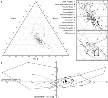
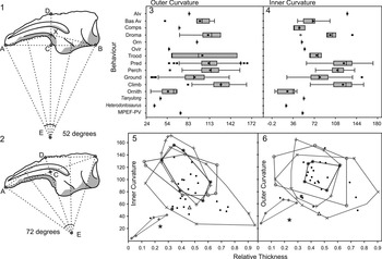
In this communication, we describe a new ornithischian specimen from the Cañadón Asfalto Formation (MPEF-PV 3826), also found in the same locality and horizon as Manidens condorensis. MPEF-PV 3826 includes both metatarsi associated with several disarticulated pedal phalanges and caudal vertebrae with chevrons. This small-bodied specimen shows a combination of plesiomorphic features among ornithischians with heterodontosaurid apomorphies, some of which have been noted by Santa Luca (Reference Santa Luca1980) for Heterodontosaurus tucki Crompton and Charig, Reference Crompton and Charig1962, and others are new characters identified in this study. The comparison of MPEF-PV 3826 with other ornithischian material is focusing mostly on small sized and closely related ornithischians. We also describe the histology of metatarsal IV and compare the overall similarity of the metatarsus with other ornithischians through a cluster analysis. Also, we add new information to a previous phylogenetic analysis (re-scoring of some heterodontosaurid taxa and adding 18 characters identified by Sereno [Reference Sereno2012], and 11 new characters), including the addition of Pegomastax africanus Sereno, Reference Sereno2012 and MPEF-PV 3826 to test the affinities of the latter specimen. To infer possible aspects of mode of life of this specimen we also use the length proportion of pedal phalanges of digit III to perform a ternary diagram and a principal component analysis, and a comparison of the inner and outer curvature in pedal ungual phalanges with other dinosaurs and recent birds.
Materials and methods
The performed statistical analyses were used to provide complementary comparisons to those of the morphological description. The length of the metatarsals in MPEF-PV 3826 were measured using a digital caliper (accuracy 0.01 mm, all measurements presented at Table 1 in Supplemental Data 1), while all measurements on comparative material where taken from photographs or the literature. The cluster analysis and claw curvature/behavior box plots were realized using the Infostat free version software (Di Rienzo et al., Reference Di Rienzo, Casanoves, Balzarini, Gonzalez, Tablada and Robledo2011). Ternary diagram and principal component analysis on length proportions of phalanges of digit III, and the claw-curvature/relative-thickness diagrams were performed using the software PAST, version 2.10 (PAlaeontological STastics, Hammer et al., Reference Hammer, Harper and Ryan2001), with data presented by Hopson (Reference Hopson2001) and Birn-Jeffery et al. (Reference Birn-Jeffery, Miller, Naish, Rayfield and Hone2012) and adding some ornithischian taxa. Although the database of Hopson (Reference Hopson2001) was used without changes, several species sampled in the Birn-Jeffery et al. (Reference Birn-Jeffery, Miller, Naish, Rayfield and Hone2012) database were excluded because they lack the relevant information for the diagrams. With these data, box plots include 742 specimens for the inner curvature, 763 for the outer curvature, 742 specimens for the inner-curvature/relative-thickness graph, and 649 for the outer-curvature/relative-thickness graph. Of these, 22 specimens correspond to Mesozoic theropods, eight to ornithischians, including MPEF-PV 3826, and the remaining specimens represent several families of recent birds and lizards (Birn-Jeffery et al., Reference Birn-Jeffery, Miller, Naish, Rayfield and Hone2012).
To test the taxonomic affinities of this new specimen a cladistic analysis was performed, using the data matrix originally compiled by Butler et al. (Reference Butler, Upchurch and Norman2008), and including the changes in characters proposed by Pol et al. (Reference Pol, Rauhut and Becerra2011) and others (see below). The data matrix includes 53 taxa and 259 characters and was analysed using equally weighted parsimony in TNT, version 1.1 (Goloboff et al., Reference Goloboff, Farris and Nixon2008a, Reference Goloboff, Farris and Nixon2008b).
To determine the growth pattern and the ontogenetic stage of the MPEF-PV 3826, a middiaphyseal cross section of metatarsal IV was taken. The cross-section was prepared using standard paleohistological techniques (Chinsamy and Raath, Reference Chinsamy and Raath1992) and studied under transmitted light, both normal and polarized. The terminology used follows the general terminology of bone histology described in Francillon-Vieillot et al. (Reference Fracillon-Vieillont, Debuffrénil, Castanet, Geraudie, Meurier, Sire, Zylberberg and De Ricqlés1990) and Chinsamy-Turan (Reference Chinsamy-Turan2005).
Abbreviations
BP, Evolutionary Studies Institute, University of the Witwatersrand, Johannesburg, South Africa; IVPP, Institute of Vertebrate Paleontology and Paleoanthropology, Beijing, People’s Republic of China; MB Museum für Naturkunde, Berlin, Germany; MCF-PVPH, Museo Carmen Funes, Paleontología de Vertebrados, Plaza Huincul, Neuquén, Argentina; MPEF, Museo Paleontológico Egidio Feruglio, Trelew, Chubut, Argentina; MUCPv, Museo de la Universidad Nacional del Comahue, Neuquen Province, Paleovertebrates; NHMUK, Natural History Museum, London, United Kingdom; PVL, Instituto Miguel Lillo, Tucumán, Argentina; SAM, Iziko South African Museum, Cape Town, South Africa; STMN, Shandong Tianyu Museum of Nature, Pingyi, Shandong Province, People’s Republic of China; YMP, Peabody Museum of Natural History, Yale University, New Haven.
Systematic paleontology
Dinosauria Owen, 1842
Ornithischia Seeley, 1887
Heterodontosauridae Kuhn, 1966
Genus and Species Indeterminate
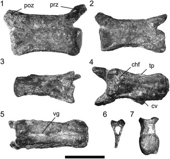
Indeterminate Heterodontosauridae MPEF-PV 3826, isolated caudal vertebrae and chevron: (1–3) caudal vertebrae in lateral view, positioned from proximal to distal elements; (4, 5) caudal vertebrae in ventral view; (6) isolated hemal arch or chevron; (7) anterior view of isolated vertebra. chf, chevron facet; cv, chevron; poz, postzygapophysis; prz, prezygapophysis; tp, transversal process vg, ventral longitudinal groove. Scale bar equals 5 mm.

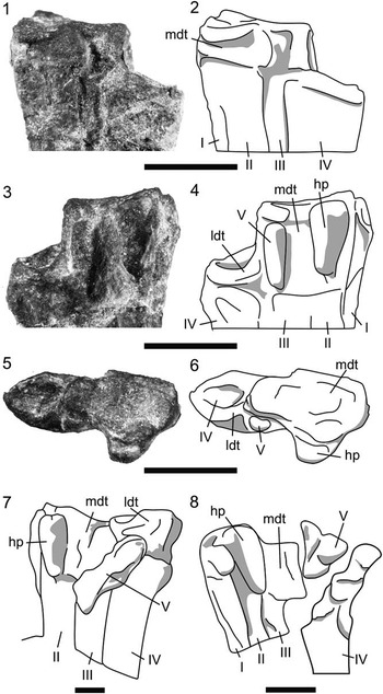
Indeterminate Heterodontosauridae MPEF-PV 3826, preserved tarsals and comparisons. (1, 3, 5) Photographs; (2, 4, 6, 7, 8) schematic line drawings. (1, 2) Anterior; (3, 4) plantar; (5, 6) proximal view of the preserved medial distal tarsal and the fragmented lateral tarsal of the left pes; (7) Heteterodontosaurus tucki, schematic line drawing of plantar view of tarsals and proximal metatarsals (modified from Santa Luca, Reference Santa Luca1980); (8) Tianyulong confuciusi, schematic line drawing of plantar view of tarsals and proximal metatarsals (modified from Zheng et al., Reference Zheng, You, Xu and Dong2009, supplementary material). I, metatarsal I; II, metatarsal II; III, metatarsal III; IV, metatarsal IV; V, metatarsal V, represented as a reduced bone; hp, “hypotarsal-like” process, resembling the feature mentioned by Santa Luca (Reference Santa Luca1980) for Heterodontosaurus; ldt, lateral distal tarsal; mdt, medial distal tarsal. Scale bars equal 5 mm.

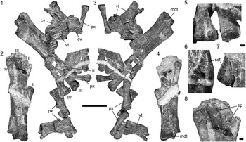
Indeterminate Heterodontosauridae MPEF-PV 3826. Metatarsals and scattered phalanges and caudal vertebrae: (1, 3) left and (2, 4) right metatarsals and fused tarsals in plantar (1, 2) and anterior (3, 4) view; (5–7) distal condyles in plantar view of metatarsal IV (5, left); metatarsal III (5, right); metatarsal II (6); and metatarsal I (7); (8) cast on rock representing the distal condyles of metatarsals II (right) to IV (left) in anterior view. I, metatarsal I; II, metatarsal II; III, metatarsal III; IV, metatarsal IV; cv, chevron; ep, extensor pit; mdt, medial distal tarsal; px, phalanx; scf, supracondylar fossa; vt, cervical vertebrate. Scale bars equal 1 cm for (1–4) and 1 mm for (5–8).

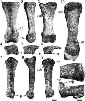
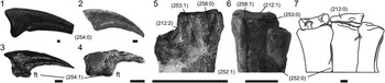
Indeterminate Heterodontosauridae MPEF-PV 3826, phalanges of the pes; (1–3, 10–12) examples of robust phalanx in dorsal (1), lateral (2) and plantar view (3); (4–6) three of four isolated ungual phalanges in lateral view; (7–9) example of slender phalanx in dorsal (7), lateral (8), and plantar view (9); (10) first phalanx of digit III; (11, 12) detail of dorsal extensor process on first phalanx of digit III (character 259:1) in dorsal (11) and lateral (12) views. dep, dorsal extensor process; ep, extensor pit; ft, flexor tubercle; lp, lateral pits; ppc, proximoplantar concavity, for interphalangeal pulley joint muscles. Scale bars equal 1 mm.

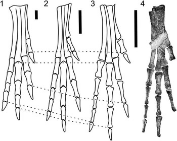
Indeterminate Heterodontosauridae MPEF-PV 3826, reconstruction of the pes and comparison with other heterodontosaurid taxa; metatarsals drawn to subequal size and aligned to compare the digit length among heterodontosaurids with the size of the pes: (1) Heterodontosaurus tucki, simplified schematic drawing modified from Sereno (Reference Sereno2012); (2) Tianyulong confuciousi, schematic drawing that combines the shape of the metatarsaus of specimen STMN 26-3 (Zheng et al., Reference Zheng, You, Xu and Dong2009) with the phalangeal measurements of the more complete specimen IVPP V17090 from Sereno (Reference Sereno2012), with metatarsal proportions re-scaled to aproximate the phalangeal measurements; (3) MPEF-PV 3826, schematic line drawing of right foot in anterior (dorsal) view; (4) reconstruction of the pes of MPEF-PV 3826, with distal portions represented by the cast preserved on rock. Phalanges I-1 and IV-1 were specularly rotated to aval orientation. For comparisons, dashed lines mark the distalmost point of metatarsal 3 and the end of each digit (without counting the ungual phalanx). Scale bars equal 2 cm.

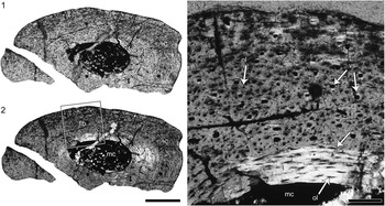
Indeterminate Heterodontosauridae MPEF-PV 3826: histological photographs of the middle shaft of left metatarsal 4. (1, 2) General view in normal (1) and polarized light with lambda compensator (2). (3) Detail of the cortical tissue in viewed under polarized light with lambda compensator (box inset in 2). Note the slight change in the intrinsic fiber arrangement at the outer cortex. flb, fibrolamellar bone; icl, inner circumferential layer; mc, medullary cavity; ol, osteocyte lacunae; lm, lamellar bone; rl, resorption line; vs, vascular spaces. Scale bar equals 0.5 mm in (1, 2) and 0.1 mm in (3).

Specimen
MPEF-PV 3826, right and left medial distal tarsals, almost complete left and right metatarsus, associated with many scattered phalanges and caudal vertebrae with chevrons. This material has features unknown in other ornithischians (e.g., proportional length of pedal phalanges) but we refrain from erecting a new species, given that this specimen cannot be compared with the holotype of Manidens condorensis (found at the same locality and horizon) because of the lack of diagnostic overlapping material, and might even represent the same specimen as the holotype of the latter (see below).
Occurrence
MPEF-PV 3826 was collected at the Queso Rayado Locality (43°24’33,55”S; 69°13’50,1”W), from outcrops that belong to the Cañadón Asfalto Formation, located at 5.5 km northwest of the village of the Cerro Condor, in Chubut province, Argentina. The vertebrate-bearing lithology at this microvertebrate locality was characterized as a silicified mudstone deposited in a shallow lacustrine environment with volcanic influences (Rougier et al., Reference Rougier, Garrido, Gaetano, Puerta, Corbitt and Novacek2007a). This locality has been interpreted as belonging to the lowermost section of the Cañadón Asfalto Formation (Rougier et al., Reference Rougier, Garrido, Gaetano, Puerta, Corbitt and Novacek2007a; Cuneo et al., Reference Cuneo, Ramezani, Scasso, Pol, Escapa, Zavattieri and Bowring2013). The base of this unit has been recently dated as latest Early Jurassic (late Toarcian; Cuneo et al., Reference Cuneo, Ramezani, Scasso, Pol, Escapa, Zavattieri and Bowring2013). The deposition of the Cañadon Asfalto Formation extended into the early Middle Jurassic, as indicated by other radiometric dates (Salani, Reference Salani2007; Cabaleri et al., Reference Cabaleri, Volkheimer, Armella, Gallego, Silva Nieto, Páez, Cagnoni, Ramos, Panarello and Koukharsky2010) and recent palynological studies (Volkheimer et al., Reference Volkheimer, Gallego, Cabaleri, Armella, Narváez, Silva Nieto and Páez2009; Zavattieri et al., Reference Zavattieri, Escapa, Scasso and Olivera2010).
Description
Vertebral column
Seventeen scattered caudal vertebrae are preserved in MPEF-PV 3826 and are closely associated with the metatarsals and phalanges, two of them being found partially articulated underneath the metatarsi (Fig. 1, see also Supplemental Data 2 and Supplemental Data 3). Some of the vertebrae are slightly crushed or distorted. All centra are spool-shaped, long and low, and vary more in their dorsoventral height than their length (Fig. 1.1–1.3). The width of the articular surface ranges between 28.6% and 36.6% of the length of each centrum (Fig. 1.7). The anterior articular surface is flat and the posterior articular surface is slightly concave, as in Fruitadens haagarorum Butler, Galton, Porro, Chiappe, Henderson, and Erickson, Reference Butler, Porro, Galton, Chiappe, Henderson and Erickson2010 (Butler et al., Reference Butler, Porro, Galton and Chiappe2012, fig. 11H–11I). The size and orientation of the pre- and postzygapophyses are similar in lateral view in all preserved vertebrae, but the prezygapophyses are more steeply dorsally inclined than the postzygapophyses (e.g., Fig. 1.1). In anterior and posterior view, the articular surfaces of the prezygapophyses are obliquely oriented (at 45° with respect to the horizontal axis), whereas the postzygapophyseal surfaces stand almost vertically. The vertebral centra have a conspicuous longitudinal ventral groove (Fig. 1.4–1.5), which is poorly developed or absent in smaller and more distal vertebrae. The lateral surface of the centrum is slightly concave in transverse section. Only the largest vertebra has a smooth ventral chevron facet on the proximal end (Fig. 1.4). There are only two vertebrae that have a small transverse process (Fig. 1.4) preserved on the left side. The small size of the transverse processes resembles the transverse processes of the middle-posterior caudal vertebrae of Heterodontosaurus Crompton and Charig, Reference Crompton and Charig1962. The neural spine is only a slightly developed longitudinal ridge in the larger, more proximal vertebrae, and is completely absent in the smaller (more distal) elements. There is no evidence of ossified tendons in the caudal vertebrae, as in Heterodontosaurus, but differing from the situation in Tianyulong confuciusi Zheng, You, Xu and Dong, Reference Zheng, You, Xu and Dong2009. In all preserved vertebrae, the sutures between the neural arches and centra are completely closed, a condition that might be related to an advanced ontogenetic stage (Brochu, Reference Brochu1996; Irmis, Reference Irmis2007).
In comparison with Heterodontosaurus (Santa Luca, Reference Santa Luca1980), the reduction of the neural spine and transverse processes described above indicates that these elements belong to the medial–distal portion of the tail, resembling the condition in Friutadens Butler, Galton, Porro, Chiappe, Henderson, and Erickson, Reference Butler, Porro, Galton, Chiappe, Henderson and Erickson2010 (Butler et al., Reference Butler, Porro, Galton and Chiappe2012). The more distal vertebrae are not only less robust and dorsoventrally lower, but their centra also become rod-shaped (rather than spool-shaped). There is only small variation in centrum length between the preserved vertebrae in MPEF-PV 3826 and the size of pre- and postzygapophyses, regardless of their position in the tail (in contrast to the development of the transverse processess and neural spines), as commonly seen in medial-distal caudal vertebrae within Dinosauria (i.e., Weishampel et al., Reference Weishampel, Dodson and Osmólska2004). The holotype (MPEF-PV 3211) of the heterodontosaurid Manidens includes one described proximal caudal, the length of which is equal to the length of the anterior-most preserved vertebrae of MPEF-PV 3826 (Pol et al., Reference Pol, Rauhut and Becerra2011; personal observation).
Chevrons
There seem to be only seven preserved chevrons, which are partially covered and scattered among the elements of MPEF-PV 3826 (Fig. 1.4, 1.6; see also Supplementary Data 2 and Supplementary Data 3). The rod-like shaft of the chevrons is laterally compressed, but the proximal expansion is weak, as in Heterodontosaurus posterior to the sixth chevron (Santa Luca, Reference Santa Luca1980). This condition is also commonly found in basal ornithopods (i.e., Hypsilophodon foxii Huxley, Reference Huxley1869 and Thescelosaurus neglectus Gilmore, Reference Gilmore1913 in the middle caudal region, proximal chevrons in Gasparinisaura cincosaltensis Coria and Salgado, Reference Coria and Salgado1996). The widest portion of the chevron corresponds is the proximal articular surface in both lateral and posterior views. Although the distal portion of the largest chevron is slightly expanded anteroposteriorly, there is no evidence of distal widening of the chevrons in MPEF-PV 3826. The comparison with Heterodontosaurus suggests that the preserved chevrons belong to the caudalmost vertebrae that have chevrons, which are usually placed at the middle portion of the caudal series. This is consistent with the inferred position of the preserved caudal vertebrae mentioned above. The chevrons of MPEF-PV 3826 are dorsoventrally higher than the neural spine of the preserved vertebrae, as in the middle section of the caudal series of Heterodontosaurus, Gasparinisaura Coria and Salgado, Reference Coria and Salgado1996, and basal ornithischians. Other taxa, such as Dryosaurus altus (Marsh, Reference Marsh1878); Thescelosaurus Gilmore, Reference Gilmore1913; Hypsilophodon Huxley, Reference Huxley1869 and more derived ornithopods, have chevrons at the middle section of the caudal series that are equal or shorter than the neural spine.
Autopodium
The autopodium is represented by complete right and left metatarsi, in both cases including the medial distal tarsals and associated phalanges. The metatarsus is composed of five metatarsals, which are proximally fused, as in the cursorial heterodontosaurids Heterodontosaurus and Tianyulong Zheng, You, Xu, and Dong, Reference Zheng, You, Xu and Dong2009 (Fig. 2.7–2.8).
Tarsus
The distal tarsus consists of two elements, the common condition in ornithischian dinosaurs (Norman et al., Reference Norman, Sues, Witmer and Coria2004), including Heterodontosaurus (Sereno, Reference Sereno2012). The medial distal tarsal is complete (Fig. 2.1–2.6) and the lateral distal tarsal is represented by a small bone fragment (Fig. 2.3–2.6). An unusual characteristic among ornithischians is that the preserved tarsals are strongly coossified to each other and to the metatarsus (Fig. 2.1–2.4). This is a widespread feature in theropods (i.e., Olmólska, Reference Osmólska1981), especially birds, but has only been reported in Heterodontosaurus (Santa Luca, Reference Santa Luca1980) and Tianyulong (at least in the medial distal tarsal [Sereno, Reference Sereno2012]) among ornithischians. Even though the distal tarsals and the metapodium are not preserved in Fruitadens Butler, Galton, Porro, Chiappe, Henderson, and Erickson, Reference Butler, Porro, Galton, Chiappe, Henderson and Erickson2010, it is worth noting that there is coossification of the distal tibia, fibula, and proximal tarsals in this taxon, as in the other heterodontosaurids Heterodontosaurus and Tianyulong (Santa Luca, Reference Santa Luca1980; Butler et al., Reference Butler, Porro, Galton and Chiappe2012; Sereno, Reference Sereno2012). This feature might be functionally related to the fusion of the tarsomentatarsi here reported for MPEF-PV 3826, but it might be worth noting that it is absent in Abrictosaurus and seems to be variable in Heterodontosaurus (Sereno, Reference Sereno2012). The medial distal tarsal is the largest element of the distal tarsus and is coossified with metatarsals I, II, III, and V (the latter being represented by a strongly reduced splint of bone positioned on the plantar side of the metatarsus, Fig. 2.3–2.6), with an obliterated sutural line. The medial distal tarsal has a squared proximal articular surface that is lateromedially concave and anteroplantarly convex (Fig. 2.5–2.6). The plantar surface of this tarsal bears a proximodistally elongated process overhanging metatarsal II (Fig. 2.3–2.4), as it is also present in Heterdontosaurus (Santa Luca, Reference Santa Luca1980; Norman et al., Reference Norman, Sues, Witmer and Coria2004; Fig. 2.7) and Tianyulong (STMN 26-3; see Zheng et al., Reference Zheng, You, Xu and Dong2009, supplementary material; Fig. 2.8). This was interpreted as a functional equivalent of the hypotarsal ridge (Baumel and Witmer, Reference Baumel and Witmer1993) of extant birds (Santa Luca, Reference Santa Luca1980), related to the attachment of the flexor tendons of the digits. In proximal view, this process forms a plantar protuberance of the medial distal tarsal. A similar morphology was described for Orodromeus makelai Horner and Weishampel, Reference Horner and Weishampel1988 and Jeholosaurus Xu, Wang, and You, Reference Xu, Wang and You2000a (Han et al., Reference Han, Barrett, Butler and Xu2012), but the development of this process is notably reduced in comparison with the condition seen in heterodontosaurids (see below: Phylogenetic Analysis).
The lateral distal tarsal is located over the proximal portion of metatarsal IV, and it is represented only by a small bone fragment (Fig. 2.3–2.6). This bone fragment is located on the plantar side of the proximal articular facet of the metatarsal IV and is likely to be fused, while the rest of the articular surface of metatarsal IV remains exposed. The lateral distal tarsal fits into the deeply concave proximal surface of metatarsal IV (Fig. 2.3–2.6). This feature (in contrast to the presence of a flat proximal articular surface of metatarsal IV), suggests that the lateral tarsal was tightly integrated in the tarsometatarsus and probably fused to the metatarsus, resembling Heterodontosaurus (Santa Luca, Reference Santa Luca1980; Sereno, Reference Sereno2012), rather than the condition described for Tianyulong (Sereno, Reference Sereno2012).
Metatarsus
The left metatarsus is complete but fractured, with slightly displaced fragments of the shafts (Fig. 3.1, 3.3), and the right metatarsus is distally incomplete (Fig. 3.2, 3.4), preserving only the cast of the distal ends of the metatarsals II, III, and IV in the matrix (Fig. 3.8). The metatarsals were found in natural articulation and are proximally coossified to each other, though with recognizable suture lines between them (Fig. 2.1–2.4). Similar to the condition in other ornithischian dinosaurs, the weight bearing elements were metatarsals II, III, and IV. Metatarsal I is considerably shorter than metatarsals II–IV, and metatarsal V is represented only by a small splint of bone, which is located proximo-plantarly on the metatarsus and fused to the plantar surface of metatarsal III and the medial distal tarsal. The reduction of metatarsals I and V is a plesiomorphic feature for Ornithischia (Weishampel et al., Reference Weishampel, Dodson and Osmólska2004), and is present in numerous representatives of this clade, including Heterodontosaurus tucki (Santa Luca, Reference Santa Luca1980), Othnielosaurus consors Galton, Reference Galton2006 (=Laosaurus consors Marsh, Reference Marsh1894), Thescelosaurus neglectus (Gilmore, Reference Gilmore1915), Agilisaurus louderbacki Peng, Reference Peng1990 (Peng, Reference Peng1992), and Gongbusaurus wucaiwanensis Dong, Reference Dong1989. In anterior view, metatarsal III is proximally pinched between metatarsals II and IV, similar to the condition described in Gasparinisaura (Coria and Salgado, Reference Coria and Salgado1996), but not to the degree seen in arctometatarsalian theropods (Holtz, Reference Holtz1994). Metatarsals II–IV remain closely appressed to each other down to the midshaft, whereas metatarsal IV deflects slightly laterally.
Metatarsal I is a slender bone that is closely appressed to metatarsal II down to the proximodistal midpoint of the latter, but its distal condyle is deflected medially (Fig. 3.2, 3.4). The distal condyle is robust and twisted along its length (approximately 80°), so that the medial surface of this bone faces anteromedially at the distal end. The proximal region of metatarsal I, which reaches the tarsus, is slender, being almost 31% of the mediolateral width and 68% of the anteroplantal depth of metatarsal II. A proximally slender metatarsal I is a widespread feature among basal ornithischians. The contact of metatarsal I with the tarsus was considered to be an autapomorphic feature of Archaeoceratops oshimai Dong and Azuma, Reference Dong and Azuma1997, by You and Dodson (Reference You and Dodson2004), but this condition is commonly found in ornithischians that have an elongated metatarsal I. For instance, a proximal contact between the medial distal tarsal and metatarsal I, as it is present in MPEF-PV 3826, is also found in other heterodontosaurids (Tianyulong and Heterodontosaurus), Anabisetia Coria and Calvo, Reference Coria and Calvo2002, and Lesothosaurus Galton, Reference Galton1978 (Sereno, Reference Sereno1991). Metatarsal I reaches the proximal-most portion of the metatarsus and probably contacted a cartilaginous continuation of the tarsals in Othnielosaurus Galton, Reference Galton2006, Tenontosaurus Ostrom, Reference Ostrom1970 and Thescelosaurus, as noted for Hypsilophodon (Galton, Reference Galton1974b). Furthermore, a contact between these two bones is a widespread condition among basal dinosaurs (basal Saurischia [Langer, Reference Langer2004]; some basal sauropodomorphs, such as Anchisaurus Marsh, Reference Marsh1885 [Galton and Upchurch, Reference Galton and Upchurch2004a] and Efraasia minor Huene, Reference Huene1908 [SMNS 17928, following Yates, Reference Yates2003]). The rudimentary metatarsal I does not contact the tarsals in Camptosaurus dispar (Marsh, Reference Marsh1879) (Gilmore, Reference Gilmore1909), Dryosaurus altus, Dysalotosaurus lettowvorbecki Virchow, Reference Virchow1919, and Iguanodon Mantell, Reference Mantell1825, and this element is completely lost in hadrosaurs (Gilmore, Reference Gilmore1909; Norman, Reference Norman1980; Galton, Reference Galton1981). The common presence of this feature among basal species of Ornithischa and other basal dinosaurs suggests that the proximal contact of the medial distal tarsal and metatarsal I is a plesiomorphic condition of Dinosauria, and its presence in Archaeoceratops oshimai should be interpreted as retention of the plesiomorphic condition rather than an autapomorphic feature, being the most parsimonious assumption.
In MPEF-PV 3826, metatarsal I is proximally fused with metatarsal II (e.g., Figs. 2.1–2.4, 3.2), as in Tianyulong and Heterodontosaurus (Sereno, Reference Sereno2012; Fig. 2.7–2.8) and the medial distal tarsal (Fig. 2.3–2.4), as in Heterodontosaurus (Santa Luca, Reference Santa Luca1980; Fig. 2.7). Distally, the trochlear portion is similar in its mediolateral width to that of all other metatarsals, but is medially deflected (approximately 15° in respect to the shaft of metatarsal II, Fig. 3.7). In distal view, the articular surface for phalanx I.1 is anteroplantarly wide, with the anteriormost portion of the trochlea exceeding the shaft in height, forming an anteroplantarly projecting articular trochlea in anterior view. This feature widens the arc of flexion of the first phalanx of digit I, a feature not mentioned previously for any heterodontosaurid. In plantar view, the intercondylar groove of the distal trochlea is less deep and the condyles are less bulbous than in other metatarsals. However, there is a deep supracondylar fossa in plantar view that is absent in all other metatarsals (Fig. 3.7). A similar fossa is present but shallower in Tianyulong (STMN 26-3). This deep fossa likely served for the attachment of flexor ligaments of digit I. In medial view, the collateral ligament groove of the distal metatarsal I for ligament attachment is replaced by a tubercle, a condition not observed in other heterodontosaurids. A similar tubercle is also present on the lateral side of distal metatarsal I, as can be observed in plantar view; both structures are here related to ligament attachment. Based on the identification of phalanx I.1 (see below) and the average length of all preserved ungual phalanges, digit I most probably exceeded the length of metatarsal II. This represents the plesiomorphic condition within Ornithischia, which is present in heterodontosaurids (e.g., Heterodontosaurus, Santa Luca, Reference Santa Luca1980; Tianyulong, Zheng et al., Reference Zheng, You, Xu and Dong2009) and other basal taxa (Othnielosaurus [Galton and Jensen, Reference Galton and Jensen1973]; Psittacosaurus Osborn Reference Osborn1923 [Osborn, Reference Osborn1924]; Archaeoceratops Dong and Azuma, Reference Dong and Azuma1997 [You and Dodson, Reference You and Dodson2003]).
Metatarsal II is of constant width throughout its length and slightly shorter than metatarsal IV, being approximately 86% the length of metatarsal III. The element is straight along its proximal half and has a slight medial deflection toward the distal condyle, a feature most noticeable in plantar view (Fig. 3.1, 3.3). The plantar surface, which contacts the proximal half of the metatarsal I, is anteroplantarly enlarged by a thin lamina that projects plantarly, but the anteroplantar width of metatarsal II in this region does not exceed that of metatarsals III and IV, contrasting with the well-developed plantar lamina of basal ornithopods, such as Anabisetia (MGB, personal observation), Othnielosaurus (Galton and Jensen, Reference Galton and Jensen1973), and Dryosaurus (Galton, Reference Galton1981, fig. 15E, 15F). A deep extensor pit is present anteriorly above the distal condyle (Fig. 3.8), in contrast to the condition in Abrictosaurus Hopson, Reference Hopson1975, and Heterodontosaurus, which only have extensor pits in metatarsal III and metatarsal III and IV, respectively (Sereno, Reference Sereno2012). The lateromedial width of the distal condyle is larger than its anteroplantar depth. A supracondylar fossa is present on the plantar surface, but it is shallower than that of metatarsal I.
Metatarsal III is the longest element of the pes. It is straight but has a slightly laterally oriented distal condyle, resembling the condition in Abrictosaurus, but differing from Heterodontosaurus (Sereno, Reference Sereno2012). The distal end of metatarsal III has a well-developed extensor pit, as in Abrictosaurus, Tianyulong (specimen IVPP V17090; Sereno, Reference Sereno2012, fig. 28) and Heterodontosaurus (Santa Luca, Reference Santa Luca1980). In contrast to the widespread condition in ornithischians, the mediolateral width at the midshaft of metatarsal III is narrower than that of metatarsals II and IV in both anterior and plantar views, being 50% narrower than metatarsal II and 17% narrower than metatarsal IV (Fig. 3.1–3.4). Moreover, metatarsal III is also narrower than metatarsal II and IV at the proximal end, and only along its distal third it becomes broader. A similar condition is present, but less developed in Heterodontosaurus and Abrictosaurus (SAM-PK-K1332, MGB personal observation; NHMUK RU B54, Sereno, Reference Sereno2012, fig. 37), and it may also be present in Tianyulong but cannot be determined with confidence based on the published material (STMN 26-3, IVPP V17090). Although this feature is more conspicuous in MPEF-PV 3826 than in other heterodontosaurids, it does not reach the arctometatarsalian condition of certain theropod clades (Holtz, Reference Holtz1994). The appressed configuration of the metatarsals seems to represent a mechanical response to cursorial bipedality (Holtz, 1995).
Finally, metatarsal IV is the second longest bone in the metapodium (approximately 96% the length of metatarsal III), as in other heterodontosaurids, but also resembling the condition of many other bipedal ornithischians (Stormbergia Butler, Reference Butler2005, Hypsilophodon, D. altus). Both the proximal and distal ends are broader than the midshaft. Slightly distal to the mid-length of the shaft, the metatarsal is slightly deflected laterally and thus diverges slightly from metatarsal III. This results in a gap between metatarsal III and IV in their distal third, although the expansion of the distal condyles of metatarsals III and IV close this gap again. The distal part of the shaft of metatarsal IV is slightly sigmoidal and the articular surface of the distal condyle faces distolaterally, a feature that resembles the condition in Abrictosaurus, but not in Heterodontosaurus (Fig. 3.1–3.4, 3.8). The anteroplantar depth of the distal condyle is smaller than its lateromedial width. However, the width of the distal condyle is larger than that of the other metatarsals.
Although the articular surfaces of the distal ends of metatarsals II, III and IV face slightly medially or laterally, the slender metatarsals diverge distally only slightly from each other along their entire length, resembling the condition of small cursorial ornithischian dinosaurs (e.g., Heterodontosaurus [Santa Luca, Reference Santa Luca1980], Lesothosaurus [Sereno, Reference Sereno1991], Gasparinisaura [Coria and Salgado, Reference Coria and Salgado1996; Salgado et al., Reference Salgado, Coria and Heredia1997], Tianyulong [Zheng et al., Reference Zheng, You, Xu and Dong2009], Agilisaurus [Peng, Reference Peng1992]). In contrast, larger ornithopods have more distally diverging metatarsals (i.e., dryosaurids [Galton, Reference Galton1981], Tenontosaurus [Ostrom, Reference Ostrom1970]) and the metatarsus is not as compactly arranged (e.g., Thescelosaurus [Gilmore, Reference Gilmore1913], Camptosaurus [Galton and Powell, Reference Galton and Powell1980], ceratopsids [Dodson et al., Reference Dodson, Forster and Sampson2004], thyreophorans [Galton and Upchurch, Reference Galton and Upchurch2004b; Vickaryous et al., Reference Vickaryous, Maryańska and Weishampel2004], hadrosaurs [Horner et al., Reference Horner, Weishampel and Forster2004]). In these more graviportal taxa, the metatarsals are more robust and proportionally shorter (see Coombs, Reference Coombs1978).
The lateromedial width of distal condyles in metatarsals II and IV is larger than their anteroplantar depth, a feature that is present in basal ornithischian taxa, such as Pisanosaurus Casamiquela, Reference Casamiquela1967 (PVL 2577, M.G.B. personal observation) and heterodontosaurids (Fruitadens [Butler et al., Reference Butler, Porro, Galton and Chiappe2012], Heterodontosaurus [SAM-PK-K1332]), and also in Stormbergia (SAM-PK-K1105), basal ceratopsids (e.g., Longrich, Reference Longrich2010), and psittacosaurids (e.g., You and Dodson, Reference You and Dodson2004). Furthermore, this is a widespread condition in basal dinosauriforms and basal saurischians as Marasuchus lilloensis (Romer, Reference Romer1972) (Sereno and Arcucci, Reference Sereno and Arcucci1994), Herrerasaurus Reig, Reference Reig1963 (Novas, Reference Novas1993), and basal sauropodomorphs (Galton and Upchurch, Reference Galton and Upchurch2004a) such as Coloradisaurus brevis (Bonaparte, Reference Bonaparte1978) (Apaldetti and Martinez, 2010; Apaldetti et al., Reference Apaldetti, Pol and Yates2013). However, the lateromedial width is smaller than or subequal to the anteroplantar depth in these distal condyles in many ornithischians, such as Lesothosaurus (SAM-PK-K426), Hypsilophodon (Galton, Reference Galton1974b), Dysalotosaurus lettowvorbecki (MBR 1540, 1541 and 1542, [Galton, Reference Galton1981]), Othnielosaurus (YPM 1882), Scelidosaurus Owen, Reference Owen1861 (NHMUK R1111) and hadrosaurs (Horner et al., Reference Horner, Weishampel and Forster2004). However, a return to the condition in basal groups is observed in pachycephalosaurids (i.e., Perle et al., Reference Perle, Maryańska and Osmólska1982; Maryanska and Osmólska, Reference Maryanska and Osmólska1974) and other more derived and usually larger ornithischians, such as thyreophorans (i.e. Gilmore, Reference Gilmore1914; Carpenter, Reference Carpenter2004), and ceratopsids (Dodson et al., Reference Dodson, Forster and Sampson2004). This subject is discussed further below. Finally, the presence of coossified tarsometatarsi is an uncommon feature for an ornithischian, but its presence in Heterodontosaurus supports the affinities of MPEF-PV 3826 with Heterodontosauridae (Santa Luca, Reference Santa Luca1980; Sereno, Reference Sereno2012).
Phalanges
All phalanges were disarticulated but closely associated with the metatarsi. There are 19 different phalanges, including four unguals (see Supplemental Data 4 and Supplemental Data 5). All non-ungual phalanges are relatively slender and elongated but vary in their proportions, with some being shorter than others in relation to their lateromedial width. These variations in proportion are interpreted as reflecting the different positions within each digit (Fig. 4.1–4.3, 4.7–4.10). All non-ungual phalanges have a subtriangular to subrhomboidal concavity on their plantar surface, located at the proximal end. These phalanges have an inverted V-shaped distal ginglymoidal facet, in which the condyles are more bulbous plantarly than dorsally and are divided by a deep groove, which matches the proximal, subtriangular articular surface. The lateral and medial ligament pits are deep and well developed in the distal phalanges, as in Tianyulong (Sereno, Reference Sereno2012, fig. 28), other heterodontosaurids (Butler et al., Reference Butler, Porro, Galton and Chiappe2012; Sereno, Reference Sereno2012), and bipedal ornithopods (Norman et al., Reference Norman, Sues, Witmer and Coria2004).
The more robust phalanges (proximal width/proximodistal length ratio varying between 0.296 and 0.467) are straight and have a gently concave proximal articular surface. The proximal and distal articular ends are either aligned with each other (phalanges associated with digit III) or slightly twisted along the proximodistal axis of the phalanx (phalanges referred to the other digits). These phalanges have a deep extensor pit above the distal condyle (Fig. 4.1, 4.7), which is absent or poorly developed on the slender-type phalanges (see below). The presence of deep extensor pits with well-marked limits contrasts with the condition in other bipedal ornithischians, such as Gasparinisaura and Anabisetia, while presenting a similar condition than Othnielosaurus and Heterodontosaurus, but present in all proximal phalanges (M.G.B. personal observation). The proximal concavity on the plantar surface of these phalanges is subtriangular and the elements have a triangular cross section at their midshaft.
The more slender phalanges have a range of proximal width/proximodistal length ratio that varies between 0.259 and 0.321 (Fig. 4.7–4.9) and are interpreted as non-proximal phalanges. These phalanges have a deeper proximal articular facet, which is bordered dorsally by a proximodorsal lip (Fig. 4.7–4.9). The distal articular surface of these phalanges allows a range of motion that is greater than in the proximal phalanges (approximately 260° and 195°, respectively), as seen in lateral view. The dorsal surface of the proximodorsal lip extends along the shaft as a wide dorsal crest that gradually decreases in prominence and disappears by the midshaft, where the phalanges are sub-circular in cross section. These phalanges have a deep concavity in the proximal region of their plantar surface, which is longer and sub-rhomboidal in comparison with the triangular depressions of the proximal phalanges. The more slender phalanges lack a distal extensor pit.
Two of the ungual phalanges have a dorsally convex and ventrally concave distal (claw) portion preserved. One of these elements is long and slender, while the other is distally incomplete (Fig. 4.5–4.6). The two other phalanges have only their proximal portion preserved (eg., Fig. 4.4). The ungual phalanges comprise a blocky proximal half and a thin and arched distal half (Fig. 4.4–4.6). The proximal articular surface is deep and sub-circular, and its margins project slightly laterally and medially with respect to the rest of the ungual. In the most complete ungual, a long and wide collateral groove extends distally from the dorsal midpoint of the phalanx but fails to reach the distal tip of the ungual. The dorsal and plantar margins of this groove are not well defined. The distal region of the ungual is ventrally rounded, with a subcircular to ellipsoidal cross section, and lacks lateral projections of the margins plantar to the collateral groove. This type of ungual phalanx resembles that of Tianyulong, but differs from the ventrally flat, arrow-shaped unguals with laterally elongated margins and a subtriangular transversal section of Heterodontosaurus, Anabisetia, Hypsilophodon, and other bipedal ornithischians. One particular feature of MPEF-PV 3826 is that the flexor tubercle is not placed directly ventral to the articular surface but is displaced distally, indicating the presence of flexor ligaments with larger leverage, as inferred for some theropod dinosaurs (Ostrom, Reference Ostrom1969). The flexor tubercle and the articular surface are separated by a lateromedially convex and anteroposteriorly flat plantar surface of the ungual, which accounts for approximately 45% of the total length of the bone. The presence of a large and ventrally projected flexor tubercle is a feature shared with the small-bodied Tianyulong, but is a condition absent in Heterodontosaurus and derived ornithopods, having a small and poorly developed flexor tubercle or completely absent (which have the claw morphology mentioned above or hoof-like unguals). Comparing the only complete ungual of MPEF-PV 3826 with those of Tianyulong, the recurved portion of the ungual is shorter relative to the rest of the element and the flexor tubercle is smaller and less displaced distally in the Chinese taxon (Fig. 4.2).
Reconstruction of the pes
All the available phalanges were disarticulated, but their position within the pes can be inferred on the basis of their relative size and morphology, as well as based on comparison with Heterodontosaurus and Tianyulong (Fig. 5). The pedal phalangeal formula is assumed to be 2-3-4-5-0, the common condition in basal dinosaurs (basal Saurischia [Langer, Reference Langer2004], basal Theropoda [Weishampel et al., Reference Weishampel, Dodson and Osmólska2004], basal Sauropodomorpha [Galton and Upchurch, Reference Galton and Upchurch2004a]), including most basal and bipedal ornithischians (i.e., Heterodontosaurus, Tianyulong, Lesothosaurus, Eocursor Butler, Smith and Norman, Reference Butler, Smith and Norman2007, Agilisaurus, Scutellosaurus Colbert, Reference Colbert1981).
The more robust and ventrally flat phalanges with a well-developed extensor pit and a limited condylar surface are interpreted as proximal phalanges that articulate with the metatarsals. Four phalanges have these features, but they are slightly different from each other, so they are interpreted as the first phalanx of each digit. One of these phalanges has a proximal articulation that is laterally offset from the shaft and is proximodistally shorter than the other elements. Its relative size resembles that of phalanx I.1 of other heterodontosaurids, and we have therefore identified it as the first phalanx of digit I (Fig. 5.4). Among the remaining proximal phalanges, one element has a straight shaft with aligned proximal and distal articular surfaces, as in phalanx III.1 of other bipedal ornithischians (e.g., Heterodontosaurus, Anabisetia, Gasparinisaura). This phalanx also has a proximodorsal process (Fig. 4.10–4.12), a feature present in phalanx III.1 in Heterodontosaurus (Santa Luca, Reference Santa Luca1980; Butler et al., Reference Butler, Porro, Galton and Chiappe2012), but unknown in other heterodontosaurids with preserved hind limb material (i.e., Fruitadens, Tianyulong, and Abrictosaurus). Based on these characters we have identified this element as the first phalanx of digit III. The two remaining proximal phalanges were identified based on their relative length, given that in Heterodontosaurus and Tianyulong phalanx II.1 is longer than the phalanx IV.1 (Fig. 5).
All other non-terminal phalanges (11 elements) are interpreted to come from more distal positions (Fig. 5.3–5.4). Among these slender phalanges, four elements have a lateromedially and anteroplantarly wider proximal articular surface than the other phalanges. Two of these are identical to each other and interpreted as the same element of the left and right pes (phalanx IV.2; see below), whereas the two others differ in their relative length and width as well as the shape of the distal articular surface. One of the latter phalanges has a markedly narrow distal articular surface that likely articulated with an ungual, which is expected for phalanx II.2. The other three phalanges have a broader distal end, with the repeated elements being proportionately narrower at their distal end than the remaining third phalanx. Based on comparisons with other heterodontosaurids, and considering their proportions and the relative development of the distal articular surface, we identify the repeated elements as IV.2 and the distally broader phalanx as III.2 (Fig. 5).
The remaining seven phalanges include six complete elements and one phalanx that is too fragmentary to be identified. The six complete phalanges can be separated into three pairs of identical elements (in terms of their proportions and morphology). These phalanges have a lateromedially and dorsoplantarly smaller proximal articular facet than the previous elements and a narrower distal articular surface (suitable for the articulation of another slender phalanx or ungual phalanges). The pair of phalanges with the narrowest distal articular surface and a deep intercondylar groove is interpreted as phalanges IV.4 of either foot. Of the two other pairs of phalanges, the longer and proximally broader elements are interpreted as III.3, whereas the relatively shorter elements with a narrower proximal articular facet are interpreted as IV.3 (Fig. 5).
The foot was clearly digitigrade. The orientation of the distal condyles of the metatarsals and the associated phalanges indicate that digit III was aligned with the pedal axis, as in Heterodontosaurus (Santa Luca, Reference Santa Luca1980). Based on the interpretation presented above, the complete digit I would be longer than metatarsal II, and digit III would be the longest in the pes, as in Heterodontosaurus and Tianyulong (Sereno, Reference Sereno2012). The pes of MPEF-PV 3826 has slightly longer pedal digits II, III, and IV (in proportion to the metatarsus) than other heterodontosaurids (Fig. 5). In comparison with other bipedal ornithischian, the preserved phalanges furthermore seem to be longer and proportionately more slender. Based on the interpretation of phalangeal identity presented above, the distalmost non-ungual phalanges are longer than the immediately preceding elements (Fig. 5.3–5.4). This feature is unusual and differs from the generalized condition of the pes in cursorial taxa, which have proportionately longer metatarsals and phalanges that become progressively shorter toward more distal positions (Coombs, Reference Coombs1978; Hopson, Reference Hopson2001).
Histological analysis
To identify the ontogenetic stage of MPEF-PV 3826, a thin section of the mid-distal portion of the shaft of the left metatarsal IV was taken and analyzed. The cross section reveals a thick compact cortex that encircles a free medullary cavity (Fig. 6, color version presented in Supplementary Data 6). The perimedullary cortex consists of an inner circumferential layer (ICL sensu Chinsamy-Turan, Reference Chinsamy-Turan2005) of endosteal lamellar tissue, which is strongly birefringent under polarized light. This tissue has numerous lamellae that are concentrically deposited and enclose strongly flattened osteocyte lacunae. The middle and outer cortex are formed by primary tissue of woven fibered matrix, mostly mono-refringent under polarized light. The primary matrix is vascularized by numerous longitudinally oriented canals. Many of these canals have a thin band of lamellar tissue internally, indicating that they are primary osteons. The presence of primary osteons embedded in a woven matrix identifies the primary tissue as a fibrolamellar tissue. The fibrolamellar tissue (primary) is clearly separated from the ICL (secondary tissue) by a resorption line. The primary tissue has a rather homogeneous aspect throughout the cortex, and becomes only slightly birefringent in some regions of the outer cortex. This slightly birefringence in the outer region is interpreted as a minor change of the fiber arrangement from woven fibered to a more parallel fibered tissue. There are no visible growth marks in the cortical tissue. Secondary (Haversian) osteons are also absent in the sampled bone. The sub-periosteal surface seems to be smooth, indicating that the externally opening vascular canals were closed.
Available information of comparable thin sections of other ornithischian dinosaurs is limited. Although Horner et al. (Reference Horner, De Ricqlés, Padian and Scheetz2009) sectioned several bones (including metatarsals) of various ontogenetical stages of Orodromeus makelai, Dryosaurus altus, and Tenontosaurus tilletti Ostrom, Reference Ostrom1970, only femora and tibiae sections were discussed. Similarly, histological sections of metatarsals were performed but not described in Dysalothosaurus lettowvorbecki Virchow, Reference Virchow1919 and in several other studies on the bone histology of ornithischian dinosaurs (e.g., Chinsamy, Reference Chinsamy1995; Hayashi et al., Reference Hayashi, Carpenter and Suzuki2009; Woodward et al., Reference Woodward, Rich, Chinsamy and Vickers-Rich2011; Hübner, Reference Hübner2012; Werning, Reference Werning2012; Redelstorff et al., Reference Redelstorff, Hübner, Chinsamy and Sander2013). The thin sections of the metatarsals of Gasparinisaura cincosaltensis (Cerda and Chinsamy, Reference Cerda and Chinsamy2012) and Maiasaura Horner and Makela, Reference Horner and Makela1979 (Horner et al., Reference Horner, De Ricqlés and Padian2000) are the only available source for direct comparison.
The juvenile stages of Maiasaura already show Haversian tissue in the massive primary fibrolamellar cortex, which continues to develop from the inner region of the cortex toward the external region during ontogeny. The secondary bone tissue in the medullary cavity and the endosteal margin around it also start to grow at early juvenile stages. This type of secondary tissue development could not be identified in MPEF-PV 3826, nor could any sign of bone absorption and reconstruction or Haversian tissue development. The Harvesian tissue growing during the first stages of ontogeny might respond to the elevated growth rates and/or (more probably) the need to accomplish mechanical and support requirements. Because MPEF-PV 3826 and Maiasaura are significantly different in several respects (e.g., size, locomotory habits) and they are also phylogenetically distant, their histological differences are not unexpected. The available metatarsal thin sections of Gasparinisaura, a small-sized ornithischian, are more similar in terms of the histological features. Gasparinisauria has a transition from endosteal lamellar tissue surrounding the medullary cavity, via compacted coarse cancellous and fibrolamellar, to parallel-fibered bone in the shaft of metatarsal IV. Similarly, in MPEF-PV 3826 there is endosteal lamellar tissue covering the medullary cavity and fibrolamellar bone in the cortex (indicating a high growth rate; Francillon-Vieillot et al., Reference Fracillon-Vieillont, Debuffrénil, Castanet, Geraudie, Meurier, Sire, Zylberberg and De Ricqlés1990; Reid, Reference Reid1996; Chinsamy-Turan, Reference Chinsamy-Turan2005), but there are no lines of arrested growth (LAGs), no secondary Haversian tissue, or secondary osteons in this portion.
Morphometrics of metatarsals in bipedal ornithischians
Here we use a cluster analysis as a comparative tool, to test the overall similarity of the proportions of the metatarsals of MPEF-PV 3826 to other ornithischian taxa with bipedal locomotion. This analysis has no systematic purposes, as the phenetic similarity is not necessarily indicative of phylogenetic affinity (Farris, Reference Farris1981). We perform a cluster analysis, using the average linkage as a distance measure between specimens and a hierarchical classification algorithm to maximize the differences between clusters in relation to the variation within each cluster. In the results, the cophenetic correlation or coefficient of linear correlation (of Pearson) is given, which refers to the approximation of the distance matrix to an ultrametric matrix based on the proximity criterion taken. This coefficient is equal to 1 if both matrices are proportional.
We use the proportional length of metatarsals I, II, and IV compared to the length of metatarsal III as variables that can be scored in MPEF-PV 3826 and a broad sample of bipedal ornithischian taxa. Metatarsal V is not included in this analysis because of the highly variable length and presence among ornithischians which might influence the resulting clusters. The metric measurements are presented in Supplementary Data 7, whereas the resulting euclidean and ultrametric matrix are presented in Supplementary Data 8 and 9, respectively.
Results
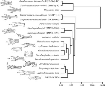
The results obtained in the hierarchical cluster analysis places MPEF-PV 3826 together with heterodontosaurids, which is recovered as a unique and well differentiated cluster (Fig. 7). In general terms, this analysis clusters heterodontosaurids with other small bipedal taxa, such as basal ornithopods (e.g., Anabisetia, Hypsilophodon, Thescelosaurus, Othnielosaurus, Agilisaurus) and basal ornithischians (e.g., Lesothosaurus, Stormbergia). However, other bipedal taxa, such as dryosaurids and Gasparinisaura, are placed outside this cluster. The high cophenetic correlation coefficient (CCC=0.946) indicates an overall good correlation between the distance and the ultrametric matrices.
Cluster analysis results, including schematic representation of the pes of included taxa.

The recovery of MPEF-PV 3826 within the heterodontosaurid cluster reflects the overall similarity in metatarsal proportions. Furthermore, the proportions of the metatarsus within Heterodontosauridae are relatively homogeneous, as represented by the small Euclidean distances between taxa (in comparison with those of other bipedal ornithischians). Given that all included taxa in this analysis are small bipedal species which probably share similar locomotory biomechanics, it is likely that the different clusters found for heterodontosaurids and basal ornithopods reflect some phylogenetic signal. The variability in the proportions of the metatarsals of these taxa were incorporated in the phylogenetic analysis (as continuous characters, given the nature of their variation; see below).
Phylogenetic analysis
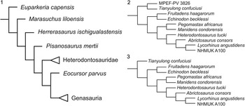
The phylogenetic affinities of MPEF-PV 3826 were tested using a modified version of the data matrix originally compiled by Butler et al. (Reference Butler, Upchurch and Norman2008). We integrated the subsequent modifications made by several authors to this original dataset (Zheng et al., Reference Zheng, You, Xu and Dong2009; Butler et al., Reference Butler, Porro, Galton, Chiappe, Henderson and Erickson2010, Reference Butler, Liyong, Jun and Godefroit2011; Pol et al., Reference Pol, Rauhut and Becerra2011; Han et al., Reference Han, Barrett, Butler and Xu2012), the new information provided by Sereno (Reference Sereno2012) on heterodontosaurids, and added new data.
Subsequent modifications of the dataset of Butler et al. (Reference Butler, Upchurch and Norman2008) have added taxa (e.g., Fruitadens haagarorum, Tianyulong confuciusi, Eocursor parvus Butler, Smith and Norman, Reference Butler, Smith and Norman2007, Yinlong downsii Xu, Forster, Clark and Mo, Reference Xu, Forster, Clark and Mo2006 [Butler et al., Reference Butler, Porro, Galton, Chiappe, Henderson and Erickson2010, Reference Butler, Liyong, Jun and Godefroit2011]), re-scored cells for some taxa (e.g., Wannanosaurus yansiensis Hou, Reference Hou1977, Micropachycephalosaurus hongtuyanensis Dong, Reference Dong1978, and Stenopelix valdensis Von Meyer, Reference Von Meyer1857, following Butler et al., Reference Butler, Liyong, Jun and Godefroit2011; and Jeholosaurus shanyuanensis Xu, Wang, and You, Reference Xu, Wang and You2000a, following Han et al., Reference Han, Barrett, Butler and Xu2012) and added new characters. Some of these papers were published almost simultaneously and therefore we have here integrated all of these changes.
We followed the recent anatomical interpretations presented by Sereno (Reference Sereno2012) in his comprehensive review of Heterodontosauridae and incorporated these into the updated version of the dataset of Butler et al. (Reference Butler, Upchurch and Norman2008). These include the addition of Pegomastax to the data matrix, a re-scoring of most heterodontosaurids based on the new anatomical information, and the addition of 18 new characters included in the heterodontosaurid data matrix of Sereno (Reference Sereno2012). The scores for Manidens condorensis were based on Pol et al. (Reference Pol, Rauhut and Becerra2011) and Sereno (Reference Sereno2012), with codings for the new characters being added. Only two characters were re-scored in Jeholosaurus based on the recent detailed description by Han et al. (Reference Han, Barrett, Butler and Xu2012). All changes are detailed in Supplementary Data 10.
Finally, as our phenetic analysis found informative variation in the metatarsus (see above), which may characterize heterodontosaurids among ornithischian dinosaurs, we have added 11 new characters to the data matrix (three continuous and eight discrete characters) and modified the definition of character 209 (Butler et al., Reference Butler, Upchurch and Norman2008), adding a new character state. The new characters, as well as the modified character 209, are discussed below. The final version of the data matrix includes 53 taxa and 259 characters. These modifications aim to test the affinities of the specimen MPEF-PV 3826 with Heterodontosauridae within the context of ornithischian dinosaurs.
Character descriptions: continuous characters
The results of the cluster analysis indicate that there are differences in metatarsal proportions (compared to metatarsal III) that group heterodontosaurids and basal ornithischians in different clusters, despite the fact that these species share similar size and locomotor capabilities (Fig. 8.5). The following characters capture linear morphometric ratios that vary continuously among taxa and lack clearly defined gaps in their distribution. They were therefore treated as continuous characters in the phylogenetic analysis (Fig. 8.5), and were normalized to range between 0 and 1.
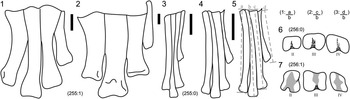
Schematic line drawings representing the new characters described: (1) Tenontosaurus tilletti, modified from Galton (Reference Galton1974a); (2) Camptosaurus dispar, modified from Galton (Reference Galton1974a); (3) Tianyulong confuciusi, based on photographs in Zheng et al. (Reference Zheng, You, Xu and Dong2009; supplementary material); (4) MPEF-PV 3826, left metatarsals; (5) the same as (4) but outlining the measurements taken to calculate the proportions for characters (1–3); (6, 7) schematic line drawing of distal view of condyles of metatarsals II–IV of MPEF-PV 3826 and Hipsilophodon foxii Huxley, Reference Huxley1869 (modified from Galton, Reference Galton1974b). The numbers represent the character and state figured as (ch:st). Scale bars equal 1 cm.

1: Metatarsal I/metatarsal III length ratio
The importance of the lateral and medial digits tends to diminish through the evolution of Ornithopoda, as indicated by the reduction and finally complete loss of medial and lateral metatarsals I and V. Several basal ornithopods lack phalanges in the reduced digit V, but only Gasparinisaura and Orodromeus also lack phalanges in digit I among non-iguanodontian ornithopods (Norman et al., Reference Norman, Sues, Witmer and Coria2004). Furthermore, complete loss of the phalanges in digits I and V is also documented in several basal iguanodontians (Dryosaurus altus, Iguanodon bernissartensis Boulenger, Reference Boulenger1881, Mantellisaurus atherfieldensis Hooley, Reference Hooley1925 [Norman, Reference Norman2004]), and hadrosaurs, which also lack metatarsals I and V (Horner et al., Reference Horner, Weishampel and Forster2004). In basal ornithischians and ornithopods, metatarsal I is present and its length in relation to metatarsal III varies markedly, with values ranging between 0.10 and 0.68. However, this ratio is relatively homogeneous among heterodontosaurids (0.55–0.58). This contrasts with the situation in ornithopods, in which the progressive reduction of metatarsals I and V results in a large interval of ratios. Thus, heterodontosaurids seem to be anatomically conservative in this respect.
2: Metatarsal II/metatarsal III length ratio
This ratio varies between 0.86 and 0.9 among heterodontosaurids. Among the other species there are two patterns. While some small-bodied basal ornithopods show smaller values of length ratio (i.e., Hypsilophodon, Anabisetia, Othnielosaurus, Stormbergia with less than 0.83), others show higher values of length ratio (as Gasparinisaura, with more than 0.9), although most of the considered species at the cluster analysis show similar values than heterodontosaurids. Among other ornithischian lineages, the acquisition of columnar limbs in the heavy-bodied thyreophorans and the retention of several adaptations to a subcursorial locomotion in large cerapodans (Coombs, Reference Coombs1978) result in metatarsal elements that are subequal in length and show a marked symmetry on both sides of digit III. In these groups, the decrease in length of all digits and the reduction of lateral digits lead to a robust pes with a large surface for support in relation to the length of the metapodium. The difference in ratio between small and large ornithischians is thus certainly related to an increase in body size (Coombs, Reference Coombs1978).
3: Metatarsal IV/metatarsal III length ratio
As in the former character, bipedal cursorial ornithischians have lower ratios than large-bodied taxa. However, in comparison with metatarsal II, metatarsal IV is usually slightly shorter in larger ornithischians, whereas it is similar (or longer) than metatarsal II in smaller ornithischians.
Character description: discrete characters
The only characters described here are those modified from the original matrix of Butler et al. (Reference Butler, Upchurch and Norman2008: character 209) and eight new characters (Figs. 8–9).
Photographs illustrating new characters. (1) Lateral view of ungual phalanx of digit I of Othnielosaurus consors YMP 1882, photo courtesy of Dr. I. Diaz Martinez; (2) Heterodontosaurus tucki SAM 1332, ungual phalanx of digit IV; (3) Tianyulong confuciousi STMN 26-3, ungual phalanx of digit III (Zheng et al., Reference Zheng, You, Xu and Dong2009, supplementary material); (4) most complete ungual phalanx of MPEF-PV 3826; (5) proximoplantar view of medial distal tarsal and proximal metatarsals of MPEF-PV 3826; (6) proximoplantar view of medial distal tarsal and metatarsals of Gasparinisaura cincosaltensis (MUCPv-208); (7) anteroproximal view of medial and lateral tarsals of Camptosaurus dispar, modified from Gilmore (Reference Gilmore1909). ft, flexor tubercle. The numbers represent the character and state figured (ch:st). Scale bars equal 1 mm in (1–4) and 1 cm in (5–7).

212: Medial distal tarsal
(0) articulates with metatarsal III only; (1) articulates with metatarsals II and III; (2) articulates with metatarsals I, II and III. (modified from character 209 of Butler et al. [Reference Butler, Upchurch and Norman2008] by adding character state 2). The distal tarsals of Heterodontosaurus tucki were first described as three fused elements, which would be an unusual condition among ornithopods, which commonly retain only two elements, and to which clade this species was referred at that time (Santa Luca, Reference Santa Luca1980; Norman et al., Reference Norman, Sues, Witmer and Coria2004). In this interpretation, distal tarsal 1 was said to contact metatarsals I and II, distal tarsal 2 contacted metatarsal III, and distal tarsal 3 contacted metatarsal IV (also in Norman et al., Reference Norman, Sues, Witmer and Coria2004). Butler et al. (Reference Butler, Porro, Galton, Chiappe, Henderson and Erickson2010) scored Heterodontosaurus as having the articulation of the medial distal tarsal with metatarsal III only, and Zheng et al. (Reference Zheng, You, Xu and Dong2009) scored the same character state for the heterodontosaurid Tianyulong (Zheng et al., Reference Zheng, You, Xu and Dong2009). However, Sereno (Reference Sereno2012) argued that there are only two distal tarsals in the right pes of Heterodontosaurus (SAM-PK-K1332), and that the putative contact between distal tarsal 1 and 2 (Santa Luca, Reference Santa Luca1980) is indeed a crack. Therefore, he concluded that this taxon has the common condition of only two distal tarsals (as in Tianyulong). Based on this interpretation, Heterodontosaurus and Tianyulong are here regarded as having an enlarged medial distal tarsal that articulates with metatarsals I, II and III. This character state also is present in MPEF-PV 3826, and might be a common feature among heterodontosaurids.
252 (new character)
Metatarsals proximally coossified to each other (in sub-adult to adult specimens): (0) absent; (1) present. Santa Luca (Reference Santa Luca1980) considered the metatarsals of Heterodontosaurus to be closely appressed to each other (a common condition among ornithischians [Norman et al., Reference Norman, Sues, Witmer and Coria2004]). However, Sereno (Reference Sereno2012) argued that the metatarsals were in fact fused, not only in Heterodontosaurus but also in Tianyulong. Furthermore, he interpreted the absence of a coossified tarsometatarsus and the absence fused metatarsals in Abrictosaurus (NHMUK RU B54) to reflect the juvenile ontogenetic stage of this specimen. In MPEF-PV 3826 the metatarsals are coossified proximally.
253 (new character)
Presence of a tarsometatarsus, with at least metatarsals I-III being partially or totally coossified with medial distal tarsal (in sub-adult to adult specimens): (0) absent; (1) present. The presence of a tarsometatarsus (fusion of metatarsals and distal tarsals) in Heterodontosaurus was regarded as a distinctive feature among ornithischian dinosaurs (Santa Luca, Reference Santa Luca1980). Although the lateral distal tarsal is missing in Tianyulong, the medial distal tarsal is completely fused to the metatarsals also in this taxon (Sereno, Reference Sereno2012). In MPEF-PV 3826, the lateral distal tarsal is also missing, but the medial distal tarsal is fused to metatarsals I-III. The coossification of the lateral distal tarsal and metatarsal IV is highly likely in MPEF-PV 3826 (see above), but the character definition is here restricted to the medial distal tarsal, given that it is the only element preserved in both Tianyulong and MPEF-PV 3826. The character was coded as uncertain in Abrictosaurus, due to the immature ontogenetic stage of the type specimen of this taxon. A tarsometatarsus is also present in some theropods (some coelophysoids [Tykoski and Rowe, Reference Tykoski and Rowe2004], oviraptorids [Olmólska et al., Reference Osmólska, Currie and Barsbold2004], troodontids [Makovicky and Norell, Reference Makovicky and Norell2004], dromaeosaurids [Norell and Makovicky, Reference Norell and Makovicky2004] and birds [Padian, Reference Padian2004]), but in heterodontosaurids it does not reach the degree of coossification as observed in some of these lineages.
254 (new character)
Ventral flexor tubercle in pedal unguals: (0) absent or developed as a low mound near the articular surface of the ungual phalanx; (1) present, well-developed protuberant process that is displaced distally, resulting in a flat ventral space between the tubercle and the articular surface. Although the presence of a flexor tubercle in pedal ungual phalanges is a widespread feature among archosaurs and plantarly prominent and rounded flexor tubercles commonly seen among Theropoda, the presence of a plantarly prominent distally displaced flexor tubercle is an unreported feature within Ornithischia. However, this feature is present in both MPEF-PV 3826 and Tianyulong (IVPP V17090, Sereno, Reference Sereno2012, fig 28–29; STMN 26-3, Zheng et al., Reference Zheng, You, Xu and Dong2009, supplementary material). As in some extant birds, MPEF-PV 3826 has a highly developed flexor tubercle, which is also present but less pronounced in Tianyulong (if compared to the total length of the ungual). This phylogenetic character might also be informative from an ecological point of view, as it was interpreted as an acquisition to assist grasping in bipedal cursorial dinosaurs with small forelimbs (Sereno, Reference Sereno2012).
255 (new character)
Subarctometatarsalian condition (sensu White, Reference White2009): (0) absent, no proximal overlapping of metatarsals II and IV over metatarsal III and metatarsal III being wider lateromedially than the other metatarsals in anterior view (representing more than 30% of total width of the shaft in anterior view); (1) incipient subarctometatarsalian condition, with the proximal part of metatarsal III being partially covered by metatarsals II and IV, and the lateromedial width of metatarsal III is notably less than that of the other metatarsals (being less than 30% of the width of the shaft proximally and until the mid-length in anterior view). In the subarctometatarsalian condition the proximal end of metatarsal III is equally visible in both anterior and plantar views. In plantar view, metatarsal III is visible along its entire length. Proximally, metatarsal III is lateromedially pinched between metatarsals II and IV, a feature that extends at least down to the midshaft, while the distal end of metatarsal III broadens (White, Reference White2009). This condition was described for the basal tyrannosaurid Dilong paradoxus Xu, Norell, Kuang, Wang, Zhao, and Jia, Reference Xu, Norrel, Kuang, Wang, Zhao and Jia2004 (IVPP V11579). The specimen MPEF-PV 3826 has an incipient subarctometatarsal condition, which is markedly different from the generalized ornithischian morphology. The metatarsals are tightly packed, and metatarsal III is partially covered by metatarsals II and IV along its proximal half in both anterior and plantar views. Metatarsal III has a trapezoidal cross section (with the narrower end pointing plantarly, instead of being wedge-shaped as in Dilong). This condition is also present in Heterodontosaurus (SAM-PK-K 1332), and might be present in Tianyulong (specimen STMN 26-3), for which no anterior view has been published, but mentatarsal III is characteristically pinched in plantar view. The condition in Abrictosaurus is unclear due to the supposedly juvenile stage of the type and only known specimen (Sereno, Reference Sereno2012). All other bipedal ornithischian dinosaurs have tightly packed, cylindrical metatarsals without a pinched shaft (e.g., Lesothosaurus, Stormbergia, Scutellosaurus, basal Ornithopoda) or lateromedially expanded metatarsals that are subequal in length and adapted for graviportal locomotion in heavier and often quadrupedal animals, as mentioned above (thyreophorans, more derived ornithopods and hadrosaurs, ceratopsids).
256 (new character)
Metatarsals II and IV, mediolateral width of articular condyles in distal view: (0) larger than anteroplantar height; (1) equal to or smaller than anteroplantar height. The lateromedial width is larger than the anteroplantar height in metatarsals II and IV in several basal dinosaur groups (e.g., basal sauropodomorphs, basal saurischians, heterodontosaurids) and the robust metatarsals of more heavy-bodied ornithischians (ceratopsids, thyreophorans). This contrast with the proportionately narrow distal ends of metatarsals II and IV in cursorial ornithopods (i.e., Dysalotosaurus, Hypsilophodon, Othnielosaurus, Parksosaurus warreni Sternberg, Reference Sternberg1937 [=Thescelosaurus warreni Parks, Reference Parks1926], Lesothosaurus, Gasparinisaura, Anabisetia), as well as in large ornithopods, iguanodontids and hadrosaurs. In these species, the anteroplantar height is larger than the lateromedial width in the distal ends of metatarsals II and IV. The presence of lateromedially wider condylar surfaces in heterodontosaurids can be considered to be the plesiomorphic condition of this character that remained unchanged in heterodontosaurids throughout most of the Mesozoic.
257 (new character)
Extensor pits in distal ends of proximal phalanges of pedal digits II–IV: (0) absent; (1) present. This character was discussed by Sereno (Reference Sereno1986), who proposed it as a synapomorphy of Heterodontosauridae, and briefly mentioned by Butler et al. (Reference Butler, Porro, Galton and Chiappe2012), but it has not been included in a phylogenetic analyses. We include this feature as a phylogenetic character.
258 (new character)
“Hypotarsal-like” proximodistally oriented process on the plantar surface of the medial distal tarsal, above metatarsal II: (0) absent; (1) present. Santa Luca (Reference Santa Luca1980, fig. 10C) described a proximodistally oriented process or flange exposed plantarly in tarsal 1 (medial distal tarsal sensu Sereno, Reference Sereno2012) in Heterodontosaurus. This process was argued to serve for the attachment of flexor tendons of the pedal digits, as it is the case in the hypotarsal ridge of extant birds (Santa Luca, Reference Santa Luca1980; Norman et al., Reference Norman, Sues, Witmer and Coria2004). The plantar side of both pedes is exposed in the holotype specimen of Tianyulong (STMN 26-3), showing the same vertically oriented process above metatarsal II (Zheng et al., Reference Zheng, You, Xu and Dong2009; supplementary material). Finally, MPEF-PV 3826 also shows this hypotarsal ridge (Fig. 2.3–2.6, 3.1–3.2). This feature cannot be scored for Fruitadens and Abrictosaurus, and it has not been described in any other ornithischian. The only possible exception was noted by Han et al. (Reference Han, Barrett, Butler and Xu2012), who described an L-shaped medial distal tarsal in Jeholosaurus shangyuanensis (horizontal and vertical portion of the “L” corresponding to distal tarsals 1 and 2, respectively), with a proximally placed ridge marking the suture line between the fused tarsals. A similar condition may also be present in Orodromeus makelai, as described by Scheetz (Reference Scheetz1999). In these two species, this particular tarsal seems to expose a protuberant structure plantarly (see Han et al., Reference Han, Barrett, Butler and Xu2012, fig. 12), resulting in an L-shaped medial distal tarsal in proximal view (with the horizontal portion of the L being formed by this structure), but there is no further description of this structure. The condition in Orodromeus and Jeholosaurus might be morphologically and functionally comparable to the hypotarsal-like process of the medial distal tarsal in heterodontosaurids, but the lack of an a detailed description, including the orientation and development of this process, does not allow to score character state 1 for these two species with certainty, so they were scored as ambiguous in the data matrix.
259 (new character)
Extensor process located dorsally at the proximal end of the first phalanx of pedal digit III: (0) absent; (1) present. The presence of an extensor process above the proximal articular surface of pedal phalanx III.1 was described for Heterodontosaurus by Santa Luca (Reference Santa Luca1980). This process can also be recognized in MPEF-PV 3826 (Fig. 4.10–4.12), and is subtly developed in Abrictosaurus (NHMUK RU B54; Sereno, Reference Sereno2012, fig. 37). The presence of such a process cannot be determined for Tianyulong (specimen IVPP V17090, Sereno, Reference Sereno2012, fig. 28) and Fruitadens (Butler et al., Reference Butler, Porro, Galton and Chiappe2012), because the respective phalanx is not preserved. The process is absent in Pisanosaurus (PVL 2577, personal observation; Bonaparte, Reference Bonaparte1976), indicating that this taxon, wich was argued to be a heterodontosaurid by Casamiquela (Reference Casamiquela1967) and Bonaparte (Reference Bonaparte1976), lies outside of this clade or might represent one of its most basal members.
Considerations on the phylogenetic analysis
The vertebrate-bearing layer of the Queso Rallado locality consists of a hard, strongly silicified limestone, which makes it impossible to excavate specimens in situ. Vertebrate material from Queso Rallado is therefore collected by separating large blocks of matrix in the quarry and subsequently splitting these blocks in camp. Both the holotype of Manidens condorensis (MPEF-PV 3211) and the specimen MPEF-PV 3826 were found in smaller matrix slabs resulting from this process, but no information about a potential original association of these remains is available, as only preparation in the lab revealed the identity of the remains. Based on morphologic features, MPEF-PV 3826 surely represents a small heterodontosaurid, the specimens MPEF-PV 3826 and 3211 are of matching size, and it is conspicuous that the former specimen only contains elements that are missing in the type of M. condorensis, indicating that both might represent a single individual. On the other hand, the specimen MPEF-PV 3826 might also represent another individual of Manidens condorensis. However, given the lack of overlap in the specimens and the missing information about a possible association of these remains, none of the possibilities can be proven at the moment. Therefore, a referral of MPEF-PV 3826 to Manidens condorensis may be probable, but is not conclusive. Thus, in the phylogenetic analysis we have alternatively included Manidens condorensis and MPEF-PV 3826 as independent OTUs and as a single OUT (combining the character scorings).
The taxonomic affinity of the specimen NHMUK A100 with Lycorhinus angustidens Haughton, Reference Haughton1924 (=Lanasaurus scalpridens Gow, Reference Gow1975) is still debated (Butler et al., Reference Butler, Upchurch and Norman2008; Sereno, Reference Sereno2012). Butler et al. (Reference Butler, Upchurch and Norman2008) included NHMUK A100 as a separate OTU in their phylogenetic analysis, with some of the characters scored for NHMUK A100 being based on the identifications of Thulborn (Reference Thulborn1970), who misinterpreted some of the skull bones (e.g., frontal, parietal). The reinterpretation of these elements by Sereno (Reference Sereno2012) was followed here and NHMUK A100 was therefore rescored for these features. The referral of NHMUK A100 to Lycorhinus Haughton, Reference Haughton1924, as Thulborn (Reference Thulborn1970) and Sereno (Reference Sereno2012) suggested, was tested by considering the former as a separate OTU from the latter taxon. Lycorhinus angustidens was coded based on the holotype SAM-PK-K3606 and the referred specimens BP/1/4244, and BP/1/5253. The expanded dataset presented here provides a broader empirical basis to test these issues.
Phylogenetic results
The data matrix was analyzed using equally weighted parsimony in TNT, version 1.1 (Goloboff et al., Reference Goloboff, Farris and Nixon2008a, Reference Goloboff, Farris and Nixon2008b). Although the phylogenetic position of Stenopelix Von Meyer, Reference Von Meyer1857, Yandusaurus He, Reference He1979 and Talenkauen Novas, Cambiaso and Ambrosio, Reference Novas, Cambiaso and Ambrosio2004, varies among the most parsimonious trees (MPTs), they were not excluded from the strict consensus tree, because the resolution of the nodes involved was not relevant for our goals (Fig. 10.1; see Supplemental Data 11). The number of MPTs in both analyses does not vary, as both yielded 120 MPTs of 682 steps using Manidens and MPEF-PV 3826 as independent OTUs, and 683 steps considering Manidens and MPEF-PV 3826 as the same taxon. The global topology of the strict consensus tree does not significantly differ from that of Butler et al. (Reference Butler, Upchurch and Norman2008) and successive analyses. All species previously referred to Heterodontosauridae are clustered in a monophyletic group (Fig. 10), and Heterodontosaurinae is recovered with the same taxonomic content as found by Sereno (Reference Sereno2012). The topology within Heterodontosauridae in the strict consensus tree supports a clade of South African heterodontosaurids (Heterodontosaurus, Abrictosaurus, Lycorhinus, and NHMUK A100), as in Pol et al. (Reference Pol, Rauhut and Becerra2011), but differing from the hypothesis of Sereno (Reference Sereno2012), who retrieved Lycorhinus as a more basal taxon. Lycorhinus and NHMUK A100 were found to be sister taxa, based on a character reversal (absence of considerable size variation in crowns along the cheek toothrow [character 236]), providing support for the identification proposed by Thulborn (Reference Thulborn1970) and Sereno (Reference Sereno2012). This clade was found to be more closely related to Abrictosaurus than to Heterodontosaurus. The sister group relationship of Pegomastax Sereno, Reference Sereno2012 and Manidens retrieved by Sereno (Reference Sereno2012) was corroborated in our analysis.
Phylogenetic analysis results: (1) summarized overall topology showing the placement of the taxa of interest, (2) topology of Heterodontosauridae considering MPEF-PV 3826 and Manidens condorensis as independent taxa, (3) topology of Heterodontosauridae considering MPEF-PV 3826 and Manidens condorensis as the same taxon.

The inclusion of MPEF-PV 3826 within Heterodontosauridae is supported by the presence of seven of 17 synapomorphies of the group (Medial distal tarsal articulates distally with metatarsals I, II and III [character 212]; Metatarsals proximally coossified (character 252) and fused with the medial distal tarsal forming a tarsometatarsus (character 253); Subarctometatarsalian condition [character 255]; Extensor pits present at the distal end of proximal phalanges of pedal digits [character 257]; proximodistally oriented “Hypotarsal-like” process on the plantar side of the medial distal tarsal above metatarsal II [character 258]; and an extensor process located dorsally at the proximal end on the first phalanx of digit III [character 259]). When analyzed as a separate OTU, MPEF-PV 3826 is recovered as sister taxon to the small and basal Chinese taxon Tianyulong, based on the presence of two synapomorphies (similar metatarsal IV / metatarsal III length ratio [character 3], Flexor tubercle in ungual phalanges of pes [character 254]). The referral of MPEF-PV 3826 to Manidens condorensis (including combined character scorings) does not change the position of Manidens within Heterodontosauridae (Fig. 10.3).
Discussion
Histology
While the presence of some external osteological features (e.g., complete fusion of the neurocentral sutures of all caudal vertebrae, coossification of the tarsometatarsi [Brochu, Reference Brochu1996; Horner et al., Reference Horner, De Ricqlés and Padian2000; Cerda and Chinsamy, Reference Cerda and Chinsamy2012; Sereno, Reference Sereno2012]) might suggest that MPEF-PV 3826 was a fully-grown individual, histological data supports a subadult ontogenetic stage for this specimen. The subadult condition for the individual is based on the absence of an external fundamental system (EFS or outer circumferential layer) in the outermost cortex. The presence of an EFS indicates that the individual has reached somatic maturity (Chinsamy-Turan, Reference Chinsamy-Turan2005). Regarding sexual maturity, this parameter was historically inferred from a marked transition of the primary cortical bone from fibrolamellar to parallel fibred bone and/or a notable decease in the width of the zones in dinosaurs and other ornithodirans. This change results from a slow-down of bone apposition rates in response to energy being redirected from growth to reproduction (Chinsamy-Turan, Reference Chinsamy-Turan2005). Although a slight change in fiber arrangement is observed in the sectioned metatarsal, we consider that this change is not marked enough to indicate a global slow-down in periosteal bone formation and, thus, the reach of sexual maturity. The relative age of the individual could not be assessed because the sampled material lacks growth marks (annuli or lines of arrested growth). This might be related to different causes. If the growth of the element was cyclical, growth marks could be absent because of (1) destruction of early formed growth lines through bone resorption from the medullary cavity, or (2) the individual died before it reached its first annual cycle. On the other hand, if the specimen had continuous growth, LAGs and/or annuli might never have been formed, even in individuals older than 1 year. As noted above in the comparisons with Gasparinisaura, the dominance of fibrolamellar tissue in the middle–outer cortex, could be seen as evidence for a high growth rate (Francillon-Vieillot et al., Reference Fracillon-Vieillont, Debuffrénil, Castanet, Geraudie, Meurier, Sire, Zylberberg and De Ricqlés1990; Reid, Reference Reid1996; Chinsamy-Turan, Reference Chinsamy-Turan2005). The presence of external osteological features of maturity that would be expected in adult individuals in a histologically subadult specimen suggests that the former might not be conclusive evidence for ontogenetic interpretations in heterodontosaurids, and the size reached by the taxon represented by MPEF-PV 3826 might be larger than currently documented, as recently found in Heterodontosaurus tucki (Porro et al., Reference Porro, Butler, Barrett, Moore-Fay and Abel2011). As in MPEF-PV 3826, the Heterodontosaurus specimen SAM-PK-K 1332 also showed the osteological features of individual maturity mentioned (Santa Luca, Reference Santa Luca1980; Sereno, Reference Sereno2012), but the find and description of NM QR 1788 (Porro et al., Reference Porro, Butler, Barrett, Moore-Fay and Abel2011) reveal that adult sizes of Heterodontosaurus tucki could reach at least twice of that expected previously.
Cladistic analysis
Although a possible referral of MPEF-PV 3826 to Manidens cannot be evaluated, due to the lack of overlapping material, both the discrete and continuous characters relate this specimen to the family Heterodontosauridae, with some derived features being shared with the small basal heterodontosaurid Tianyulong. The small size and shared features with Tianyulong (including the inferred grasping function of the pes) may reflect similarities of both taxa in terms of their ecological niche or habits. A referral of MPEF-PV 3826 to M. condorensis would increase the anatomical knowledge of this species, but does not affect the phylogenetic position of this taxon. Thus, MPEF-PV 3826 is a small form of Heterodontosauridae that might be identified as Manidens condorensis. The placement of Lycorhinus angustidens and NHMUK A100 as sister taxa support the hypothesis that all specimens (the holotype SAM-PK-K3606, the referred specimens BP/1/4244 and BP/1/5253, and the controversial NHMUK A100) can be referred to the same species, as argued by Thulborn (Reference Thulborn1970) and Sereno (Reference Sereno2012). Concerning the new characters added here, most of these features are present only in Heterodontosauridae: six of the discrete characters were retrieved as synapomorphies for this family (see above), and character 254 (flexor tubercle in ungual phalanges) links MPEF-PV 3826 with Tianyulong. In the new character 256 (lateromedial width in relation to anteroplantar height of distal condyles of metatarsals II and IV) character state “0” (condyles wider lateromedially than anteroplantarly) was found to represent the plesiomorphic condition in dinosaurs, as it was observed in Marasuchus, basal saurischians, basal sauropodomorphs, Pisanosaurus, and heterodontosaurids. The derived condition is present in pachycephalosaurids, basal ornithopods and ankylopolexians. The presence of lateromedially wider distal condyles of metatarsals II and IV is therefore a plesiomorphic feature in heterodontosaurids (present in MPEF-PV 3826) that supports their basal phylogenetic position within Ornithischia. The plesiomorphic condition is present in some derived lineages (e.g., psittacosaurids, ceratopsids, thyreophorans). As with other morphological features of the hindlimb (i.e., robust bones forming a columnar limb, short inner metatarsals that are subequal in length, short digits with discoidal phalanges, etc.), the reversal to the plesiomorphic condition in heavy-bodied lineages of ornithischians might be a response to biomechanic constraints.
Cursorial capabilities
In comparison with other phylogentically close taxa the relative length of the metatarsus in relation to the mediolateral width of the proximal articular surface shows that the material describe here belongs to a taxon with a gracile pes (more similar to Heterodontosaurus and Gasparinisaura than to Dryosaurus and Thescelosaurus). The closely appressed configuration of the fused tarsometatarsi present in MPEF-PV 3826 probably represents an adaptation to resist the bending stress experienced during the step more efficiently (Holtz, 1995), and, together with the slender and proximodistally elongated morphology, suggest cursorial locomotion with running capabilities in this specimen. Thus, the presence of well–co-ossified tarsometatarsi might be a response to mechanic stress during cursorial movement independently of ontogeny, and although the specimen may represent a histologically advanced growth stage, fusion of the metatarsals and tarsals is not conclusive for inferring somatic maturity of individuals. The flat surface on the plantar region of the unguals (between the flexor tubercle and the proximal articular surface) probably contacted the ground when walking, standing, or running, with the pointed tip of the ungual not participating in the step cycle. The pointed claw was probably used only for a grasping function (as in extant birds).
Grasping capabilities
The plantarly curved and lateromedially slender claw with an anterior displacement of the flexor tubercle in the pedal ungual phalanges of MPEF-PV 3826 is a combination of features that is absent in other ornithischian dinosaurs, except for Tianyulong. This feature is not present in Heterodontosaurus, which has claw-like, plantarly flat unguals in the pes, which are lateromedially wide and distally elongated, with poorly developed flexor tubercles (SAM-PK-K1332), a common condition in basal ornithischians. Although digit I of MPEF-PV 3826 was certainly too short to reach the ground during normal locomotion, the presence of a deep distal plantar supracondylar fossa in metatarsal I (a feature only described in this specimen among Heterodontosauridae), an extensor pit that is located distally on phalanx I.1, and an anteroplantarly protuberant articular trochlea (allowing a wider range of motion of the phalanx) indicate that this digit might have had a grasping function. Furthermore, the lateromedial width of the distal trochlea of metatarsal I, which is similar to that of all other metatarsals (rather than reduced as in other ornithischians), together with the anteroplantarly wide range of motion and the lateral tubercles for ligament attachment (instead of grooves) on both sides of the distal trochlea are features not described in any other heterodontosaurid that also suggest a grasping role of digit I. In summary, the presence of a protuberant, “hypotarsal-like” process in the tarsus for attachment of flexor ligaments of the pes, the deep anterior extensor pits for tendon attachments (as found in a wide range of archosauromorph taxa by Hutchinson [Reference Hutchinson2002]) on the distal ends of metatarsals II and III and the first phalanges of all digits (features shared with other heterodontosaurids), the proximally directed and slightly prominent dorsal portion of the proximal articular facet in slender and distally placed phalanges for the attachment of extensor ligaments, elongate penultimate pedal phalanges, the presence of curved and slender claws with strongly developed, distally offset flexor tubercles in ungual phalanges (shared with Tianyulong), and the morphology of the distal trochlea of metatarsal I are all features that strongly suggest a grasping function for the pes in MPEF-PV 3826.
Ecological inferences
The inclusion of MPEF-PV 3826 and the new characters resulting from this study in the phylogenetic analysis results in the identification of some features (i.e., hypotarsal-like process, subarctometatarsalian condition, claw-like unguals with a prominent flexor tubercle, coossified tarsometatarsi) as specializations of heterodontosaurids (or a subclade thereof) among ornithischians. Although there are many differences to birds (metatarsal I reaches the tarsus and fuses with the tarsals and does not reach the level of the distal end of the other metatarsals in Heterodontosauridae, excluding perching capabilities) some of the features mentioned are convergently present in extant birds (Santa Luca, Reference Santa Luca1980). The penultimate phalanges on digits II-IV of the pes in MPEF-PV 3826 are longer proximodistally than all other, more proximally placed phalanges of the same digit, a feature not described in any other ornithischian, but shared with several species of perching and climbing arboreal birds, non-avian theropods, pterosaurs and lizards (Fisher, Reference Fisher1946; Zhou and Hou, Reference Zhou and Hou1998; Clark et al., Reference Clark, Hopson, Hernandez, Fastovsky and Montellano1998; Hopson Reference Hopson2001; Zhou and Farlow, Reference Zhou and Farlow2001; Kambic, Reference Kambic2008). To characterize digit morphology of MPEF-PV 3826 (and its possible relation to an ecological niche), a comparative analysis of the length of phalanges of digit III was performed. The specimen MPEF-PV 3826 was compared with extant birds of arboreal, terrestrial and undetermined habits (animals displaying both arboreal and terrestrial habits), following the work of Hopson (Reference Hopson2001). We furthermore included basal birds (Rahonavis Forster, Sampson, Chiappe and Krause, Reference Forster, Sampson, Chiappe and Krause1998, Sinornis Sereno and Rao, Reference Sereno and Rao1992, and some species of the families Archaeopterygidae and Confuciusornithidae), non-avian theropods for which arboreal habits have been suggested (Epidendrosaurus Zhang, Zhao, Xu and Wang, Reference Zhang, Zhou, Xu and Wang2002 [=Scansoriopteryx Czerkas and Yuan, Reference Czerkas and Yuan2002], Microraptor Xu, Zhou and Wang, Reference Xu, Zhou and Wang2000b), terrestrial theropods, and several ornithischians, using bibliographic data (see Supplementary Data 12). The data was plotted on a ternary diagram subjected to a Principal Component Analysis. The results reveal that the lengths of the phalanges of digit III of MPEF-PV 3826 lie outside of the proportions seen in other ornithischians. Specimen MPEF-PV 3826 shares the morphospace in the ternary diagram and PCA graphics with extant birds that display arboreal and undetermined habits (both arboreal and terrestrial; Hopson, Reference Hopson2001). Among extinct taxa, MPEF_PV 3826 shares the morphospace with confuciusornithids, and is close to Microraptor and archaeopterygids, which have been considered as species with arboreal habits by some authors (Feduccia, Reference Feduccia1993; Hopson, Reference Hopson2001). Thus the placement of MPEF-PV 3826 in the morphospace is suggestive (albeit not conclusive) for an interpretation of arboreal habits in this specimen (Fig. 11.1–11.4; PCA results in Supplemental Data 13). Regarding the anatomy of ungual phalanges, the combination of well-developed flexor tubercles and the claw curvature is not commonly present among ornithischians (see above). Thus, claw morphology may also be indicative of the habits of this specimen, as these features vary among extant birds with different habits (Feduccia, Reference Feduccia1993; Pike and Maitland, Reference Pike and Maitland2004; Burnham et al., Reference Burnham, Feduccia, Martin and Falk2011). Following the functional inferences of bony claws derived from their shape by Ostrom (Reference Ostrom1974), the form of the main body with a rounded and distally offset flexor tubercle in the preserved unguals of Tianyulong (Zheng et al., Reference Zheng, You, Xu and Dong2009, supplementary material; Sereno, Reference Sereno2012, fig. 28–29) and MPEF-PV 3826 resembles the condition found in predatory and perching birds. On the other hand, the unguals of Tianyulong and MPEF-PV 3826 share a low curvature with ground birds and ground predators, but with a slightly more robust bony claw, more similar to Archaeopteryx than to both volant and non-volant predatory birds (Ostrom, Reference Ostrom1974, fig. 6). Based on quantitative analyses of bony claw curvature, several authors interpreted ecological aspects of Mesozoic dinosaurs, although the shape of the horny sheath of the claw might be more informative to test hypotheses of lifestyle (Burnham et al., Reference Burnham, Feduccia, Martin and Falk2011). Following Birn-Jeffery et al. (Reference Birn-Jeffery, Miller, Naish, Rayfield and Hone2012), three measurements were taken and several ornithischians were included in their dataset (measure references in Fig. 12.1 and 12.2; included ornithischians and modified data from Birn-Jeffery et al. [Reference Birn-Jeffery, Miller, Naish, Rayfield and Hone2012], are presented in Supplementary Data 14). The resulting box plots based on outer and inner curvatures (inner curvature=52°; outer curvature=72°; CX/CD=0.14), find all ornithischians, including Heterodontosaurus, Tianyulong, and MPEF-PV 3826, in the ecomorphological space of ground-dwelling animals (with the latter being close to the outer curvature of predatory forms; Fig. 12.3 and 12.4), such as Compsognathus, Sinornithomimus, and Caudipteryx (Birn-Jeffery et al., Reference Birn-Jeffery, Miller, Naish, Rayfield and Hone2012). A ground-dwelling lifestyle is also indicated for Tianyulong by the relation of claw thickness to inner and outer curvature (Fig. 12.5 and 12.6), whereas MPEF-PV 3826 shares an inner-curvature/relative-thickness relation with ground-dwelling forms and an outer-curvature/relative-thickness relation (which is similar to volant and non-volant predatory birds and Mesozoic theropods) with predatory taxa, such to Compsognathus (instead of taxa with a perching/climbing/arboreal behavior; Fig. 12.5 and 12.6). For all other ornithischians and Heterodontosaurus, a ground-dwelling lifestyle is inferred, based on the outer-inner curvature/relative-thickness relations. The morphological features together with the quantitative analyses in comparison with extant birds and Mesozoic theropods indicate cursorial habits, with probably some arboreal capabilities (based on the analysis of non-ungual phalanges of digit III). The latter might also be supported by the probable grasping abilities of the pes inferred from the morphological comparisons presented above.
Ternary diagram of ecomorphological spaces of several theropods and ornithischians, using measurements of proportions of non-ungual phalanges of digit III: (1) complete diagram; (2) detail of “uncertain” zone, that includes confuciusornithids, archaeopterygids, arboreal, terrestrial birds and birds of uncertain habits (Following Hopson, Reference Hopson2001), MPEF-PV 3826, several ornithischians, non-avian theropods and other taxa; (3) clean diagram showing convex hulls in the “uncertain zone”, summarizing the most important points; (4) principal component analysis using the proportions of non ungual phalanges of digit III, convex hulls of diagram result, note the clustering of MPEF-PV 3826 with extant birds of uncertain habits, actual arboreal birds and confuciusornithids. Symbol references in figure, D1-D2-D3 axes represent the contribution of each original variable to the ternary diagram; D1, first phalanx of digit III axis; D2, second phalanx of digit III axis; D3, third phalanx of digit III axis.

Claw curvature comparison including several actual birds and lizards, with Mesozoic theropods (following Birn-Jeffery et al., Reference Birn-Jeffery, Miller, Naish, Rayfield and Hone2012), and several ornithischian taxa (Scelidosaurus, Othnielosaurus, Hadrosaurine indet., Anabisetia, Gasparinisaura, Tianyulong): (1) inner claw curvature and relative thickness (CX/CD) measured on most complete ungual phalanx of MPEF-PV 3826 (modified from Birn-Jeffery et al., Reference Birn-Jeffery, Miller, Naish, Rayfield and Hone2012); (2) Outer claw curvature of the same phalanx (modified from Birn-Jeffery et al., Reference Birn-Jeffery, Miller, Naish, Rayfield and Hone2012); (3) Box plots for outer claw curvature distinguished by behavioral category, including all data presented by Birn-Jeffery et al. (Reference Birn-Jeffery, Miller, Naish, Rayfield and Hone2012), and adding several ornithischians; (4) as in 12.3 but resampling the inner curvature; (5) inner claw curvature plotted against relative thickness of the claw; (6) outer claw curvature plotted against relative thickness of the claw. For (3) and (4), both box plots were performed following specifications by Birn-Jeffery et al. (Reference Birn-Jeffery, Miller, Naish, Rayfield and Hone2012), with the addition of the average information as a dot within the shaded boxes and considering also the information of Mesozoic theropods. In (5) and (6), convex hulls summarizing the obtained results, configuration of convex hulls and Mesozoic theropods were obtained similar to Birn-Jeffery et al. (Reference Birn-Jeffery, Miller, Naish, Rayfield and Hone2012). References at family level, for species references see Birn-Jeffery et al. (Reference Birn-Jeffery, Miller, Naish, Rayfield and Hone2012): Alv, Alvaresaurid; Bas Av, basal Avialae; Climb, Cimbing extant birds and lizards; Comps, Compsognathidae; Drom, Dromaesauridae; Ground, ground-dwelling extant birds and lizards; Orn, Ornithomimosauria; Orn, Ornithischia; Ovir, Oviraptorosauria; Perch, perching extant birds and lizards; Pred, predatory extant birds and lizards; Tianyulong specimen IVPP V17090; Trood, Troodontidae. *, climbing species; +, predatory species; ○, perching species; x, ground-dwelling species; ◊, ornithischians; ●, Mesozoic theropods; ∆, Tianyulong IVPP V17090;
 $$ \raster="rg1"$$
Heterodontosaurus SAM-PK-K 1332; □, MP EF-PV 3826.

Conclusions
The specimen MPEF-PV 3826 from the late Toarcian Queso Rallado Locality (Cañadón Asfalto Formation) includes two complete metatarsi associated with scattered phalanges and caudal vertebrae. The detailed description, comparison, and morphometric analysis show overall similarities of MPEF-PV 3826 with representatives of the family Heterodontosauridae. Although the referral of MPEF-PV 3826 to Manidens condorensis (found in the same locality and horizon) is likely, there is no osteological evidence to identify this specimen as this taxon, as no overlapping elements are preserved. The phylogenetic results relate MPEF-PV 3826 to Heterodontosauridae, and if these remains are scored as part of Manidens condorensis the phylogenetic position of this taxon does not change in the resulting trees (as the sister taxon of Pegomastax, corroborating the hypothesis of Sereno, Reference Sereno2012).
The histological thin sections and comparisons with the metatarsi of Heterodontosaurus indicate that the remains described here probably pertain to a sub-adult individual rather than a full-grown specimen (as for Heterodontosaurus specimens SAM-PK-K1332 and NM QR 1788 mentioned by Porro et al., Reference Porro, Butler, Barrett, Moore-Fay and Abel2011).
The morphological comparison of the pes with that of other ornithischian taxa reveals the presence of new apomorphic features for Heterodontosauridae that distinguish this family even more from other ornithischians.
The reconstruction of the pes shows long and tightly appressed metatarsals with digits that are elongated in comparison with other ornithischian taxa (including other heterodontosaurids). A comparative analysis of the length ratio of non-ungual phalanges of digit III denotes similarities with both arboreal and ground-dwelling bird taxa. In contrast, comparative analysis of the pedal ungual phalanx reveals similarities of this specimen with predatory forms. These results might indicate cursorial, ground-dwelling behavior, but with a grasping pes and even possibly facultative arboreal capabilities in MPEF-PV 3826. Nonetheless, more information is needed to thoroughly test these hypotheses, including articulated remains that will allow testing the putative referral of this material to Manidens condorensis, as well as remains of the forelimb that is completely unknown for this taxon up to now.
Acknowledgments
Fieldwork and research were funded by ANPCyT PICT 1756, 0808 (to D.P.), DFG RA1012/9-1 (to O.W.M.R.), National Science Foundation grants DEB 0946430 and DEB 1068089 (to Dr. Guillermo Rougier), and international collaboration grant CONICET-Chinese Academy of Sciences to DP. We thank R. Coria for the measurements of the pes of Anabisetia saldiviai, I. Diaz Martinez for the help with character setup and the ornithischian photographic material he shared freely, Lic. D. Perez for the help in searching bibliography, L. Canessa for the mechanical preparation of the specimen MPEF-PV 3826, and M. Caffa for the thin section of metatarsal IV. We furthermore thank J. M. Leardi, C. Marsicano, and L. Gaetano for information on South African heterodontosaurids and basal ornithischians. S. Kaal is thanked for access to comparative specimens of Heterodontosaurus at the Iziko-South African Museum to DP. X.-T. Zheng is thanked for access to comparative specimens of Tianyulong at the Shandong Tianyu Museum of Nature to D.P. X. Zhang is also thanked for her help, and X. Xu is thanked for providing access to other relevant comparative material. Fieldwork was made possible by the authorities of the Secretaría de Cultura de la Provincia del Chubut.
Accessibility of supplementary data
Data available from the Dryad Digital Repository: http://datadryad.org/review?doi=doi:10.5061/dryad.gn960