

Introduction
The pantropical aquatic flowering plant family Podostemaceae has morphological features that differ markedly from the usual structures of other angiosperms (Engler, Reference Engler, Engler and Prantl1930; Cook & Rutishauser, Reference Cook, Rutishauser and Kubitzki2007). These peculiarities are due to the habitat in which the plants grow, i.e. rocks in waterfalls (Jäger-Zürn, Reference Jäger-Zürn2003, Reference Jäger-Zürn2005a). Such a habitat seems to necessitate adaptations to protect growing areas and young flower buds from the turbulent water. So-called dithecous leaves, which have two sheaths (theca), one on either side (adaxial and abaxial) of the leaf base (Engler, Reference Engler, Engler and Prantl1930), surround the shoot tip and the terminal flower bud, like bracts. Their obvious protective function enables the plants to resist the impact of the water and they have presumably evolved as a response to the specialised habitat. The occurrence of dithecous leaves is widespread among members of subfamily Podostemoideae of the Old and New World but their morphological nature has remained poorly understood since they were first described by Warming (Reference Warming1888). In many members of the subfamily leaf bases are fused to a compressed shoot axis (Jäger-Zürn, Reference Jäger-Zürn2005a) and the dithecous leaves are fused to neighbouring leaves. Both features obscure the position of dithecous leaves within the branched shoot system, and it is therefore not surprising that the interpretation of dithecous leaves has remained controversial (see Jäger-Zürn, Reference Jäger-Zürn2009).
It has become evident that dithecous leaves occur only at branching (Jäger-Zürn, Reference Jäger-Zürn1994), i.e. within the inflorescences in most species as branching in vegetative shoots is rare. However, branching in vegetative shoots does occur, for example in Zeylanidium subulatum (Gard.) C.Cusset. Investigations into branching in this species have revealed that the dithecous leaf is the first in a succession of leaves that develop. Accordingly, the dithecous leaf arises from the branch (Jäger-Zürn, Reference Jäger-Zürn1994: 399, as Podostemum subulatum Gard.).
However, as dithecous leaves are usually prominent and develop large sheathing wing-like expansions towards the terminal flower of the main shoot axis, these leaves appeared to arise from the main shoot axis, not the branch. This appearance has resulted in the view that the branch is subfoliar, i.e. below, instead of above, the leaf (Jäger-Zürn, Reference Jäger-Zürn1999, Reference Jäger-Zürn2000).
The placement of the branch in a subfoliar position has been shown not to apply to the elongated flowering (i.e. branched) shoot axis of Apinagia riedelii (Bong.) Tul. (Jäger-Zürn, Reference Jäger-Zürn2009). In this species, at branching, two leaves arise one above the other on the same side of the distichously leafy shoot instead of the more typical alternating pattern (only one leaf alternating on either side). Surprisingly, Apinagia riedelii possesses a subtending leaf (a so-called monothecous leaf with one adaxial sheath) below the floriferous branch (flower bud), and a dithecous leaf above the flower bud. Consequently, the hitherto accepted view of the dithecous leaf is not supported. It is the branch, not the main shoot axis, from which the dithecous leaf arises as the first leaf (addorsed hypsophyll) (Jäger-Zürn, Reference Jäger-Zürn2009). The lower sheath of the dithecous leaf is thus the real (adaxial, ventral) sheath that envelops the branch bud. An interpretation in which the branch is thought to be subfoliar in position is therefore incorrect and only valid in a descriptive sense: the true branch position is axillary and above the subtending leaf and not leaf-borne and subfoliar, below the dithecous leaf. Ramification conforms to the angiosperm norm. However, subtending leaves are absent in most species with dithecous leaves. In addition, the abaxial (dorsal) sheath of the dithecous leaf, in the form of dorsal wings, is directed towards the flower of the main shoot. These sheath-like wings are seen as novel structural outgrowths. In Apinagia riedelii the base of the dithecous leaf is fused to the main shoot axis and subsequently included in the forward growth of the shoot axis. This means that, in mature parts of the shoot, the blade of the dithecous leaf apparently arises from the main shoot axis. In the neotropical Podostemum rutifolium subsp. ricciiforme (Liebm.) Novelo & C.T.Philbrick, however, there is no such fusion. Study of the tiny plants of this species can thus reveal more clearly the position of dithecous leaves.
Podostemum rutifolium subsp. ricciiforme has only recently been rediscovered and redescribed, as Podostemum ricciiforme (Liebm.) P.Royen, by Novelo & Philbrick (Reference Novelo and Philbrick1997). These authors have subsequently included the taxon as a subspecies of Podostemum rutifolium Warm. (Philbrick & Novelo, Reference Philbrick and Novelo2004). The purpose of the present paper is to investigate the position of the dithecous leaf of Podostemum rutifolium subsp. ricciiforme in order to confirm the more recent interpretation of the nature of the dithecous leaf.
Materials and Methods
Fixed material (formaldehyde, propionic acid, ethanol) of Podostemum rutifolium subsp. ricciiforme was collected on 15 November 1967 in Colombia, Prov. Nariño, Rio Arrayan near to the confluence with Rio Guiza, by Wolfgang Hagemann, Heidelberg, Germany (Hagemann 19081). Voucher specimens are in the herbarium of the Senckenberg Museum, Frankfurt am Main, Germany. Microtome sections (10 μm; suitable for cell size) were stained with gentian violet. The illustrations are camera lucida drawings.
Results
General description
The thread-like roots of Podostemum rutifolium subsp. ricciiforme intertwine plagiotropously and densely cover the rocky substratum of the river bed. The twine-like roots consist of thin-walled parenchyma cells filled with starch grains. The roots contain a central vascular strand of prosenchymatous cells that do not show further differentiation. Occasionally, rhizodermal cells of the lower root surface develop into adhesive hairs which attach to the biofilm of the substratum (Jäger-Zürn & Grubert, Reference Jäger-Zürn and Grubert2000). Root surface cells and subrhizodermal cells are filled with sharp-edged silica bodies. These inclusions occupy the whole vacuole of the cells.
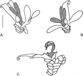
Many root-borne shoots, up to 6 mm long, arise laterally from the root surface. They are densely distichously covered with small, simple, entire or lobed leaves (Fig. 1A–C). The cells of the leaf base also have silica bodies, in the epidermal and subepidermal layer and occasionally in the parenchyma cells. The leaf blades largely lack silica bodies. The leaf lobes consist of only a few cell layers (Fig. 4). The leaf blades in mature plants tear off (Fig. 1C), whereas the basal parts of the leaves persist on the lower part of the shoot. The tight succession of leaves, with silica bodies in the basal leaf parts, forms a stiff cover around the shoot axis (Fig. 2A). Such specimens have only a few complete leaves, distichously arranged, at the top of the shoot, mostly with a central leaf in between (Fig. 1). The position of the latter cannot be observed without dissection.
Podostemum rutifolium subsp. ricciiforme (Liebm.) Novelo & C.T.Philbrick. Root-borne shoots with complete leaves present on top of densely leafy shoots, proximal parts of shoots only with leaf bases; note central dithecous leaf (in grey). Scale bar = 1 mm.

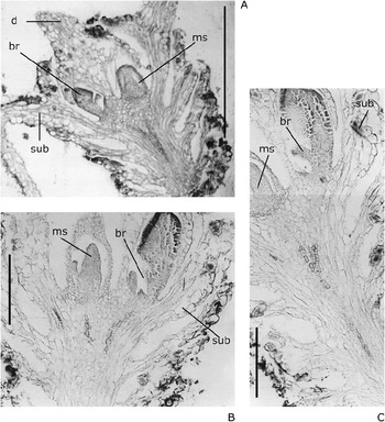
Podostemum rutifolium subsp. ricciiforme. Longitudinal sections of distichously leafy branched shoots showing flower bud of main shoot and branch in axil of subtending leaf; leaves forming a cortex around central part of shoot axis; upper parts of leaves torn off, lower parts with silica inclusions in epidermal cells; note dithecous leaf is first leaf of branch. br = branch; d = dithecous leaf; ms = main shoot; sub = subtending leaf. Scale bars: A = 500 μm; B–C = 200 μm.

Ramification
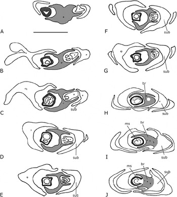
Ramification, which is usually associated with flowering in Podostemoideae, is observed in Podostemum rutifolium subsp. ricciiforme (Fig. 2). The structural pattern of the ramified shoot is best seen from serial microtome cross-sections that trace the succession of lateral appendages, i.e. leaves and branches. At the branching point appears a dithecous leaf (Fig. 1). The branch commonly develops leaves below the terminal flower (Fig. 2A–B). The specimens investigated here have a terminal flower on the leafy main shoot (Figs 2, 3, 4D–H) and a branch with several leaves (Figs 3F–H, 4).
Podostemum rutifolium subsp. ricciiforme. A–G: Line drawings of serial cross-sections of branching region (apical to proximal) of the photographs shown in Figs 4 and 5: dithecous leaf (grey) with two sheaths, one (left side) pointing towards terminal flower of main shoot surrounded by spathella (in black), the other towards branch (right side). Note double T-shape of dithecous leaf in cross-section. A–E, branch with two young leaves sheathed by dithecous leaf; F, youngest leaf fused to dithecous leaf; G, dithecous leaf and other young leaves of branch fused. H–J: Line drawings of serial cross-sections of the photographs shown in Fig. 6 (branch in grey). H, branch with dithecous leaf; I, branch in axil of subtending leaf, showing tracheary elements of dithecous leaf (left side) and juxtaposed to next younger leaf in distichous order (right side), tracheary elements of axis of branch in the centre; note dithecous leaf sheathing flower bud of main shoot; J, branch axillary between main shoot axis and subtending leaf. br = branch; ms = main shoot; sub = subtending leaf. Distance between each microtome section H–I = 30 μm, I–J = 20 μm. Scale bar = 500 μm.

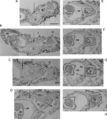
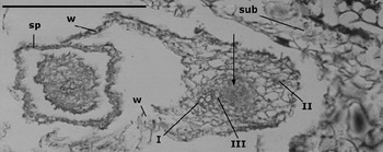
Podostemum rutifolium subsp. ricciiforme. Photographs of the branched shoot explained in the line drawings in Fig. 3 (apical to proximal). A, petiole and two sheaths of dithecous leaf; B, dithecous leaf sheathing terminal flower (surrounded by spathella) of main shoot (left), as well as branch (right side); C, dithecous leaf sheathing younger leaf of branch; note subtending leaf; D–E, double T-shaped dithecous leaf sheathing two young leaves of branch; F–G, younger leaves of branch fused to dithecous leaf; H, dithecous leaf sheathing younger leaves around apical furrow. I, II, III = numbered leaves of branch; dithecous leaf marked by arrow; sp = spathella of flower; sub = subtending leaf. Scale bar = 200 μm.

Serial cross-sections (Figs 3, 4) show the sheathing leaf bases of two leaves in a distichous position, one on either side of the main shoot axis, as well as a prominent leaf petiole between them (Figs 3A, 4A). The dithecous shape of the latter becomes visible on cross-sections made lower down (Figs 3A–G, 4B–H). Since, unlike most other members of Podostemoideae, no fusion occurs between the dithecous leaf and neighbouring appendages or to the shoot axis, the typical double T-shape of the dithecous leaf is visible in cross-section. One sheath is directed towards the terminal flower of the main shoot, while the other is directed towards a sequence of several young leaves assumed to belong to a branch of the main shoot (Figs 3E–G, 4E–G). The evidence for this assumption is in the position of the young shoot in the axil of one of the two uppermost leaves of the main shoot (Fig. 2). The plane of distichy of the leafy branch is the same as that of the main shoot. The branch base fuses to the (subtending) leaf in the area of its midrib, as seen on sections lower down (Figs 2B–C, 3H–I, 6C). The dithecous leaf is integrated within the sequence of distichous leaves along the branch (Figs 3E–G, 4F–H, 6A). Since the dithecous leaf is not fused to other organs of the main shoot, its association with the branch is apparent (Figs 3F–H, 4F–H). This shows that the dithecous leaf is clearly the first leaf of the branch (Figs 3F–G, 4G, 5). The sheath towards the axis of the branch is thus the adaxial (ventral) sheath of the dithecous leaf. The other sheath, towards the flower of the main shoot, occurs on the abaxial (dorsal) side of the dithecous leaf (Fig. 3G). The two wings of the dorsal (abaxial) sheath half surround the flower. There is no ligule around the flower of the main shoot as there is in Apinagia riedelii. The margins of the uppermost leaves of the main shoot cohere below the branch.
Podostemum rutifolium subsp. ricciiforme. Branch showing shoot apex, surrounded by three leaves in distichous order conforming to the plane of distichy of main shoot, leaves identifiable by median vascular strands; note dithecous leaf is first leaf of branch, half surrounding the terminal flower of main shoot by two wings. I, II, III = numbered leaves of branch, identified by tracheary elements of vascular strands; apex of branch marked by arrow; sp = spathella of flower; sub = subtending leaf; w = wing of abaxial sheath. Scale bar = 200 μm.

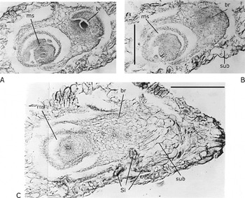
The shoot apex of the branch occurs as an apical furrow (Fig. 4) surrounded by leaves (Fig. 2G). The position of leaves is indicated by the single vascular strand of each leaf: the first, dithecous, leaf and the two subsequent leaves (Fig. 5). The dithecous leaf appears as one of the leaves around the shoot axis of the branch. The dorsal wings of the dithecous leaf are already present at the base of the branch and the branch is visible in the axil of the subtending leaf (Figs 3H–J, 5, 6C).
Podostemum rutifolium subsp. ricciiforme. Photographs of serial cross-sections depicted as line drawings in Fig. 3H–J (apical to proximal). A, branch with dithecous leaf and three younger leaves partly fused to each other; note dithecous leaf sheathing flower bud of main shoot and branch; B, apex of branch, leaves fused around branch axis; C, branch between main shoot and subtending leaf in axillary position; note vascular strands of the branch axis and two leaves in distichous phyllotaxy. br = branch; ms = main shoot; sub = subtending leaf; Si = silica bodies, rupturing the tissue when cut. Distance between each microtome section A–B = 20 μm, B–C = 50 μm. Scale bars = 200 μm.

Discussion
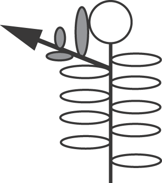
Flowering is reported to be rare in Podostemum rutifolium subsp. ricciiforme, but does occur (Philbrick & Novelo, Reference Philbrick and Novelo2004, and this study). Since flowering is typically connected with branching, the presence of dithecous leaves (that are present only at branching) is thus expected (Moline et al., Reference Moline, Les, Philbrick, Novelo, Pfeifer and Rutishauser2006: 445, fig. 36). It has hitherto been reported as self-evident that the dithecous leaf is positioned on the axis of the main shoot (Rutishauser et al., Reference Rutishauser, Pfeifer and Moline2003). The prominent size of the dithecous leaf, i.e. resembling leaves of the main shoot, has been a factor in its misinterpretation. However, an alternative interpretation has recently been reported for Apinagia riedelii in which the dithecous leaf is the first leaf of the branch (Jäger-Zürn, Reference Jäger-Zürn2009). In Apinagia riedelii the base of the dithecous leaf and the axis of the main shoot are fused, making interpretations difficult. However, the presence of subtending leaves in Apinagia riedelii makes the nature of the dithecous leaf apparent, and it is clear that the branch develops from an axillary meristem and not from the dithecous leaf. Fortunately, the fusion of vegetative structures which occurs in Apinagia riedelii does not occur in Podostemum rutifolium subsp. ricciiforme. In the latter taxon a subtending leaf is also present, confirming the axillary position of the branch (Figs 2, 3I–J, 6C). It is also clear in Podostemum rutifolium subsp. ricciiforme that the dithecous leaf is aligned within the sequence of leaves that develop from the branch (Figs 3H, 4F–H) and that it arises from the branch rather than the axis of the main shoot (Fig. 7). Both Apinagia riedelii and Podostemum rutifolium subsp. ricciiforme show that branching conforms to the typical axillary ramification pattern of angiosperms. Since the plane of distichy of the main shoot and branch is the same in both species, the first (dithecous) leaf of the branch is in an addorsed position.
Diagrammatic illustration of branched shoot (leaves of branch in grey); note first leaf of branch is dithecous.

The new interpretation is fortunately quite apparent in the simple shoot architecture of Podostemum rutifolium subsp. ricciiforme. Although tiny, this taxon has elongated – but not compressed – shoots with a dense succession of leaves that are not fused (Figs 1, 2A). Thus, unlike many other related species that have a compressed ‘thalloid’ plant body, shoots of this species are not modified. In contrast, the ramification pattern of the congeneric Podostemum weddellianum (Tul.) C.T.Philbrick & Novelo is obscure due to fusion of the dithecous leaf to the compressed shoot axis (Jäger-Zürn, Reference Jäger-Zürn2002a, as Crenias weddelliana). In this species, vascular strands diverge from the central area of the branch towards either side, and one of the strands seems to belong to the dithecous leaf (Jäger-Zürn, Reference Jäger-Zürn2002a: 69, fig. 5D–E). However, it is not possible to clearly associate the dithecous leaf either with the main shoot or with the branch. The morphological structure of the dithecous leaf in Podostemum weddellianum could be assumed to be the same as in Podostemum rutifolium subsp. ricciiforme. Since the shape of dithecous leaves is uniform throughout, the same development is predicted for all species of Podostemoideae which bear them.
The new interpretation also explains the nature of the single vascular strand which regularly occurs at the base below the branching point of the branch and the dithecous leaf. This vascular strand divides higher up into two strands supplying the branch and the dithecous leaf. It has so far been unclear whether the single strand (below the point of separation) is a leaf bundle (meaning the branch has to be viewed as leaf-borne) or the central strand of a shoot axis (the branch). Such a common vascular strand has been described in detail in Zeylanidium subulatum (Jäger-Zürn, Reference Jäger-Zürn1994: 401, as Podostemum subulatum) and Sphaerothylax abyssinica (Wedd.) Warm. (Jäger-Zürn, Reference Jäger-Zürn2000: 207, 215), but is also observed in other species with dithecous leaves, for example Apinagia multibranchiata (Matthiesen) P.Royen, Apinagia goejei Went, Marathrum foeniculaceum Humb. & Bonpl. (Jäger-Zürn, Reference Jäger-Zürn2002b, Reference Jäger-Zürn2005a, Reference Jäger-Zürn2005b) and Marathrum utile Tul. (unpubl. data). According to the new interpretation, the single vascular strand below the separation of branch and dithecous leaf is the central strand of the branch, not of the leaf. The central strand gives rise to the vascular bundles of the dithecous leaf and to the next younger leaf of the branch. Such structures also occur in Podostemum rutifolium subsp. ricciiforme (Figs 5, 6C).
The dorsal (abaxial) sheath of the dithecous leaf towards the main shoot varies in size between species in subfamily Podostemoideae, from a slight concave mould (as in Zeylanidium subulatum; Jäger-Zürn, Reference Jäger-Zürn1999) to prominent median stipule-like structures with elongated, but thin, wings at the sides (as in Apinagia riedelii; Jäger-Zürn, Reference Jäger-Zürn2009). In Apinagia riedelii the lateral wings forming the dorsal sheaths of juxtaposed dithecous leaves cohere and form a ligule-like or ochrea-like cover, and even a hood, over the flower bud of the main shoot.
Although the idea of the branch being subfoliar (Jäger-Zürn, Reference Jäger-Zürn1999) no longer holds true, the concept has been maintained by Imaichi et al. (Reference Imaichi, Hiyama and Kato2005) for some Asian members of subfamily Podostemoideae. Their work includes the assumption that a new leaf arises from the next older leaf separated by programmed cell death (Koi et al., Reference Koi, Imaichi and Kato2005). This view has been challenged (Jäger-Zürn, Reference Jäger-Zürn2007). According to Imaichi et al. (Reference Imaichi, Hiyama and Kato2005) and Koi et al. (Reference Koi, Imaichi and Kato2005) the shoot is suggested to be a chain of leaves without an apical meristem. In this interpretation, based on an investigation of Zeylanidium subulatum, the branch arises in a subfoliar position from the ‘dorsal’ (abaxial) side of the dithecous leaf. The species investigated by these authors lack an elongated and distinctly visible shoot axis, unlike Podostemum rutifolium subsp. ricciiforme which has a distinct shoot axis from which many leaves arise in distichous phyllotaxy. Leaf tissues are distinguishable as they form a cortex around the central part of the shoot axis. Similarly, the shoot axis of the branch is visible on the meristematic tissue which keeps the two youngest leaves apart from each other in a juxtaposed position (Figs 5, 6C). The meristematic tissue is visible as the apex of the shoot axis of the branch. Interpretation of the dithecous leaf generally depends on whether it is regarded as an appendage of the main shoot axis (e.g. Imaichi et al., Reference Imaichi, Hiyama and Kato2005) or of the branch (this study). The plane of distichy is the same in the main shoot and in the branch, leading to a view of the dithecous leaf being an appendage of the main shoot. If the first case were true the branch would be sheathed by the dorsal sheath of the dithecous leaf in a subfoliar position, below it on its dorsal side. If the second case were true the branch would be sheathed by the ventral sheath of the dithecous leaf.
The present study confirms the second case. The dithecous leaf is the first leaf of a sequence of leaves on the branch (Figs 3G–H, 4G, 6, 7). The leafy branch is adnate to the next lower leaf belonging to the respective orthostichy of the main shoot (Figs 3I–J, 6C). It is located in the axil of that leaf which is thus the subtending leaf. The position of the leafy branch (including the dithecous leaf) is clearly between the shoot axis of the main shoot (flower bud) and the subtending leaf. The tissue of the (older) subtending leaf differs from that of the leafy branch (Fig. 6C).
It should further be mentioned that programmed cell death has not been observed during leaf development in Podostemum rutifolium subsp. ricciiforme. In the species of subfamily Podostemoideae investigated so far the apical meristem is mostly very small, consisting of only a few meristem cells. These few cells (in the form of a meristème d'attente) may be easily overlooked (Jäger-Zürn, Reference Jäger-Zürn2007: 388). The absence of a prominent plumula in tunica corpus figuration thus does not convincingly point to the absence of an apical meristem.
In another interpretation both sheaths of the dithecous leaf are viewed as having leaf axils (like the axil of the subtending leaf) from which shoots or branches develop (Rutishauser et al., Reference Rutishauser, Pfeifer and Moline2003). This view is suggested by the flattening of the leaf perpendicular to the plane of leaf insertion, as well as by young leaf tips curling fern-like towards the so-called ‘front side’ of the dorsiventral plant body found in many species of subfamily Podostemoideae. The apical curl of the leaf tips is not directed towards the shoot axis from which the leaf develops, i.e. towards one of the two sheaths, as would be expected. However, leaves of these species are ensiform not bifacial in structure. The ensiform blade thus spreads between the two sheaths which are located basally at both sides of the flattened dithecous leaf. This has been described, for example, in Apinagia multibranchiata (Jäger-Zürn, Reference Jäger-Zürn2002b: 396) and Podostemum ceratophyllum Michx. (Rutishauser et al., Reference Rutishauser, Pfeifer and Moline2003: 341). The dithecous leaf appears to have a central position between two shoot axes arising from the two sheaths of the dithecous leaf, thereby leading to the interpretation above. The present findings refute this concept (see also Jäger-Zürn, Reference Jäger-Zürn2009).
Given that dithecous leaves are such specialised structures, one may speculate as to how they have evolved. The fact that when the subtending leaf is absent the specialised prominent hypsophyll develops a sheath-like new structure on its dorsal side, in addition to the true ventral sheath as in the species of subfamily Podostemoideae that have dithecous leaves, points to an evolutionary process that may only have occurred once. The wing-like expansions on the dorsal side of the dithecous leaf may have been formed as protective outgrowths for the terminal bud. This character is found in only some genera of subfamily Podostemoideae and may be of taxonomic value.
Acknowledgements
I am very much indebted to Prof. Dr Wolfgang Hagemann, Heidelberg, Germany, for the material of Podostemum rutifolium subsp. ricciiforme collected in Colombia. I also thank Prof. C. T. Philbrick (Danbury, Connecticut, USA) and Mr William B. Trott (London, UK) for reading the English version of the manuscript, and gratefully acknowledge the comments of the two reviewers.