1. Introduction
The first half of the Cambrian period witnessed the emergence and rapid diversification of biomineralizing metazoans (Bengtson, Reference Bengtson, Lipps and Waggoner2004). Many of the earliest skeletal fossils are found as problematic spines, shells or sclerites among the so-called small shelly fossils (SSFs), which are commonly retrieved from acid-resistant residues of marine limestone samples. Although some SSFs remain problematic in terms of their function and biological affinity, it has been discovered that others represent stem group members of different modern lineages, including molluscs (Vinther, Reference Vinther2009), lobopods (Liu et al. Reference Liu, Steiner, Dunlop, Keupp, Shu, Ou, Han, Zhang and Zhang2011; Caron, Smith & Harvey, Reference Caron, Smith and Harvey2013) and chaetognaths (Szaniawski, Reference Szaniawski1982, Reference Szaniawski2002).
Tommotiids are among the most common and conspicuous SSFs (Matthews & Missarzhevsky, Reference Matthews and Missarzhevsky1975; Bengtson et al. Reference Bengtson, Conway Morris, Cooper, Jell and Runnegar1990). Their organophosphatic, cone-shaped sclerites with distinct co-marginal growth are found throughout the first half of the Cambrian Period and on all Cambrian palaeocontinents. Recently, tommotiids have been extensively studied and discussed as potential members of the stem group of the phyla Brachiopoda and Phoronida (Williams & Holmer, Reference Williams and Holmer2002; Balthasar et al. Reference Balthasar, Skovsted, Holmer and Brock2009; Kouchinsky, Bengtson & Murdock, Reference Kouchinsky, Bengtson and Murdock2010; Skovsted et al. Reference Skovsted, Brock, Topper, Paterson and Holmer2011, Reference Skovsted, Clausen, Álvaro and Ponlevé2014, Reference Skovsted, Betts, Topper and Brock2015; Murdock et al. Reference Murdock, Donoghue, Bengtson and Marone2012, Reference Murdock, Bengtson, Marone, Greenwood and Donoghue2014; Kouchinsky et al. Reference Kouchinsky, Holmer, Steiner and Ushatinskaya2015; Devaere et al. Reference Devaere, Clausen, Monceret, Vizcaïno, Vachard and Genge2014, Reference Devaere, Holmer, Clausen and Vachard2015). The phosphatic sclerites and rare partially articulated specimens of tommotiids preserve both morphological and ultrastructural features that allow a potential reconstruction of the lophophorate body plan evolution (Skovsted et al. Reference Skovsted, Brock, Topper, Paterson and Holmer2011, Reference Skovsted, Clausen, Álvaro and Ponlevé2014). In particular, the so-called eccentrothecimorph tommotiids show great similarities to paterinid brachiopods and other members of the earliest-known brachiopod communities (Balthasar et al. Reference Balthasar, Skovsted, Holmer and Brock2009; Larsson et al. Reference Larsson, Skovsted, Brock, Balthasar, Topper and Holmer2014). However, a large remaining problem is our very poor understanding of the function and morphology of the camenellans, a large group of tommotiids previously considered as basal members of the phoronid-brachiopod total group (Skovsted et al. Reference Skovsted, Balthasar, Brock and Paterson2009).
Based on articulated specimens, the eccentrothecimorph tommotiids are known to have a tube-shaped scleritome structure where the tube is constructed by multiple ring-shaped units formed by several sclerites (Holmer et al. Reference Holmer, Skovsted, Brock, Valentine and Paterson2008; Skovsted et al. Reference Skovsted, Brock, Topper, Paterson and Holmer2011, Reference Skovsted, Clausen, Álvaro and Ponlevé2014; Larsson et al. Reference Larsson, Skovsted, Brock, Balthasar, Topper and Holmer2014). The scleritome structure of camenellans is not known from articulated material but has generally been assumed to be radically different from its eccentrothecimorph counterpart; almost all proposed reconstructions are based on a slug-like animal with an imbricated array of dorsal sclerites (Bengtson, Reference Bengtson1970, Reference Bengtson1977; Evans & Rowell, Reference Evans and Rowell1990; McMenamin, Reference McMenamin1992; Demidenko, Reference Demidenko2004; Skovsted et al. Reference Skovsted, Balthasar, Brock and Paterson2009, Reference Skovsted, Betts, Topper and Brock2015). To a large degree, the current uncertainties concerning camenellans and their relationships to other tommotiids is a consequence of our lack of understanding of the lapworthellids, the largest and most diverse group of tommotiids. Lapworthellids are among the first tommotiids to occur in the fossil record and have the longest stratigraphical range (Maloof et al. Reference Maloof, Porter, Moore, Dudás, Bowring, Higgins, Fike and Eddy2010; Kouchinsky et al. Reference Kouchinsky, Bengtson, Runnegar, Skovsted, Steiner and Vendrasco2012). Their sclerites are often simple cones with distinct co-marginal ribs and are highly variable in shape, dimensions and ornament. The sclerites may or may not bear internal septa. Unlike most other camenellans where the sclerites occur in two or three recurrent sclerite types, no fixed sclerite types have been identified in lapworthellids. Here we describe sclerites of Lapworthella shodackensis (Lochman, Reference Lochman1956) from the Bastion Formation of NE Greenland. The recovered sclerites are few, but the excellent preservation allows us to contribute to a revision of both the lapworthellid scleritome structure and the mode of formation of Lapworthella sclerites.
2. Geological setting
The lower Cambrian Bastion Formation is part of a late Proterozoic – Ordovician sedimentary sequence cropping out in the Caledonian region of NE Greenland (Stouge et al. Reference Stouge, Boyce, Christiansen, Harper and Knight2001). The clastic-dominated Bastion Formation contains mainly sand- and siltstones in the lower part and dark shales with minor limestone layers and nodules in the upper part (Cowie & Adams, Reference Cowie and Adams1957). Based on well-preserved olenellid trilobites in the upper Bastion Formation, the age of this unit is likely within Cambrian Stage 4; a similar age is also likely for the overlying carbonate-dominated Ella Island Formation (Stouge et al. Reference Stouge, Boyce, Christiansen, Harper and Knight2001; Skovsted, Reference Skovsted2006; Stein, Reference Stein2008). In addition to trilobites, the upper Bastion Formation of Albert Heim Bjerge and CH Ostenfeld Nunatak has yielded a rich shelly fauna including brachiopods (Skovsted & Holmer, Reference Skovsted and Holmer2003, Reference Skovsted and Holmer2005), hyoliths (Malinky & Skovsted, Reference Malinky and Skovsted2004), molluscs (Skovsted, Reference Skovsted2004) and diverse small shelly fossils including the lapworthellid specimens described here (Skovsted, Reference Skovsted2006). The stratigraphical succession of the upper Bastion Formation and the geographical location of the Albert Heim Bjerge and CH Ostenfeld Nunatak sections were summarized by Skovsted (Reference Skovsted2006).
3. Materials and methods
The available material of Lapworthella from the Bastion Formation of NE Greenland is limited to 34 more or less complete sclerites and numerous shell fragments. The specimens come from acid-resistant residues of limestone samples digested in buffered 10% acetic acid at the Geological Museum in Copenhagen and Uppsala University. Selected specimens were coated by gold-palladium alloy and studied using a scanning electron microscope at Uppsala University and the Swedish Museum of Natural History in Stockholm. Lapworthellid specimens were recovered from GGU samples 314809, 314837 and 314838 of the Albert Heim Bjerge region and GGU samples 314901, 314902, 314904, 314908, 314910, 314931 and 314933 of CH Ostenfeld Nunatak (Skovsted, Reference Skovsted2006). However, the majority of complete sclerites (N=25) were found in a single sample, GGU sample 314837 from Albert Heim Bjerge. Illustrated specimens of Lapworthella shodackensis are deposited in the collections of the Geological Museum of Copenhagen (MGUH) and the Swedish Museum of Natural History in Stockholm (SMNH).
4. Systematic palaeontology
Phylum uncertain
Class uncertain
Order TOMMOTIIDA Missarzhevsky, Reference Missarzhevsky1970 emend. Matthews, Reference Matthews1973
Family LAPWORTHELLIDAE Missarzhevsky in Rozanov & Missarzhevsky, Reference Rozanov and Missarzhevsky1966 emend. Landing, Reference Landing1984
Genus Lapworthella Cobbold, Reference Cobbold1921
Lapworthella schodackensis (Lochman, Reference Lochman1956)
Figures 1–4
Reference Lochman1956 Stenothecopsis schodakensis Lochman, p. 1394–1395, pl. 4, figs 1–2.
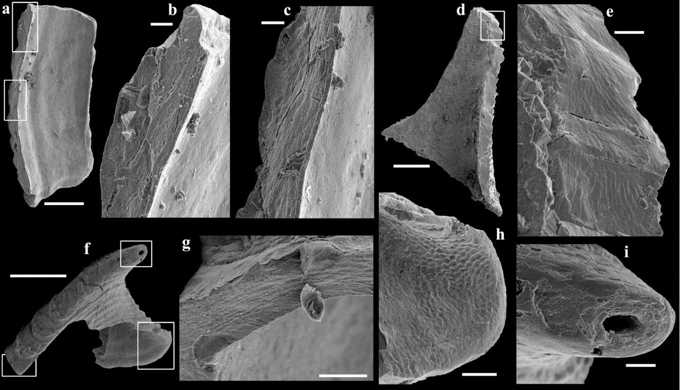
Lapworthella schodackensis (Lochman, Reference Lochman1956) from the upper Bastion Formation of Albert Heim Bjerge and CH Ostenfeld Nunatak. (a–c) Specimen SMNH X6119. (a) Internal view of a sclerite fragment, upper boxed area magnified in (b) and lower boxed area magnified in (c). (b, c) Details of the finely laminar shell microstructure. (d, e) Specimen SMNH X6120. (d) Internal view of a sclerite fragment, boxed area magnified in (e). (e) Detail of the finely laminar shell microstructure. (f–i) Specimen SMNH X6121. (f) Broken sclerite of morphotype B, lower left boxed area magnified in (g), lower right boxed area magnified in (h) and upper boxed area magnified in (i). (g) Detail of the finely laminar shell microstructure. (h) Detail of the fine polygonal imprints organized into a reticulate pattern on the internal surface of the sclerite. (i) Detail of the apical oval cross-section with finely laminar shell microstructure visible. Scale bars: (a, d, f) 200 µm; (b, c, e, g, i) 20 µm; (h) 50 µm.

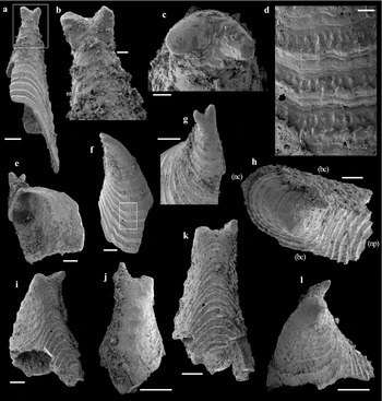
Morphotype A of Lapworthella schodackensis (Lochman, Reference Lochman1956) from the upper Bastion Formation of Albert Heim Bjerge and CH Ostenfeld Nunatak. (a–g) Specimen SMNH X6122. (a) General view, boxed area magnified in (b). (b, c, g). Close-up at the two apices of the sclerite. (d) Detail of co-marginal growth sets typical ornamentations. (e) Apertural view. (f) Lateral view, boxed area magnified in (d). (h–l) Specimen MGUH 26760. (h) Apical view. (i, l) Lateral views. (j) Anterior view. (k) Posterior view. Scale bars: (a, e–g, i, j, l) 200 µm; (b–d) 50 µm; (h, k) 100 µm.

?1962 Stenothecopsis schodakensis Lochman – Fisher, p. W138, fig. 83.
?1966 Lapworthella ex. gr. schodaka – Rozanov & Missarzhevsky, p. 92, pl. XIII, fig. 3.
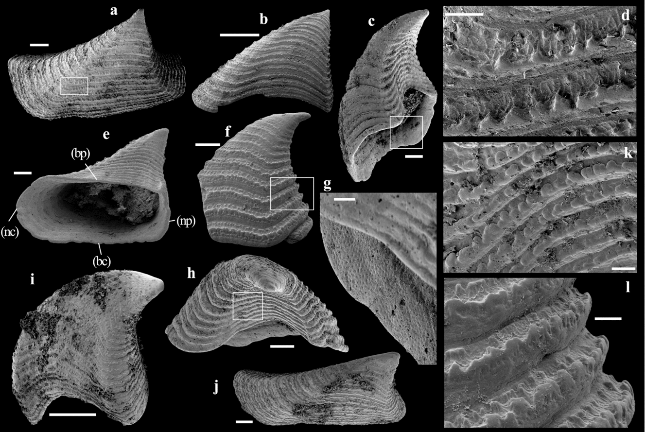
Morphotype B of Lapworthella schodackensis (Lochman, Reference Lochman1956) from the upper Bastion Formation of Albert Heim Bjerge and CH Ostenfeld Nunatak. (a, b, d) Sinistral sclerite, specimen SMNH X6123. (a) Apical view, boxed area magnified in (d). (b) View of the broad convex field of the sclerite. (d) Detail of co-marginal growth sets typical ornamentations: apical inter-rib groove covered with a polygonal network and tubercles, rib with denticles and fine transverse striations parallel to the rib on the apertural sub-rib surface. (c, e, g, h, k) Dextral sclerite, specimen MGUH 27605. (c) Lateral view showing the undulating apertural margin, boxed area magnified in (g). (e) Apertural view showing the subquadrangular cross-section; (np) – portion of the apertural margin corresponding to the narrow, planar field of the sclerite; (nc) – portion of the apertural margin corresponding to the narrow, convex field; (bp) – portion of the apertural margin corresponding to the broad, planar to concave field; and (bc) – portion of the apertural margin corresponding to the broad, convex field. (g) Detail of the polygonal imprints organized into a reticulate pattern on the internal surface of the sclerite. (h) View of the broad, concave field of the sclerite, boxed area magnified in (k). (k) Detail of the ornamentation with rounded fused denticles and tubercles. (f, l) Sinistral sclerite, specimen SMNH X6124. (f) View of the broad, convex field of the sclerite, boxed area magnified in (l). (l) Detail of co-marginal growth sets typical ornamentations in three zones. (i, j) Sinistral sclerite, specimen SMNH X6125. (i) View of the broad, convex field of the sclerite. (j) Apical view showing the subquadrangular apertural cross-section. Scale bars: (a, c, e, f, h, j) 200 µm; (b, i) 500 µm; (d, g, k) 50 µm; (l) 100 µm.

?1969 Lapworthella dentata Missarzhevsky – Rozanov et al., p. 164, pl. VI, figs 9, 14, 19.
?1975 Lapworthella dentata Missarzhevsky – Matthews & Missarzhevsky, pl. 3, fig. 12.
?1983 Lapworthella dentata Missarzhevsky – Bengtson, fig. 6.
?1984 Lapworthella schodakensis (Lochman) – Landing, p. 1395, figs 2a–o, 3d, 5a–e
?1996 Lapworthella schodakensis (Lochman) – Landing & Bartowski, fig. 9.18–19
2006 Lapworthella schodakensis (Lochman) – Skovsted, p. 1103, fig. 10.14–10.17.
2007 Lapworthella schodakensis (Lochman) – Skovsted & Peel, p. 742–743, fig. 6G.
2010 Lapworthella schodakensis (Lochman) – Rozanov et al., p. 88, pl. 58, figs 1–7.
Emended diagnosis. The sclerites are completely hollow and exhibit at least two morphotypes. The most numerous morphotype (B) is elongated, asymmetrically pyramidal with a single apex. Its cross-section is subquadrangular due to the organization of the sclerite into four surfaces or fields: (1) a narrow, planar field, perpendicular (with marked angular ridges) to (2) a planar to slightly concave broad field and (3) a convex broad field, and opposite to (4) a narrow, convex field. The rarest morphotype (A) is elongated, subsymmetrically conical to pyramidal with two apices. The external ornamentation of both morphotypes consists of co-marginal growth sets constituted of densely denticulate (>25 denticles mm−1) co-marginal ribs, apertural sub-rib slopes with fine transverse striations parallel to the rib and apical inter-rib grooves covered with a polygonal network variably associated with tubercles.
Remarks. The specimens described in this study are assigned to Lapworthella schodackensis because they exhibit densely denticulate (>25 denticles mm−1) co-marginal ribs, a character that is considered diagnostic of the species by Landing (Reference Landing1984) and Landing, Johnson & Geyer (Reference Landing, Johnson and Geyer2008). However, the diagnosis of the species is emended due to new observations on the ornamentation that are based on the present study and illustrations of specimens assigned to L. schodackensis by Rozanov et al. (Reference Rozanov, Parkhaev, Demidenko, Karlova, Korovnikov, Shabanov, Ivancov, Luchinina, Malakhovskaya, Melnikova, Naimark, Ponomarenko, Skorlotova, Sundukov, Tokarev, Ushatinskaya and Kipriyanova2010). The newly observed polygons sometimes associated with tubercles in the apical inter-rib groove, the densely denticulate co-marginal rib and the striated apertural sub-rib slope are restricted to and therefore diagnostic of this species of Lapworthella. We note that previous descriptions of lapworthellids have not reported morphologically distinct sclerite morphs and most researchers have emphasized continuous variation of a single sclerite type for Lapworthella. We define the bilaterally symmetrical sclerites in our collection with twinned apices as a specific sclerite morph. The sclerites described by Landing (Reference Landing1984), Landing & Bartowksi (Reference Landing and Bartowski1996) and Landing, Johnson & Geyer (Reference Landing, Johnson and Geyer2008) as L. schodackensis exhibit variable cross-sections with the smallest specimens having a circular cross-section. Those differences may be related to the size (and therefore growth stage, as suggested by Lochman in 1956) as the sclerites from Greenland are larger than those described by Landing (Reference Landing1984). In any case, the lack of data on the inter-rib groove ornamentation prevents definite synonymization with the specimens presented here and therefore the inclusion of the variability of sclerite cross-section in the emended diagnosis of L. schodackensis. The morphological terms used to describe L. schodackensis follow the terminology introduced by Skovsted et al. (Reference Skovsted, Betts, Topper and Brock2015) for Dailyatia Bischoff, Reference Bischoff1976 when applicable.
Description. The completely hollow sclerites exhibit two morphotypes that are differentiated in the following. In both morphotypes, the sclerite walls are variable in thickness depending on the specimen and location, ranging from 20 to 60 µm (Fig. 1a–e, g, i). Composition is phosphatic and the microstructure is finely laminar (Fig. 1b, c, e–g, i) when not completely recrystallized. The outermost and innermost shell laminae are thickest (Fig. 1b, c).
Two specimens among the 34 recovered exhibit two smooth, conical apices (Fig. 2a–c, e–l). These are referred to as sclerite morphotype A. They form subsymmetrical pyramidal sclerites, the axis of symmetry passing between the two apices (Fig. 2a, h, j, k) with an elongation parallel to the axis of symmetry (Fig. 2h). The apices are coiled towards the lateral extremities of a slightly planar to concave surface or field (Fig. 2f, h, i, l), the other fields being slightly convex (Fig. 2a, e, f, h–l). Breakage of the apertural margin of the two specimens prevents determination of the accurate dimensions of the sclerites (Fig. 2a, e, h, i–l), but the cross-section can be estimated as ovoid with a truncated extremity corresponding to the planar to concave field through the observation of the shape of the distalmost preserved growth set (Fig. 2h).
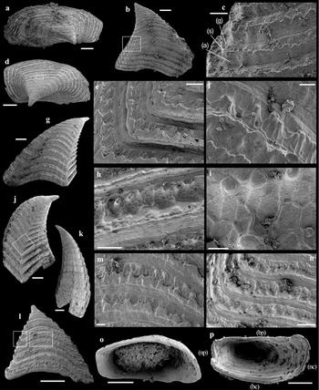
The second and most common sclerite type (32 specimens out of 34), denoted B, has a single apex and an asymmetrical, elongated pyramidal shape (Figs 3a–c, e, f, h, j, 4a, b, d, g, j–l). The height (maximal distance between the apertural margin and the apex) ranges from 660 µm to 2.23 mm (mean height 1.5 mm); the width (maximal distance between the two broadest fields of the sclerite) ranges from 265 µm to 840 µm (mean width is 500 µm); and the length (maximal distance between the two narrowest fields of the sclerite) ranges from 610 µm to 2.10 mm (mean length is 1.27 mm). The apical angle, measured on the broadest field, varies between c. 30° (Fig. 4g) and 75° (Fig. 4l). The apex is a simple smooth cone with circular to oval cross-section (Figs 1f, i, 3c, e, f, i, 4g, j, l). The apertural cross-section is subquadrangular (Figs 3a, e, j, 4a, d, o, p). The subquadrangular shape of the cross-section results from the presence of roughly four surfaces or fields of the sclerites organized into a pair of opposite, rather broad fields and a pair of opposite, rather narrow fields. Paired opposite fields are dissimilar and angles between the fields variable, which contribute to the asymmetry of the sclerites. One of the narrow fields is almost planar, corresponding to a rectilinear portion of the apertural margin (np on Figs 3e, 4p) while the other is convex, corresponding to a curved portion of the apertural margin (nc on Figs 3e, 4p). The transition between the narrow, planar field and neighbouring broad fields is abrupt, almost orthogonal, with a marked angular ridge (Fig. 4j), while the transition between the other, narrow, convex field with the broad fields is more transitional, without any angle (Fig. 4k), which gives a curved shape to the corresponding portion of the apertural cross-section (Figs 3e, 4o, p). One of the broad fields is convex (bc on Figs 3e, 4p) while the other is planar to slightly concave (bp on Figs 3e, 4p). The apertural margin is undulating in lateral view due to differences in growth rates (Figs 3b, c, h, i, 4b, g, l). The apertural margins of the two broad fields are arched apically and the two narrow fields are consequently longer in comparison with the medial part of the broad fields (Figs 3b, i, h, 4b, g, l). The sclerites are asymmetrically curved from a few degrees (Fig. 4l) for the smallest sclerites up to 70° (Fig. 3i) and invariably coiled towards the planar narrow field of the sclerite, which is either located on one side of the sclerite (Figs 3a, j, 4d) or the other (Figs 3e, 4a). Variations in the position of the narrow, planar field either on one side of the sclerite or the opposite (relative to the broad planar and concave field) and the direction of the coiling towards the narrow, planar field allow differentiation of sinistral and dextral sclerites. Of the 32 sclerites of this morphotype, it was possible to identify the orientation in 16 sclerites, of which 9 are sinistral and 7 dextral. Some specimens exhibit roughly polygonal imprints organized into a reticulate pattern on the internal surface of the sclerite (Figs 1h, 3g). The internal cavity is otherwise not subdivided by any structure (Figs 3e, 4o, p).
Morphotype B of Lapworthella schodackensis (Lochman, Reference Lochman1956) from the upper Bastion Formation of Albert Heim Bjerge and CH Ostenfeld Nunatak. (a–c) Dextral sclerite, specimen SMNH X6126. (a) Apical view showing the subquadrangular apertural cross-section. (b) View of the broad, convex field of the sclerite, boxed area magnified in (c). (c) Detail of co-marginal growth sets typical ornamentations: apical inter-rib groove (g) covered with a polygonal network and tubercles, rib (s) with denticles and fine transverse striations parallel to the rib on the apertural sub-rib surface (a). (d–k) Sinistral sclerite, Specimen MGUH 27604. (d) Apical view showing the subquadrangular apertural cross-section, boxed area magnified in (h). (e) Detail of co-marginal growth sets typical ornamentations. (f) Close-up at the ornamentation showing the tubercles associated with the polygonal network. (g) View of the broad, convex field of the sclerite. (h) Apical view of the typical ornamentation of the co-marginal ribs. (i) Close-up of the polygonal network and tubercles. (j) Lateral view centred on the angular ridge between the narrow, planar field and the broad convex field, boxed area magnified in (e). (k) View of the narrow, convex field. (l–o) Dextral sclerite, specimen SMNH X6127. (l) View of the broad, convex field of the sclerite, right boxed area magnified in (m) and left boxed area magnified in (n). (m, n) Detail of co-marginal growth sets typical ornamentations. (o) Apertural view. (p) Apertural view of sinistral sclerite, specimen SMNH X6128 (see legend of Fig. 3e for annotations). Scale bars: (a, b, d, g, j–l, o, p) 200 µm; (c) 100 µm; (e, h, n) 50 µm; (f, m) 20 µm; (i) 10 µm.

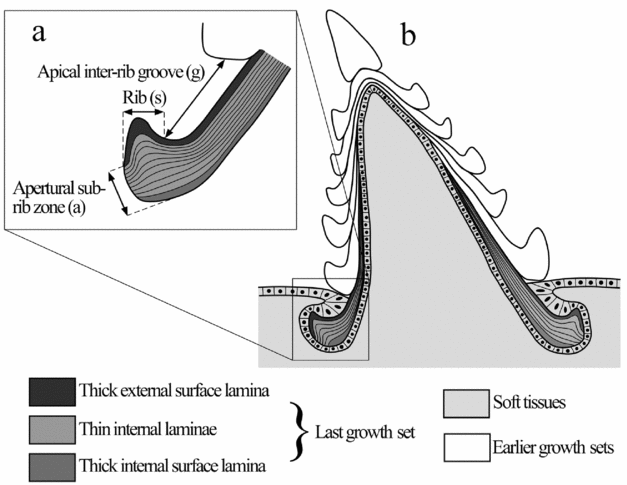
Although slightly variable, depending on preservation and/or intraspecific variability, both sclerite morphotypes are ornamented externally according to the same general pattern. The ornamentation consists of 8–29 coarse annulations parallel to the apertural margin that have been referred to as co-marginal growth sets (cf. Skovsted et al. Reference Skovsted, Betts, Topper and Brock2015). Each co-marginal growth set comprises three zones characterized by different ornamentations (defined in Fig. 5a; reported in Fig. 4c partly using the terminology introduced for Dailyatia by Skovsted et al. Reference Skovsted, Betts, Topper and Brock2015 when applicable): (1) the rib, located in the medial part of the co-marginal growth set and denoted s; (2) the apertural sub-rib zone, located on the apertural slope of the rib and denoted a; and (3) the apical inter-rib groove, located apically of the rib crest and denoted g. Varying in density from 5.6 to 28.8 mm–1, the ribs are transverse, undulating parallel to the apertural margin and bear apically directed denticles with a density ranging from 25 denticles mm–1 to 77 denticles mm–1. The denticles vary in size and shape from rounded (Figs 2d, 3d, l) to pointed (Fig. 4c, e, m, n). Fine transverse striations parallel to the rib are present at the apertural sub-rib slope (Figs 2d, 3d, l, 4c, e, h, m, n) which is inclined at 5–47° to the growth axis. The surface of the apical inter-rib groove is generally covered with a polygonal network (Figs 2d, 3d, l, 4c, e, h, i, m, n), the polygons comprising small walls surrounding depressions with an average diameter of 11.3 µm (Fig. 4i). The inter-rib groove is separated from the apertural sub-rib zone of the preceding growth set by a narrow, depressed, smooth furrow (Figs 3l, 4c, e, f, h, m, n). On the distal surface of the apical inter-rib groove, tubercles can be seen on some specimens (Figs 3d, h, l, 4c, e, f, h, i, m, n). They can be large, globular and fused with the denticles, obscuring the polygonal network (Fig. 3k), or high and grouped or dispersed along a line parallel to the rib (Figs 2d, 3d, l, 4c, e, f, h, i, m, n).
(a) Proposed nomenclature to describe the ornamentations in lapworthellids with references to the terminology proposed for Dailyatia Bischoff, Reference Bischoff1976 in Skovsted et al. (Reference Skovsted, Betts, Topper and Brock2015). (b). Hypothetical reconstruction (modified from Wrona, Reference Wrona2004) of the relationship between Lapworthella schodackensis (Lochman, Reference Lochman1956) sclerite and secretory tissues during growth, based on the present observation of surface sculpture of the co-marginal ribs and cross-section of the shell of Lapworthella dentata (Bengtson, Reference Bengtson1983).

Comparisons. Several lapworthellid species can be directly differentiated from Lapworthella schodackensis (Lochman, Reference Lochman1956) on the presence/absence of fused sclerites in the scleritome. In the genus Lapworthella, fused sclerites have only been reported in Lapworthella schodackensis by Landing (Reference Landing1984) and Lapworthella fasciculata Conway-Morris & Bengtson in Bengtson et al. Reference Bengtson, Conway Morris, Cooper, Jell and Runnegar1990, the ornamentation of which otherwise completely differs from the specimens described here by the presence of multiple fine longitudinal ribs on the entire surface. However, identification of fused sclerites in the scleritome of Lapworthella species is probably greatly dependent on taphonomic conditions, and is insufficient to confidently identify and differentiate species at the moment; other characters therefore need to be considered. The presence/absence of denticles/tubercles/nodes on the co-marginal rib is a useful character for discriminating lapworthellid species. The specimens described here exhibit denticles on the co-marginal rib that differentiate it from species that lack any denticles (also referred to as tubercles or nodes) on the rib such as Lapworthella rete Yue, Reference Yue1987, L. bornholmiensis (Poulsen, Reference Poulsen1942), L. ludvigseni Landing, Reference Landing1984, L. puttapensis Bengtson & Conway Morris in Bengtson et al. Reference Bengtson, Conway Morris, Cooper, Jell and Runnegar1990 and L. bella Missarzhevsky in Rozanov & Missarzhevsky, Reference Rozanov and Missarzhevsky1966, although the latter is too poorly illustrated and most probably preserved to conclude the validity of the species. Among the species that have a denticulate rib, the differentiation is based on the ornamentation of the apical inter-rib groove and apertural sub-rib zones when available. The fine transverse growth lines parallel to the rib seen on the apertural sub-rib surface of the specimens described here, corresponding to growth laminae of the wall, are observed in several other Lapworthella species (i.e. L. cornu (Wiman, Reference Wiman1903), L. filigrana Conway Morris & Fritz, 1984; L. tortuosa Missarzhevsky in Rozanov & Missarzhevsky, Reference Rozanov and Missarzhevsky1966). On the other hand, the polygons associated with the tubercles in the apical inter-rib groove seem to be restricted to the specimens described here. A roughly similar ornamentation of the apical inter-rib groove is observed in the specimens assigned to Lapworthella schodackensis (Lochman, Reference Lochman1956) figured by Rozanov et al. (Reference Rozanov, Parkhaev, Demidenko, Karlova, Korovnikov, Shabanov, Ivancov, Luchinina, Malakhovskaya, Melnikova, Naimark, Ponomarenko, Skorlotova, Sundukov, Tokarev, Ushatinskaya and Kipriyanova2010; pl. 58, figs 1–7); here, polygons are weakly expressed and the presence of associated tubercles is difficult to assess as this character is not well-figured. Similarities in the organization and ornamentation of the apertural sub-rib and apical inter-rib grooves of Lapworthella specimens with a denticulate transverse rib are often encountered. In this case, Bengtson (Reference Bengtson1980), Landing (Reference Landing1984) and Bengtson et al. (Reference Bengtson, Conway Morris, Cooper, Jell and Runnegar1990) proposed considering the number of denticles per millimetre for differentiation, although the taxonomic significance of this character has been legitimately questioned by Hinz (Reference Hinz1987). On this basis, Landing (Reference Landing1984) and Landing, Johnson & Geyer (Reference Landing, Johnson and Geyer2008) proposed L. schodakensis as senior synonym of L. dentata Missarzhevsky in Rozanov et al. Reference Rozanov, Missarzhevsky, Volkova, Voronova, Krylov, Keller, Korolyuk, Lendzion, Mikhnyar, Pykhova and Sidorov1969 due to their similar dense denticulation. The specimens assigned to L. schodackensis by Rozanov et al. (Reference Rozanov, Parkhaev, Demidenko, Karlova, Korovnikov, Shabanov, Ivancov, Luchinina, Malakhovskaya, Melnikova, Naimark, Ponomarenko, Skorlotova, Sundukov, Tokarev, Ushatinskaya and Kipriyanova2010) exhibit between 28 and 43 denticles mm–1 on the transverse rib, which is within the range of variation observed in the present specimens but is slightly less than the variation described for the species in the emended diagnosis of Landing (Reference Landing1984).
Based on this new, well-preserved material, an emendation of the diagnosis of L. schodackensis, to which the specimens studied here have been (Skovsted, Reference Skovsted2006) and are being assigned, is proposed in order to take into account the new data. Apart from the denticulate L. schodackensis and L. dentata, the similarly ornamented denticulate L. cornu (Wiman, Reference Wiman1903), L. nigra Cobbold, Reference Cobbold1921 and the four morphotypes of Lapworthella described by Hinz (Reference Hinz1987) have been proposed by Hinz (Reference Hinz1987) and Landing, Johnson & Geyer (Reference Landing, Johnson and Geyer2008) as synonyms due to their overlapping scattered number of denticles per mm (12–25 mm–1). Although the number of denticles per millimetre seems to be relatively significant for the taxonomy of denticulate lapworthellids, we also recommend the use of apical inter-rib groove surface ornamentation for identification, and therefore the sampling and observation of a large number of well-preserved specimens. We also note that in the camenellan genus Dailyatia, closely related species can be differentiated based on small differences in both morphology and sclerite ornament, including the presence/absence and distribution of denticles (pustules sensu Skovsted et al. Reference Skovsted, Betts, Topper and Brock2015) that are only expressed in some of the characteristic sclerite morphotypes (Skovsted et al. Reference Skovsted, Betts, Topper and Brock2015). Consequently, species identification in camenellans should ideally be based on different lines of evidence from large numbers of sclerites or multiple sclerite morphs, rather than a single characteristic.
It should be noted that accurate comparisons with Lapworthella marginita Meshkova, Reference Meshkova and Zhuravleva1969, L. cancellata Jiang in Luo et al. Reference Luo, Jiang, Wu, Song and Ouyang1982, L. hubeiensis Qian & Zhang, Reference Qian and Zhang1983, L. sinensis Duan, Reference Duan1984 (synonym with L. ablorta Duan, Reference Duan1984 and L. subrectangulata Duan, Reference Duan1984 according to Qian & Bengtson, Reference Qian and Bengtson1989), L. annulata Qian & Yin, Reference Qian and Yin1984 and L. gezhongwuensis Qian & Yin, Reference Qian and Yin1984 are difficult due to poor illustrations and preservation. Bengtson et al. (Reference Bengtson, Conway Morris, Cooper, Jell and Runnegar1990) provided an exhaustive list of other specimens that have, previously to their study, been referred to Lapworthella but reassessed as non-Lapworthella species.
5. Ornament and the process of shell formation
The surface of the sclerites of all camenellans, including lapworthellids, is characterized by repeated growth sets. Each growth set consists of a distinct annulation that is parallel to the apertural margin. It comprises co-marginal ribs and inter-rib grooves (Skovsted et al. Reference Skovsted, Balthasar, Brock and Paterson2009, Reference Skovsted, Betts, Topper and Brock2015), complemented by the differentiation of a distinct sub-rib zone on the apertural side of the rib crest (see Figs 4c, 5a). Growth sets are typically separated by narrow furrows, and details of the surface ornament of consecutive growth sets is typically identical on each sclerite. Each co-marginal rib is interpreted to correspond to the apertural inflation of selected shell laminae of an internal conical/pyramidal growth element. All growth elements and shell laminae are interpreted by Bengtson (Reference Bengtson1983), Landing (Reference Landing1984) and later authors to be produced by basal internal accretion.
Authors noted very early (Landing, Reference Landing1984; Hinz, Reference Hinz1987; Qian & Bengtson, Reference Qian and Bengtson1989; Bengtson et al. Reference Bengtson, Conway Morris, Cooper, Jell and Runnegar1990) the importance of the surface microstructure of tommotiid sclerites for their taxonomy and for interpreting their growth. Following the interpretations of the growth and ornamentation of lapworthellids, a reconstruction of the organization of soft tissues secreting the sclerites was first proposed for Lapworthella rete by Conway Morris & Chen (Reference Conway Morris and Chen1990) and then extended to Kennardiidae (Dailyatia) by Wrona (Reference Wrona2004). According to this model new growth increments (growth sets) were covered by epithelium during formation, reflected in the fossils by the external reticulate network representing moulds of epithelial cells.
The new data from the Greenland specimens of Lapworthella schodackensis and comparison with the recently described shell structure of Dailyatia (Skovsted et al. Reference Skovsted, Betts, Topper and Brock2015) enable us to complement the hypothetical reconstruction of the association of soft tissues with the sclerites and their growth for L. schodackensis (Fig. 5). Skovsted et al. (Reference Skovsted, Betts, Topper and Brock2015) showed that in Dailyatia ajax Bischoff, Reference Bischoff1976, where the entire surface of the sclerites is covered by a reticulate network, the surface of each growth set is formed by a single shell lamina while subsequent shell laminae reinforce the shell by adding thickness without incremental shell growth. Our interpretation of the shell growth of the Greenland specimens of L. schodackensis is similar in that a single shell lamina appears to form the surface of the apical inter-rib area and the rib crest itself (Figs 1b, c, 5a). A similar organization of the shell laminae is also observed in the section of a sclerite assigned to L. dentata (possibly synonym with L. schodackensis) by Bengtson (Reference Bengtson1983, figs 6D, E), although the type of associated ornamentation is difficult to assess. In the present specimens of L. schodackensis, these areas are covered by the reticulate network and denticles. The apertural sub-rib area with its fine concentric striations represents conventional co-marginal accretion of shell material, and no traces of epithelial imprints can be found here. Consequently, reticulate external surface patterns in Lapworthella, as well as in kennardiids, appear to represent the elongation of single surface forming laminae. As discussed by Skovsted et al. (Reference Skovsted, Betts, Topper and Brock2015), this could be an adaptation to facilitate rapid shell growth. The elongated laminae would have been formed under an epithelium, while the growing edge of the sclerite was embedded in soft integument (Fig. 5B). During growth, the sclerites are interpreted to have periodically erupted from the shell forming tissue, presumably in concert with the formation of new growth sets. The formation of subsequent shell laminae supporting the surface-forming lamina and forming the apertural inter-rib zone could have been secreted either while the sclerite margin was embedded in soft tissue or after the eruption. The polygons observed on the apical inter-rib zone would correspond to imprints of large epithelial cells (Conway Morris & Chen, Reference Conway Morris and Chen1990; Wrona, Reference Wrona2004) and interpreted, in L. schodackensis with the help of the tubercles, as ‘anchorage’ devices for the sclerites in the soft tissues. The reticulate network present on the sclerite interior of L. schodackensis from Greenland is composed of smaller polygonal cells compared to the external reticulate ornament and is interpreted to be only involved in secreting the shell material.
6. Scleritome construction and sclerite fusion in lapworthellids
Bilaterally symmetrical sclerites (A sclerites) constitute a large percentage of all sclerites in the camenellans Kennardia Laurie, Reference Laurie1986 and Dailyatia (Bischoff, Reference Bischoff1976; Laurie, Reference Laurie1986; Evans & Rowell, Reference Evans and Rowell1990) and, at least in Dailyatia, the symmetrical A sclerites always exhibit twin apices (Skovsted et al. Reference Skovsted, Betts, Topper and Brock2015). The A sclerites in Dailyatia constitute c. 12–25% of all sclerites in assemblages from South Australia (Skovsted et al. Reference Skovsted, Betts, Topper and Brock2015) and were interpreted by Evans & Rowell (Reference Evans and Rowell1990) and Skovsted et al. (Reference Skovsted, Betts, Topper and Brock2015) as straddling the midline of the dorsal scleritome of a slug-like animal. The asymmetrical B and C sclerites of Dailyatia occur in sinistral and dextral symmetry variants and were interpreted to be arranged symmetrically along the midline of the same scleritome (i.e. equal numbers of opposing sinistral and dextral sclerites on the left- and right-hand sides of the scleritome). If other camenellan tommotiids (mainly Camenella Missarzhevsky in Rozanov & Missarzhevsky, Reference Rozanov and Missarzhevsky1966, Kelanella Missarzhevsky in Rozanov & Missarzhevsky, Reference Rozanov and Missarzhevsky1966 and Lapworthella) were also slug-like animals with a dorsal scleritome, the most likely arrangement of their asymmetrical sclerites would be forming symmetrical sclerite pairs along the midline of the scleritome.
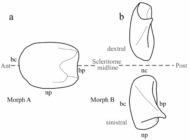
The two specimens of sclerite morph A in Lapworthella schodackensis are characterized by two equal apices and appear to be bilaterally symmetrical. The twin apices in each specimen are of the same morphology but appear to have been formed independently, and were fused to form a united sclerite during early ontogeny. Similar lapworthellid specimens showing ontogenetic sclerite fusion have been documented before (Landing, Reference Landing1984) but have usually been interpreted as fortuitous examples of sclerite malformation. However, such examples have been used as evidence for a bilaterally symmetrical multi-component scleritome in tommotiids (Landing, Reference Landing1984; Qian & Bengtson, Reference Qian and Bengtson1989; Demidenko, Reference Demidenko2004; Li & Xiao, Reference Li and Xiao2004). The bilaterally symmetrical sclerite composites (morph A) in L. shodackensis documented here could be interpreted to represent an early ontogenetic fusion of two adjacent sclerites of sinistral and dextral morph B on either side of the midline of the scleritome (Fig. 6). The more numerous B sclerites were likely oriented in symmetrical pairs along the midline of the scleritome (Fig. 6b). Both sclerites of morph A and B are oriented with their apices directed posteriorly following assumptions for Dailyatia in Skovsted et al. (Reference Skovsted, Betts, Topper and Brock2015). Growth sets on the concave sub-apical field of the A sclerite (bp) curve strongly in the apical direction, corresponding to a deep arch in the apertural margin. We interpret this arch as an accommodation of the curved surface of the animal body in transverse section. Similar arched apertural margins are present on the broad fields of the B sclerites described here. Based on these observations we suggest that broad B sclerites were arranged in pairs on either side of the midline of the scleritome with the convex broad fields (bc) oriented forwards and the convex narrow sides (nc) meeting at the midline (Fig. 6b). In this configuration the arched apertural margins of the broad fields in B sclerites would accommodate the curved body of the animal and the apices would be directed backwards and laterally, in a presumably defensive stance (Fig. 6).
Hypothetical reconstructions of the orientation of (a) moph A (outline based on specimen MGUH 26760) and (b) morph B sclerites (outline based on specimens SMNH X6126 and MGUH 27604) of Lapworthella schodackensis (Lochman, Reference Lochman1956) relative to the midline and anterior and posterior parts of a slug-like animal (see legend of Fig. 3e for annotations). Note that the relative position of A and B sclerites and the distance between morph B sclerites and the scleritome midline are not possible to determine from the present material and are completely artificial.

Although the exact structure of the scleritome remains elusive, we consider symmetrical sclerite composites (morph A) to be a normal part of the scleritome of Lapworthella schodackensis. The relative position of A and B sclerites is not possible to determine. By comparison to the proposed scleritome of Dailyatia (Skovsted et al. Reference Skovsted, Betts, Topper and Brock2015), the A and B sclerites of L. schodackensis would, in terms of position in the scleritome, correspond to A and B sclerites of Dailyatia. It is most likely that other types of asymmetrical sclerites, such as described by Landing (Reference Landing1984), could occupy other positions in the scleritome, but this is presently difficult to assess as only 34 specimens were recovered, although delicately preserved. Ontogenetically fused sclerites assigned to L. schodackensis by Landing (Reference Landing1984) have been described from the Taconic allochthon, eastern New York. The assignment of those specimens to L. schodackensis needs to be assessed through additional data on the surface sculpture of the sclerites. The synonymy with the present specimens is therefore uncertain. One specimen is bilaterally symmetrical and comparable to the fused specimens described here, whereas the other is asymmetric and could possibly correspond to a fusion of sclerites in a different part of the scleritome of L. schodackensis or to sclerite fusion in a different species.
The ubiquitous presence and large number of symmetrical sclerites with twinned apices in Dailyatia suggest that sclerite fusion in this tommotiid occurred as a programmed event early in sclerite ontogeny. By comparison, this may suggest that sclerite fusion in Lapworthella shodackensis was also a normal phenomenon in sclerite ontogeny in a particular part of the scleritome. Similar bilaterally symmetrical sclerite composites (albeit with more than two apices) were documented by Demidenko (Reference Demidenko2004) for Lapworthella fasciculata from Horse Gully in the Stansbury Basin of South Australia. Fused sclerite composite specimens of L. fasciculata with two apices only have been discovered in the Donkey Bore Syncline of the Arrowie Basin in South Australia (T. P. Topper, unpubl. thesis, Macquarie University, 2006; T. P. Topper, pers. comm. 2015). Consequently, it is not unlikely that given sufficient sample size, bilaterally symmetrical sclerite morphs could perhaps be discovered in other lapworthellids in the future. However, it may be worth considering that fused sclerite composites of the Greenland L. schodackensis, as well as the Australian L. fasciculata from both the Stansbury Basin (Demidenko, Reference Demidenko2004) and the Arrowie Basin (T. P. Topper pers. comm. 2015) are derived from single samples and the observed pattern could possibly represent differences between populations or even individuals.
7. Conclusions
Excellently preserved specimens of Lapworthella schodackensis from the Cambrian Stage 4 upper Bastion Formation at Albert Heim Bjerge and CH Ostenfeld Nunatak, NE Greenland provide significant novel data on the species taxonomy for comprehension of the scleritome structure of lapworthellids and the mode of formation of their sclerites. In the Greenland assemblage, two sclerite morphotypes of L. schodackensis were identified: one with simple apex, occurring in sinistral and dextral forms; and one bilaterally symmetrical with two apices. The excellent preservation provides exquisite new details on the sclerite ornamentation which is constructed of repeated growth sets comprising a reticulate inter-rib groove with tubercles, a densely denticulate rib and a striated sub-rib area. The new data on the ornamentation and shell microstructure of L. schodackensis, in combination with the results of Skovsted et al. (Reference Skovsted, Betts, Topper and Brock2015) on Dailyatia, also enabled us to propose that the surface of the apical inter-rib area and the rib crest itself is formed of a single shell lamina while subsequent shell laminae reinforce the shell by adding thickness and laminar structures in the sub-rib area. Finally, new information for the reconstruction of bilaterally symmetrical multi-component lapworthellid scleritome were deduced from the recovered L. schodackensis sclerites with twin apices that formed independently and fused to form a united sclerite during early ontogeny. The orientation of the ontogenetical fusion suggests that, at least, a certain number of L. schodackensis sclerites formed symmetrical pairs along the midline of the scleritome.
Acknowledgements
The authors thank Prof. John S. Peel (Uppsala) for access to collections and geological information from NE Greenland. Dr Timothy P. Topper is thanked for valuable discussions on sclerite morphology and ultrastructure. This work was supported by the European Community, Research Infrastructure Action under the FP7 ‘Capacities’ Specific Programme (LD, SYNTHESYS grant SE-TAF-4164 performed at the Swedish Museum of Natural History). LD is also indebted to Drs Daniela Kalthoff and Thomas Mörs for their hospitality and the Swedish Museum of Natural History staff for support. We are grateful to the editor and the external reviewers whose reviews greatly improved the quality of the final manuscript.