
Introduction
Trypanosomes (Kinetoplastida, Trypanosomatidae) are vector-borne haemoflagellate protozoa affecting humans and several animal species, mainly in tropical regions (Radwanska et al., Reference Radwanska, Vereecke, Deleeuw, Pinto and Magez2018). Among several trypanosomes species identified, the zoonotic ones represent a serious public health concern, due to their morbidity and mortality rate (Dunn et al., Reference Dunn, Wang and Adigun2020). For instance, Trypanosoma cruzi is responsible of the American trypanosomiasis, also known as Chagas disease (Golding, Reference Golding2013), whereas Trypanosoma brucei rhodesiense and Trypanosoma brucei gambiense of the human African trypanosomiasis, sleeping sickness, in sub-Saharan Africa (Dunn et al., Reference Dunn, Wang and Adigun2020). Some trypanosomes species are of concern to animals, such as in the case of T. brucei, Trypanosoma equiperdum and Trypanosoma evansi (etiological agent of ‘Surra’), with a high productivity loss and global socio-economic impact (Aregawi et al., Reference Aregawi, Agga, Abdi and Büscher2019).
In recent years, many studies have focused on the role of domestic animals as reservoirs of trypanosomes, such as dogs and cats for T. cruzi (Eloy and Lucheis, Reference Eloy and Lucheis2009; Elmayan et al., Reference Elmayan, Tu, Duhon, Marx, Wolfson, Balsamo, Herrera and Dumonteil2019; Murphy et al., Reference Murphy, Macchiaverna, Cardinal, Bhattacharyya, Mertens, Zeippen, Gustin, Gilleman, Gürtler and Miles2019), camels for T. evansi (Aregawi et al., Reference Aregawi, Agga, Abdi and Büscher2019), cattle for T. b. rhodesiense (Waiswa et al., Reference Waiswa, Olaho-Mukani and Katunguka-Rwakishaya2003) and pigs for Trypanosoma vivax, Trypanosoma congolense and Trypanosoma simiae (Hamill et al., Reference Hamill, Kaare, Welburn and Picozzi2013). In wildlife, opossums (Didelphis marsupialis), armadillos (Dasypus novemcinctus) and rodents are the main sylvatic reservoir of T. cruzi (Gürtler and Cardinal, Reference Gürtler and Cardinal2015; Bezerra-Santos et al., Reference Bezerra-Santos, Nascimento Ramos, Campos, Dantas-Torres and Otranto2021a), whereas capybaras (Hydrochoerus hydrochaeris), vampire bats (Desmodus rotundus), white tail deer (Odocoileus virginianus chiriquensis) and wild boars (Sus scrofa) of T. evansi (Desquesnes, Reference Desquesnes2004; Radwanska et al., Reference Radwanska, Vereecke, Deleeuw, Pinto and Magez2018). The role of wildlife as reservoirs of trypanosomatids deserves further investigations mainly in illegally imported animals (Bezerra-Santos et al., Reference Bezerra-Santos, Mendoza-Roldan, Thompson, Dantas-Torres and Otranto2021b, Reference Bezerra-Santos, Mendoza-Roldan, Thompson, Dantas-Torres and Otranto2021c). Furthermore, evidence of new clades of anuran trypanosomes highlighted the wide morphological and genetic diversity of these parasites in amphibians (da S. Ferreira et al., Reference da S. Ferreira, da Costa, Ramirez, Roldan, Saraiva, da S. Founier, Sue, Zambelli, Minervino, Verdade, Gennari and Marcili2015).
In Europe, trypanosomes have been described in domestic and wild animal species, such as Trypanosoma lewisi in rats (Rattus norvegicus, Karbowiak et al., Reference Karbowiak, Wita and Czaplińska2009), Trypanosoma pestanai in badgers (Meles meles) (Peirce and Neal, Reference Peirce and Neal1974) and in a dog (Dyachenko et al., Reference Dyachenko, Steinmann, Bangoura, Selzer, Munderloh, Daugschies and Barutzki2017), Trypanosoma vespertilionis and Trypanosoma dionisi in bats (Pipistrellus pipistrellus, Linhart et al., Reference Linhart, Bandouchova, Zukal, Votypka, Kokurewicz, Dundarova, Apoznanski, Heger, Kubickova, Nemcova, Piacek, Sedlackova, Seidlova, Berkova, Hanzal and Pikula2020), Trypanosoma theileri and Trypanosoma melophagium in domestic and wild ruminants (Buscher and Friedhoff, Reference Buscher and Friedhoff1984; Villa et al., Reference Villa, Gutierrez, Gracia, Moreno, Chacon, Sanz, Buscher and Touratier2008; Neumüller et al., Reference Neumüller, Nilsson and Påhlson2012).
However, few data are available on the occurrence of trypanosomes in wild animals in Europe, as well as on their role in the circulation of these parasites, including those of zoonotic concern. In this context, the environmental expansion of badgers in European countries has spurred the scientific interest in their role as hosts of ectoparasites and pathogens they transmit (Baker and Harris, Reference Baker and Harris2007; Otranto et al., Reference Otranto, Cantacessi, Dantas-Torres, Brianti, Pfeffer, Genchi, Guberti, Capelli and Deplazes2015).
Indeed, this animal species may harbour different ticks of the genus Ixodes, such as Ixodes canisuga, Ixodes hexagonus, Ixodes crenulatus and Ixodes frontalis (Estrada-Peña et al., Reference Estrada-Peña, Mihalca and Petney2017), which usually complete their biological life cycles on small mammals, such as reptiles and small rodents on reptiles (Mendoza-Roldan et al., Reference Mendoza-Roldan, Ribeiro, Castilho-Onofrio, Grazziotin, Rocha, Ferreto-Fiorillo, Pereira, Benelli, Otranto and Barros-Battesti2020). To date, the vectoral capacity of VBPs is demonstrated only for I. hexagonus, acting as a vector of Borrelia burgdorferi sensu lato complex (Gern et al., Reference Gern, Toutoungi, Hu and Aeschlimann1991). In addition, badgers may be infested by Paraceras melis, known as the badger flea and recognized as a vector of T. pestanai (Lizundia et al., Reference Lizundia, Newman, Buesching, Ngugi, Blake, Sin, Macdonald, Wilson and McKeever2011). The occurrence of this haemoflagellate has been retrieved only rarely in badgers from France (Rioux et al., Reference Rioux, Albaret, Bres and Dumas1966) and the UK (Lizundia et al., Reference Lizundia, Newman, Buesching, Ngugi, Blake, Sin, Macdonald, Wilson and McKeever2011; Ideozu et al., Reference Ideozu, Whiteoak, Tomlinson, Robertson, Delahay and Hide2015) and in a dog from Germany (Dyachenko et al., Reference Dyachenko, Steinmann, Bangoura, Selzer, Munderloh, Daugschies and Barutzki2017). Although European badgers can be infected by several VBPs, such as Anaplasma phagocytophilum, Babesia badger type A and B, B. burgdorferi s. l. complex and Leishmania infantum (Hofmeester et al., Reference Hofmeester, Krawczyk, van Leeuwen, Fonville, Montizaan, van den Berge, Gouwy, Ruyts, Verheyen and Sprong2018; Battisti et al., Reference Battisti, Zanet, Khalili, Trisciuoglio, Hertel and Ferroglio2020), the occurrence of trypanosomes and their arthropod vectors involved under reported.
The present study highlights T. pestanai infection in two badgers with severe clinical manifestations (i.e. ataxia and paralysis) and in their ticks (I. canisuga and Ixodes ricinus) revealing, for the first time, the presence of this protozoan in Italy and discussing the potential involvement of I. canisuga and I. ricinus in its transmission.
Materials and methods
Study area and sampling
In October 2020, alive badgers (n = 2, adult females) were retrieved in two different occasions in the province of Bari (Apulia region, southeast Italy) suffering from severe paralysis and the ectoparasites infestation.
Animals were hospitalized in the Osservatorio Faunistico Regionale della Puglia (OFR), the Apulian Regional Wildlife Rescue Centre and subsequently were moved to the Department of Veterinary Medicine of the University of Bari, Italy, for further investigations.
Anamnestic data (i.e. gender and estimated age) were obtained and blood and serum samples were collected for routine analysis (i.e. complete blood count and biochemical parameters). A blood smear was performed and stained by using the May-Grünwald-Giemsa technique (Piaton et al., Reference Piaton, Fabre, Goubin-Versini, Bretz-Grenier, Courtade-Saïdi, Vincent, Belleannée, Thivolet, Boutonnat, Debaque, Fleury-Feith, Vielh, Cochand-Priollet, Egelé, Bellocq and Michiels2015).
From the first badger, alive ticks (n = 31) were collected and incubated at 25°C and 75% of relative humidity for 5 days, whereas two dead fleas were stored in 70% ethanol. Two additional tick specimens were collected from the second badger and stored in 70% ethanol.
Five and 15 days later, respectively, the two badgers died. A complete necropsy was performed from both animals. Organs and tissues (i.e. spleen, liver, brain, heart, kidney, diaphragm, bone marrow and skeletal muscle tibialis) were sampled for molecular investigations. The present study was performed in accordance to the protocols provided by the EU Directive 2010/63/EU for animal experiments.
Morphological identification of ectoparasites
All fleas and ticks were observed at the stereomicroscopy (Leica MS5; Leica Microsystems Ltd. Heerbrugg, Germany) and morphologically identified at species level by using keys proposed by Smit (Reference Smit1957) and Estrada-Peña et al. (Reference Estrada-Peña, Mihalca and Petney2017), respectively. Ticks were classified according to gender (male or female), developmental stage (larva, nymph, adult) and feeding status (fed or unfed). All alive ticks (n = 31) were dissected and smears of haemolymph, gut and salivary glands of each specimen were performed and microscopically observed to assess the presence of any pathogens.
DNA extraction, PCR and sequencing
DNA was extracted from ectoparasites and blood samples of both badgers as well as from tissues and organs (i.e. spleen, liver, brain, heart, kidney, diaphragm, bone marrow and skeletal muscle tibialis) from the badgers by using a commercial kit (QIAampDNA Blood & Tissue; Qiagen, Hilden, Germany), according to the manufacturer's instructions. A fragment (900 bp) of the 18S rRNA gene was amplified by using primers 609F (forward: 5′-CACCCGCGGTAATTCCAGC-3′) and 706R (reverse: 5′-CTGAGACTGTAACCTCAA-3′), according to Zeb et al. (Reference Zeb, Szekeres, Takács, Kontschán, Shams, Ayaz and Hornok2019). The PCR protocol was modified as the following: an initial denaturation step at 95°C for 10 min, followed by 40 cycles of denaturation at 95°C for 40 s, annealing at 60°C for 90 s and extension at 72°C for 60 s, and final extension at 72°C for 7 min.
The PCR reaction was performed in a final volume of 25 μL (23 μL of PCR mixture and 2 μL of the DNA sample), including 5 μL of 10× PCR buffer II, 6 μL of 25 mm MgCl2, 5μL of 1.25 mm dNTPs, 0.5 μL of 100 pmol μL−1 for each primer and 1.25 U of AmpliTaq Gold (Applied Biosystems, Foster City, CA, USA). PCR products were examined on 2% agarose gels stained with GelRed (VWR International PBI, Milan, Italy) and visualized on a GelLogic 100 gel documentation system (Kodak, New York, USA). Amplicons were then purified and sequenced in both directions using the same primers as for PCR, by the Big Dye Terminator v.3.1 chemistry in a 3130 Genetic Analyzer (Applied Biosystems). Sequences were edited and analysed by the Geneious software version 9.0 (Biomatters Ltd., Auckland, New Zealand) (Kearse et al., Reference Kearse, Moir, Wilson, Stones-Havas, Cheung, Sturrock, Buxton, Cooper, Markowitz, Duran, Thierer, Ashton, Meintjes and Drummond2012) and compared with those available in the GenBank database by the Basic Local Alignment Search Tool (BLAST; blast.ncbi.nlm.nih.gov/Blast.cgi).
Phylogenetic analysis
The phylogenetic analysis was based on 555 bp of the 18S rRNA gene sequence of Trypanosoma spp. detected from several domestic and wild animal species available from the GenBank database. Phylogenetic relationship was inferred by the Maximum Likelihood (ML) method based on Akaike information criterion (AIC) TIM3 + R4 model selected by best-fit model (Nguyen et al., Reference Nguyen, Schmidt, von Haeseler and Minh2015; Kalyaanamoorthy et al., Reference Kalyaanamoorthy, Minh, Wong, von Haeseler and Jermiin2017). Evolutionary analyses were conducted on 1000 bootstrap replications using the MEGA X software (Minh et al., Reference Minh, Nguyen and von Haeseler2013; Kumar et al., Reference Kumar, Stecher, Li, Knyaz and Tamura2018). Homologous sequences from Bodo saltans and Bodo edax were used as out-groups (accession numbers: MF000702 and AY028451, respectively).
Results
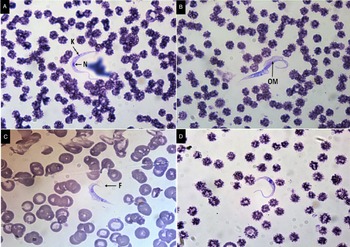
Badgers showed severe ataxia and paralysis and were infested by ectoparasites. According to the blood and serum physiological parameters, no alteration in the complete blood count, except thrombocytopenia, and biochemical analysis was found. No pathological findings in the first badger were outlined, except for a severe splenomegaly and thoracic-abdominal blood effusion. In the second one, only a slight splenomegaly was found. In the blood smears of the first and second badger, trypanosomes (i.e. nine trypomastigotes and two epimastigotes) and seven trypomastigotes were observed, respectively. Distinctive morphological features of trypomastigotes (i.e. elongated nucleus and sub-terminal kinetoplast) and epimastigotes (i.e. circular nucleus with adjacent kinetoplast) are shown in Fig. 1. Body measurements of the trypanosomes (i.e. body length, body perimeter and undulating membrane perimeter) displayed an average size of 29.0 μm (s.d. = 1.01), 38.1 μm (s.d. = 0.97) and 9.8 μm (s.d. = 0.64) for the first badger, whereas 29.6 μm (s.d. = 0.82), 38.9 μm (s.d. = 0.84) and 10.1 μm (s.d. = 0.67) for the second one, respectively; all measurements data of each trypanosome specimen examined are reported in Table 1. All tissue and organs, but not the blood samples, of the two badgers tested negative for T. pestanai DNA.
Trypomastigotes (A, B, C) and epimastigotes (D) detected in the blood smear from badgers in the present study; kinetoplast (K), nucleus (N), undulating membrane (OM) and flagellum (F) are shown. All pictures have 100× magnification.

Body measurements of T. pestanai trypanosomes (n = 18) retrieved in the blood smear of the badgers in the present study

a BL, body length.
b BP, body perimeter.
c OMP, undulating membrane perimeter.
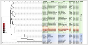
Out of 33 ticks (i.e. n = 31 I. canisuga, n = 2 I. ricinus) and two fleas (Ctenocephalides felis), 11 specimens (31.4%; 95% CI 18.5–48.0, n = 5 I. canisuga engorged nymphs, n = 4 I. canisuga engorged females and n = 2 I. ricinus engorged females) tested positive to T. pestanai DNA (Table 2). All smears of haemolymph, gut and salivary glands from alive ticks were negative for trypanosomes. The combined conventional PCR/sequencing approach revealed consensus sequences of the 18S rRNA gene displaying 100% of nucleotide identity with the sequences of T. pestanai available on the GenBank database. The 18S rRNA partial sequences of T. pestanai from ticks and badgers herein obtained clustered together with those of badgers from France (AJ009159) and in a dog from Germany (KY354582); the overall panel of phylogenetic relationships investigated is shown in Fig. 2. Representative sequences of T. pestanai from ticks (I. ricinus female – MZ144607; I. canisuga female – MZ144608; I. canisuga nymph – MZ144609) and badgers (MZ144610) herein found were submitted to the GenBank database.
Phylogenetic analysis of T. pestanai sequences herein identified with those available from the literature. The phylogenetic tree was based on 555 bp of the 18S ribosomal RNA gene sequence of Trypanosoma spp. detected in several domestic and wild animal species from different countries, including available sequences from the GenBank database. Phylogenetic relationship was inferred by the Maximum Likelihood (ML) method based on Akaike information criterion (AIC) TIM3 + R4 model selected by best-fit model (Nguyen et al., Reference Nguyen, Schmidt, von Haeseler and Minh2015; Kalyaanamoorthy et al., Reference Kalyaanamoorthy, Minh, Wong, von Haeseler and Jermiin2017). Evolutionary analyses were conducted on 1000 bootstrap replications using the MEGA X software (Minh et al., Reference Minh, Nguyen and von Haeseler2013; Kumar et al., Reference Kumar, Stecher, Li, Knyaz and Tamura2018). Homologous sequences from Bodo saltans and Bodo edax were used as out-groups (*Og).

Ectoparasites tested positive to T. pestanai DNA in the present study, via amplification of a partial fragment (900 bp) of the 18S rRNA gene

a Confidence interval 95%.
Discussion
The present study provides the first evidence of T. pestanai in badgers from Italy, as well as in I. ricinus and I. canisuga collected from them. The detection of T. pestanai in I. ricinus expanded the knowledge on the presence of trypanosomes in this tick in which other trypanosomes species have been detected (e.g. T. melophagium in fed specimens from UK – Bishop, Reference Bishop1911, Trypanosoma caninum and T. theileri in fed specimens from Switzerland – Aeschlimann et al., Reference Aeschlimann, Burgdorfer, Malile, Peter and Wyler1979, Trypanosoma sp. Bratislava1 in unfed specimens from Slovakia – Luu et al., Reference Luu, Bown, Palomar, Kazimírová and Bell-Sakyi2020). The high prevalence (i.e. 33.3%) of T. pestanai DNA in the ticks examined is not indicative for their vectorial competence, since it might simply result from a blood meal on infected hosts, without subsequent development and transmission pathways. However, despite P. melis (the only proven vector of T. pestanai – Lizundia et al., Reference Lizundia, Newman, Buesching, Ngugi, Blake, Sin, Macdonald, Wilson and McKeever2011) has a wide hosts range, being retrieved on domestic animals (i.e. dogs and cats), wildlife (i.e. red fox, hedgehog – Erinaceus europaeus, fallow deer – Dama dama, polecat – Mustela putorius, mole – Talpa europaea, beech marten – Martes foina, wolf – Canis lupus, bat – Pteropus giganteus) and even humans (Beaucournu and Launay, Reference Beaucournu and Launay1990; Ancillotto et al., Reference Ancillotto, Mazza, Menchetti and Mori2014), this flea is occasionally reported in central-northern Italy (on red fox – Mei, Reference Mei1996; on Lesser horseshoe bat, Rhinolophus hipposideros – Ancillotto et al., Reference Ancillotto, Mazza, Menchetti and Mori2014; on crested porcuspine, Hystrix cristata – Mori et al., Reference Mori, Sforzi, Menchetti, Mazza, Lovari and Pisanu2015). Therefore, the occurrence of T. pestanai in badgers from southern Italy, along with the absence of data on the presence of P. melis in this area, may suggest the existence of other arthropod vectors of this parasite.
The detection of T. pestanai in the badgers revealed the occurrence of this trypanosome species in Italy, as previously demonstrated in these carnivores only from France (Rioux et al., Reference Rioux, Albaret, Bres and Dumas1966) and UK (Peirce and Neal, Reference Peirce and Neal1974; McCarthy et al., Reference McCarthy, Shiel, O'Rourke, Murphy, Corner, Costello and Gormley2009; Lizundia et al., Reference Lizundia, Newman, Buesching, Ngugi, Blake, Sin, Macdonald, Wilson and McKeever2011; Ideozu et al., Reference Ideozu, Whiteoak, Tomlinson, Robertson, Delahay and Hide2015). Although T. pestanai has never been outlined in other wild animals, a single case was reported in a dog from Germany by the parasite isolation from blood (Dyachenko et al., Reference Dyachenko, Steinmann, Bangoura, Selzer, Munderloh, Daugschies and Barutzki2017), highlighting the potential to infect also pets.
Moreover, the similar zymography between T. theileri in Swedish cows and T. pestanai in badgers from France (Dirie et al., Reference Dirie, Wallbanks, Aden, Bornstein and Ibrahim1989) may indicate a change for this parasite to occur in domestic animals.
Therefore, more attention should be deserved by the scientific community on the epidemiology related to this trypanosome. No evidence of a relationship between neurological signs and T. pestanai infection in the badgers herein observed can be alleged, as instead demonstrated for T. evansi causing paralysis in domestic pigs (Desquesnes et al., Reference Desquesnes, Dargantes, Lai, Lun, Holzmuller and Jittapalapong2013). Thus, investigations are required to correlate symptoms and clinical findings, as for the thrombocytopenia, which was reported in both badgers infected by T. pestanai and also in a positive dog from Germany (Dyachenko et al., Reference Dyachenko, Steinmann, Bangoura, Selzer, Munderloh, Daugschies and Barutzki2017).
Additional epidemiological and clinical studies may be useful to clarify its pathogenic role. The low parasitaemia herein recorded is similar to that of previous surveys on badgers from UK (Lizundia et al., Reference Lizundia, Newman, Buesching, Ngugi, Blake, Sin, Macdonald, Wilson and McKeever2011; Ideozu et al., Reference Ideozu, Whiteoak, Tomlinson, Robertson, Delahay and Hide2015), therefore suggesting the importance of more sensitive molecular tools for the diagnosis of this infection.
The morphological and morphometrical features of the trypanosomes herein characterized are similar to those from blood of badgers and dogs (Rioux et al., Reference Rioux, Albaret, Bres and Dumas1966; Peirce and Neal, Reference Peirce and Neal1974; Lizundia et al., Reference Lizundia, Newman, Buesching, Ngugi, Blake, Sin, Macdonald, Wilson and McKeever2011; Dyachenko et al., Reference Dyachenko, Steinmann, Bangoura, Selzer, Munderloh, Daugschies and Barutzki2017). Such a similarity is also confirmed by the close phylogenetic relationship among T. pestanai sequences herein found and those retrieved in badgers from France and in a dog from Germany. Under the above circumstances, large-scale surveys may be useful to assess the existence of different strains of this trypanosome species.
Finally, the present study provides the first evidence of T. pestanai in I. ricinus and I. canisuga, as well as in badgers from Italy. Further studies are needed to assess the life cycle of T. pestanai including the vectorial competence of Ixodidae or other arthropods, as well as the wild and domestic animals as reservoirs of the parasite. The significance of ixodid ticks in the maintenance of trypanosomes and their potential impact on wildlife populations remain still under discussion.
Supplementary material
The supplementary material for this article can be found at https://doi.org/10.1017/S0031182021001190
Data
All data from the present study are available upon request to the corresponding author.
Acknowledgements
The authors wish to thank Dr Roberto Lombardi (veterinary practitioners of the CRAS OFR involved in field activities) and Dr Viviana Domenica Tarallo (Department of Veterinary Medicine, University of Bari Aldo Moro) for her kind collaboration to the lab activities.
Author contribution
Conceptualization: Giovanni Sgroi, Roberta Iatta; Methodology: Giovanni Sgroi, Riccardo Paolo Lia, Maria Stefania Latrofa; Formal Analysis: Rossella Samarelli; Data Curation: Giovanni Sgroi, Maria Stefania Latrofa, Rossella Samarelli; Writing – Original Draft Preparation: Giovanni Sgroi, Roberta Iatta; Writing – Review and Editing: Roberta Iatta, Antonio Camarda, Domenico Otranto; Supervision: Roberta Iatta, Domenico Otranto; Project Administration: Antonio Camarda, Domenico Otranto
Financial support
This research received no specific grant from any funding agency, commercial or not-for-profit sectors.
Conflict of interest
None.
Ethical standards
The present study was run under the frame of the EU Directive 2010/63/EU for animal experiments involving vertebrates.