I. INTRODUCTION
As one of the iodates (MIO3, where M = H+, NH4+, alkali ions, etc.), HIO3 has been of interest because of its potential application as an optically non-linear material (Bergman et al., Reference Bergman, Boyd, Ashkin and Kurtz1969; Naito and Inaba, Reference Naito and Inaba1972; Stahl and Szafranski, Reference Stahl and Szafranski1992). The crystal structure of α-phase of iodic acid was first reported in 1941, and crystalizes in the orthorhombic space group
 $D_2^4 $
-P212121 (Rogers and Helmholz, Reference Rogers and Helmholz1941). The structure is held together by hydrogen bonds and relatively weak intermolecular iodine oxygen bonds. More recently, neutron powder diffraction at various temperatures were able to more precisely determine the position of hydrogen atoms in the crystal structure (Stahl and Szafranski, Reference Stahl and Szafranski1992). Decreased intermolecular distances and O–I–O angles of α-HIO3 were observed with decreasing temperature. In 2005, a new metastable, centrosymmetric polymorph of iodic acid, γ-HIO3, was obtained via reaction between H5IO6 and Cr(ClO4)3 in aqueous solution, which crystalizes in the orthorhombic space group Pbca. This phase is stabilized by hydrogen-bonded (HIO3)2 dimers (Fischer and Lindsjö, Reference Fischer and Lindsjö2005).
Thermal decomposition of iodic acid in air can lead to I2O5 or HI3O8 depending on temperature (Little et al., Reference Little, Emery, Nittinger, Fantasia and Lindsay2015). Gas phase aerosol techniques offer a convenient route and potentially direct route for preparation of small particles with high purity, and is a method proven to be amenable and economical to scale-up (Jang et al., Reference Jang, Kim, Chang, Choi, Luo and Huang2013; Kaplowitz et al., Reference Kaplowitz, Jain, Gaskell, Ponce, Shang and Zachariah2013; Zhou et al., Reference Zhou, DeLisio, Li, Liu and Zachariah2015; Liu et al., Reference Liu, Taylor, Rodriguez and Zachariah2016). In our investigation of aerosol route synthesis of different iodine oxides/iodic acids using HIO3 as the precursor solution, we found a new polymorph could be obtained with silica gels regenerated at ~50 °C and the furnace temperature at ~210 °C (Wu et al., Reference Wu, SyBing, Wang and Zachariah2017). Here, its crystal structure is reported for the first time.
II. EXPERIMENTAL AND STRUCTURE DETERMINATION
Iodic acid (α-HIO3) (99.5 wt%) purchased from Sigma-Aldrich was directly used as received. All the other chemicals were of analytical grade and used as purchased without further treatment.
Small-sized particles of δ-HIO3 were obtained via aerosol spray pyrolysis (ASP). In general, 100 ml iodic acid solution (10 mg ml−1) were sprayed into small droplets (~1 µm in diameter) with a homemade pressure atomizer (~35 psi pressure air) and pass through a silica-gel diffusion drier (silica gels were regenerated at ~50 °C) to remove most of the water, and are then passed through a tube furnace for chemical conversion. The tube reactor consisted of a ~1.9 cm diameter alumina tube with a heated length of ~40 cm. The furnace temperature was set at ~210 °C to obtain δ-HIO3 with a residence time of about 1 s. We find that δ-HIO3 can be obtained within a large range of temperature from 180 to 250 °C. The final product was collected on a Millipore membrane filter (0.4 µm pore) and characterized by powder X-ray diffraction (XRD, Bruker D8 Advance using CuKα radiation).
The as-prepared samples were characterized by powder XRD. Diffraction pattern was measured using CuKα radiation in Bragg–Brentano geometry on Bruker D8 Advance powder diffractometer equipped with incident beam Soller slits, Ni β-filter and LynxEye position sensitive detector. Data were collected from 14 to 130°2θ with a step size of 0.015 78° and counting time of 1 s step−1 (total exposure time of 180 s step−1).
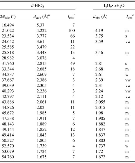
The powder pattern was indexed in the orthorhombic crystal system and reflection conditions pointed to a P212121 space-group symmetry. This phase matched a previously reported for I4O9• xH2O (PDF#00-045-0872) (Table I). In this case “I4O9• xH2O” was designated to refer to a crystal hydrate product of amorphous I4O9 according to the paper that published its XRD result (Wikjord et al., Reference Wikjord, Taylor, Torgerson and Hachkowski1980). However, the I4O9• xH2O reference provides only composition and powder pattern and motivated our consideration of determining rigorously the crystal structure.
X-ray diffraction patterns for δ-HIO3 and I4O9• xH2O (PDF#00–045–0872).

a dcalc – d-spacing calculated from lattice parameters obtained from Rietveld refinement.
b Normalized observed integrated intensities as obtained from the Rietveld refinement.
c Observed intensity denoted as m = medium, s = strong, vw = very weak, w = weak (Wikjord et al., Reference Wikjord, Taylor, Torgerson and Hachkowski1980).
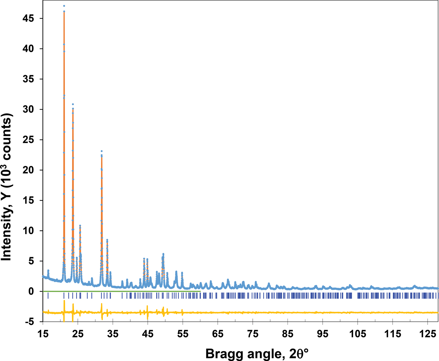
The crystal structure was solved from the integrated intensities obtained from a LeBail full pattern decomposition by a charge flipping method using an XT program from ShelX software (Sheldrick, Reference Sheldrick2015). The crystal structure (Figure 1) was refined by Rietveld method using the Topas software (Cheary and Coelho, Reference Cheary and Coelho1992). The refinement revealed a presence of preferred orientation with a March–Dollase parameter of 0.75(1). Hydrogen atom was positioned from geometric considerations at the longest I–O bond along the shortest oxygen–oxygen contact line as it was observed in other structures of iodic acid. It was refined as riding on the attached O atom.
(Colour online) The Rietveld plot of δ-HIO3 (circles – experimental profile, solid line – calculated, their difference is shown at the bottom, vertical lines show the reflection position).

III. DISCUSSION
This crystal structure determination shows that the actual chemical composition of I4O9• xH2O is HIO3, which could also be written as I4O10•2H2O. This new polymorphic modification of iodic acid is further referred as δ-HIO3 (CCDC#1526763).
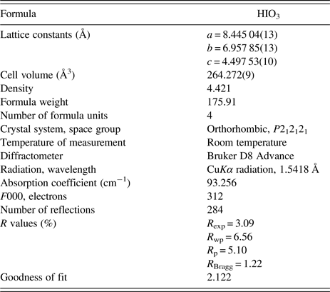
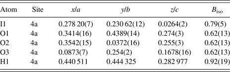
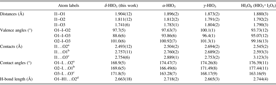
Details of the crystal structure determination are shown in Table II. The atomic parameters are listed in Table III. Selected interatomic distances and bond angles of δ-HIO3, α-HIO3 (Stahl and Szafranski, Reference Stahl and Szafranski1992), γ-HIO3 (Fischer and Lindsjö, Reference Fischer and Lindsjö2005), and HI3O8 (Fischer, Reference Fischer2005) are shown in Table IV.
Details of the crystal structure determination of δ-HIO3.

Atomic coordinates and isotropic displacement parameters (Å2) of δ-HIO3.

Selected interatomic distances (Å) and angles (°) δ-HIO3 in comparison to corresponding geometries of similar compounds.

a 1/2 − x, −y, −1/2 + z.
b 1/2 − x, 1−y, −1/2 + z.
c 1/2 + x, 1/2 − y, −z.
d 1 − x, 1/2 + y, 1/2 − z.
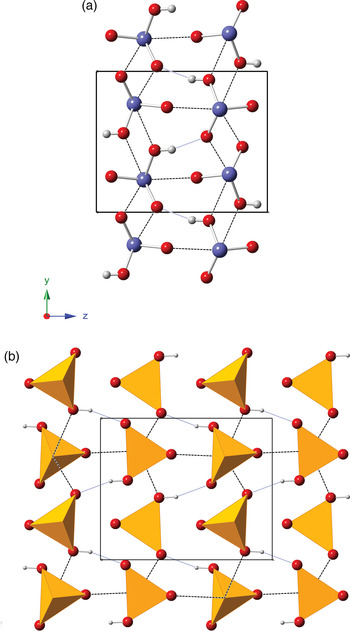
The δ-HIO3 crystalizes in the orthorhombic space group P212121. Figures 2(a) and 2(b) show the crystal structure of δ-HIO3 with each iodine atom 3 coordinated with oxygen atoms (I1–O1 = 1.904, I1–O2 = 1.811, I1–O3 = 1.741), and all four atoms form trigonal pyramid with iodine atoms in the apex and three oxygen atoms in the base. One of the I–O contacts of 2.49 ÅA between molecules make spiral chain along the z direction [Figure 2(a)], and two more long I–O contacts of ~2.76 Å bind the chains in x and y directions into a three-dimensional framework [Figure 2(b)]. O1–H1…O2 hydrogen bond links HIO3 molecules into a zig-zag chain along the y-direction, which make it unique as compared with other two polymorphs of HIO3. Two neighboring molecules are in a mirror position along with xy-plane. Three I–O bonds and three I…O contacts together make a distorted octahedra.
(Colour online) The structure of δ-HIO3 showing: (a) a layer in ab-plane with chains H-bonded along b-axis and (b) corresponding polyhedral representation. Blue dotted lines show H…O contacts and dashed black lines show I…O contacts. Red, blue, and gray balls represent oxygen, iodine, and hydrogen atoms, respectively.

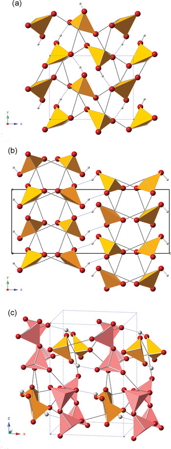
All three iodic acid structures α, γ, and δ differ by packing of the HIO3 molecules and therefore by arrangement of their hydrogen bonds. In α-HIO3, similarly δ-HIO3 structure, hydrogen bonds link molecules into a zig-zag chains [Figure 3(a)]. However, in γ-HIO3 hydrogen bonds form (HIO3)2 pair as shown in Figure 3(b). Figure 3(c) shows that HI3O8 molecule is an adduct of both HIO3 and I2O5 moieties that are connected by a hydrogen bond (Fischer, Reference Fischer2005). Interestingly, the hydrogen bond in δ-HIO3 structure is similar in length to γ-HIO3 molecule and both are slightly shorter than that of α-HIO3 and HI3O8 structures.
(Colour online) The polyhedral representation of (a) α-HIO3, (b) γ-HIO3 (both in zy plane), and (c) HI3O8 (in xz-plane) structures. Blue dotted lines show H…O contacts and dashed black lines show I…O contacts. Red and gray balls represent oxygen and hydrogen atoms, respectively. The pink and yellow pyramids in (c) represent I2O5 and HIO3, respectively.

Each IO3 pyramid forms three I…O intermolecular contacts in 2.5–3.1 Å range (Table III), which along with three I–O bonds form a distorted octahedra. Each I…O contact is positioned across I–O bond so that O–I…O angle deviates from liner for not more than 17°. This is true for all HIO3 structures as well as HIO3 and I2O5 molecules in HI3O8 structure. There is also correlation, at least qualitatively, between length of I–O bonds and opposite I…O contact distances: the shorter bond corresponds to longer contact and vice versa.
IV. CONCLUSION
A new polymorph of iodic acid, δ-HIO3, which crystallizes in the orthorhombic space group P212121, was obtained via a reproducible ASP method using iodic acid solution as the precursor. Ab initio crystal structure determination of δ-phase reveals that previously known I4O9• xH2O phase is actually HIO3.
SUPPLEMENTARY MATERIAL
The supplementary material for this article can be found at https://doi.org/10.1017/S0885715617000859.
ACKNOWLEDGEMENTS
This work was supported by DTRA. The assistance of Lu Liu in measuring the powder XRD patterns of δ-HIO3 is acknowledged.