Introduction
Parapharyngeal and retropharyngeal infections and abscess formation are common but potentially life-threatening conditions in both paediatric patients and adults. In children, these are usually caused by abscess formation within inflamed local lymph nodes during upper respiratory tract infections. With currently available radiologic equipment, diagnosis is easily established once clinical suspicion has been aroused. This can be achieved by ultrasound, computed tomography (CT), magnetic resonance imaging under sedation or a combination of these.Reference Kalmovich, Gavriel, Eviatar and Kessler1
Once a deep neck infection has been diagnosed, the question arises of whether intervention should be immediate or postponed to a later stage, after failure of medical treatment. It has frequently been proposed that small abscesses, that is with a maximum diameter of less than 1 or 1.5 cm, should be initially treated by intravenous antibiotics and close follow up, and a decision about intervention taken after clinical persistence or deterioration, airway compromise, or another complication.Reference Johnston, Schmidt and Barth2
If and when a decision is made about abscess drainage, an appropriate surgical approach must be chosen. Retropharyngeal collections can be easily and safely drained using an intra-oral approach, as can peritonsillar abscesses. For infections of the lateral neck, an external standard surgical procedure is usually inevitable, although attempts may be made to empty a pus-filled cavity using ultrasound-guided external needle aspiration.
The close proximity of the parapharyngeal space to the major vessels and most cranial nerves means that surgical approaches should avoid the blind access of the intra-oral route, even for cavities medial to the major vessels. The external approach allows anatomical dissection, preservation of the major vessels and entry to the medial parapharyngeal space; moreover, a drain may be left in place. Disadvantages of the external approach are the lengthy duration of a reasonably major surgical intervention with the possibility of morbidity and neck scarring. Thus, any minimally invasive intra-oral approach would be the procedure of choice, if proven effective. Important adjunct assistance for the surgeon in this kind of procedure would be the use of various imaging modalities as a guide.
We present a case in which neck ultrasound was used as a guide during intra-oral drainage of a parapharyngeal abscess following drainage of a retropharyngeal abscess.
Case report
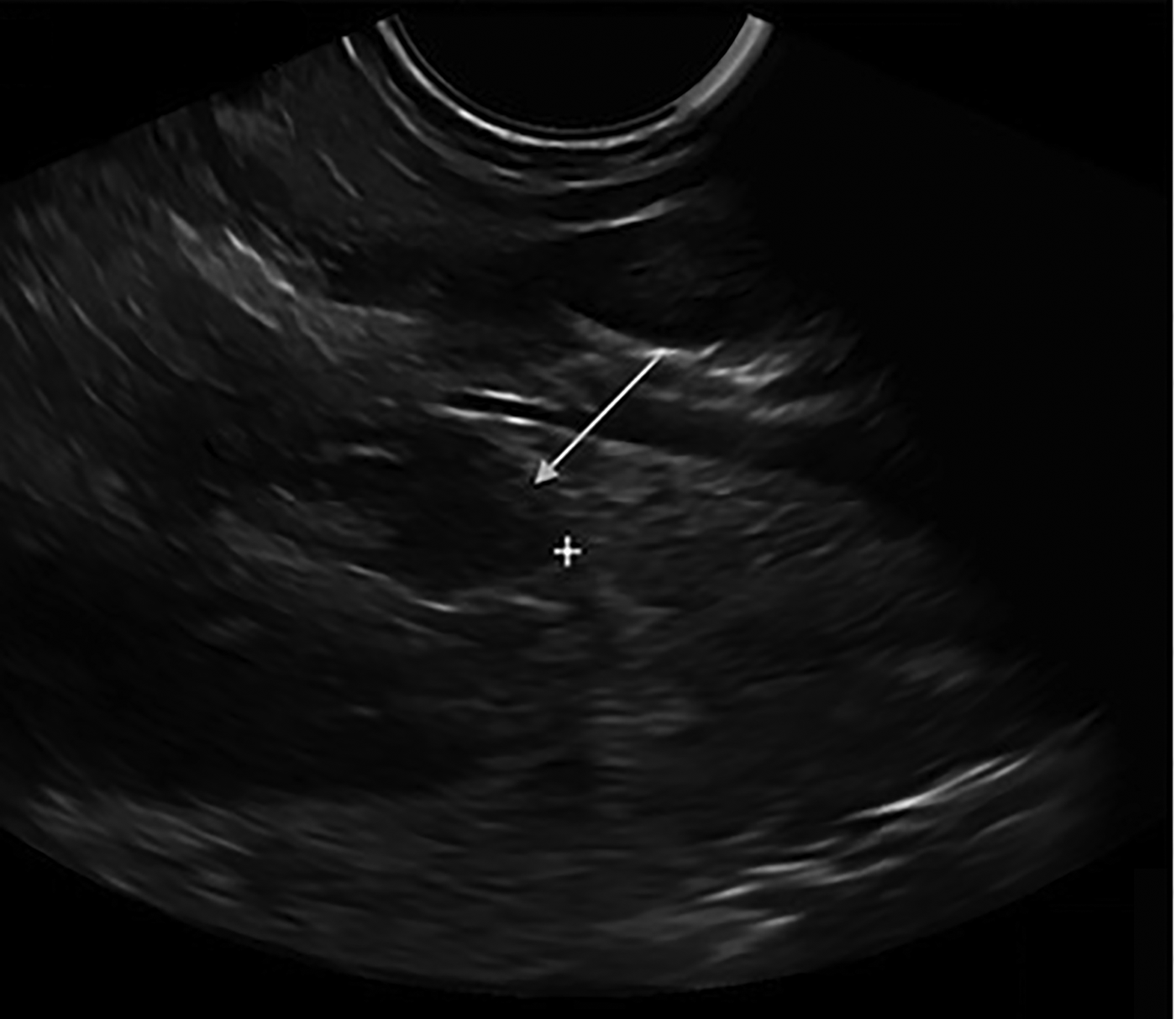
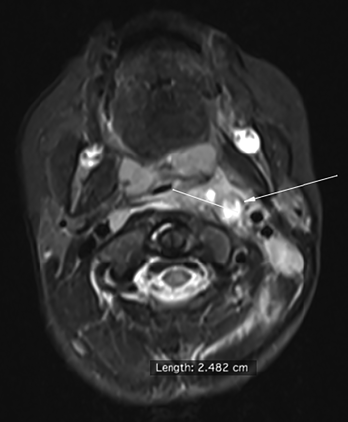
A four-year-old girl was admitted to hospital three days after her family paediatrician began treating her for an upper respiratory infection. She was febrile (39°C) and presented with neck stiffness and difficulty in swallowing. Upon physical examination, her mouth appeared normal, with no trismus and no evidence of swelling of the posterior pharyngeal wall or the tonsils. The left side of her neck had markedly enlarged, painful lymph nodes of both the anterior and posterior triangles, and there was fullness behind the angle of the mandible. The neck was firm and she could not turn her head in any direction. No other ENT pathology was present and laboratory tests showed a white blood cell (WBC) count of 26.0 × 10⁴/l, with 80 per cent neutrophils, and 16 mg/l C-reactive protein (CRP; normally 0–3 mg/l). A neck ultrasound confirmed enlarged lymph nodes with inflammatory characteristics and revealed the presence of two hypoechoic areas, consistent with abscesses within inflamed nodes (Figure 1). These were located medial to the major vessels above the level of the hyoid, and had a maximum diameter of 1.8 cm. The patient was treated with intravenous cefuroxime and clindamycin. The next day, she became afebrile with decreasing CRP and WBC count, but her symptoms remained. On the second day after admittance, she developed left-sided torticollis. Magnetic resonance imaging was performed, confirming the presence of a retropharyngeal abscess posterior to the left tonsil and a second cavity lateral to the first, located at the level of the post-styloid parapharyngeal space and medial to the major vessels (Figure 2). Surgical intervention was decided based on her clinical deterioration.
Pre-surgical ultrasound image indicating the parapharyngeal abscess (arrow) located between the calipers (shown as ‘ + ’).

T2-weighted magnetic resonance image showing both the retro- and parapharyngeal abscesses. The arrow indicates the lateral edge of the parapharyngeal cavity and the straight line indicates the dissection route. The distance between the pharyngeal wall and the great vessels was calculated to be 2.482 cm.
A DICOM Viewer (OsiriX, Pixmeo, Bernex, Switzerland) was used to calculate the distance between the lateral pharyngeal wall and major vessels to establish the safety margins for an intra-oral approach (Figure 2).
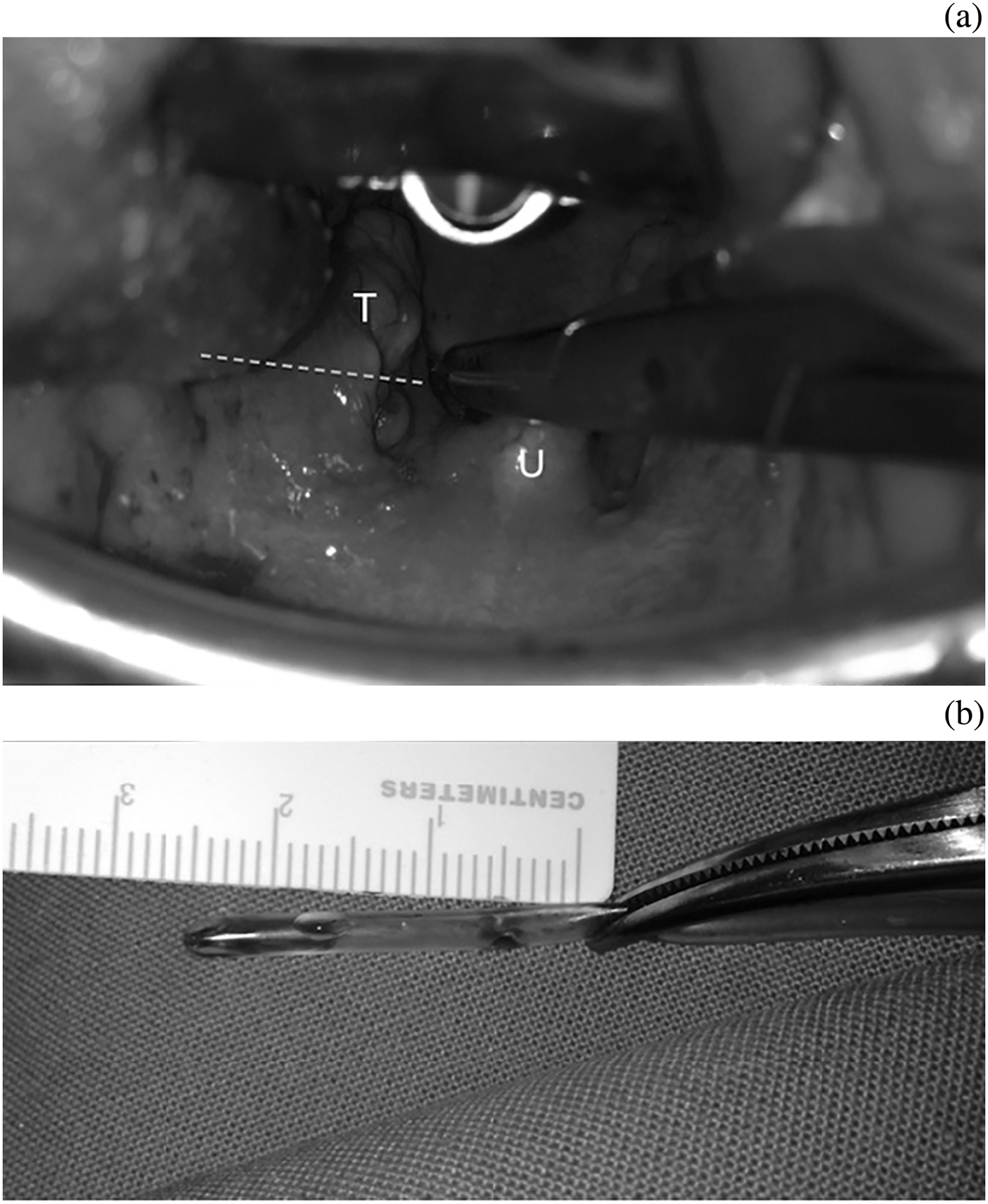
A tonsillectomy mouth dilator was inserted under general anaesthesia; when the left tonsil was pushed forward, the retropharyngeal abscess was immediately seen projecting behind it. A vertical incision was made in the mucosa using a size 15 scalpel. Blunt dissection with a haemostat revealed a small pus-containing cavity, which was drained and irrigated. The haemostat was then turned laterally and pushed into what was expected to be the parapharyngeal abscess. A small amount of pus was expelled, but it was unclear whether the lateral cavity was adequately drained. A neck ultrasound was performed, with the probe pointing toward the neck. A plastic catheter was then pushed through the oral incision into the cavity at a distance of 2.5 cm, previously decided to be sufficient to avoid the major vessels (Figure 3).
(a) Surgical view of the uvula (U) and left tonsil (T). The dashed line indicates the position of the catheter. (b) Image showing the part of the catheter that was inserted through the lateral pharyngeal wall.
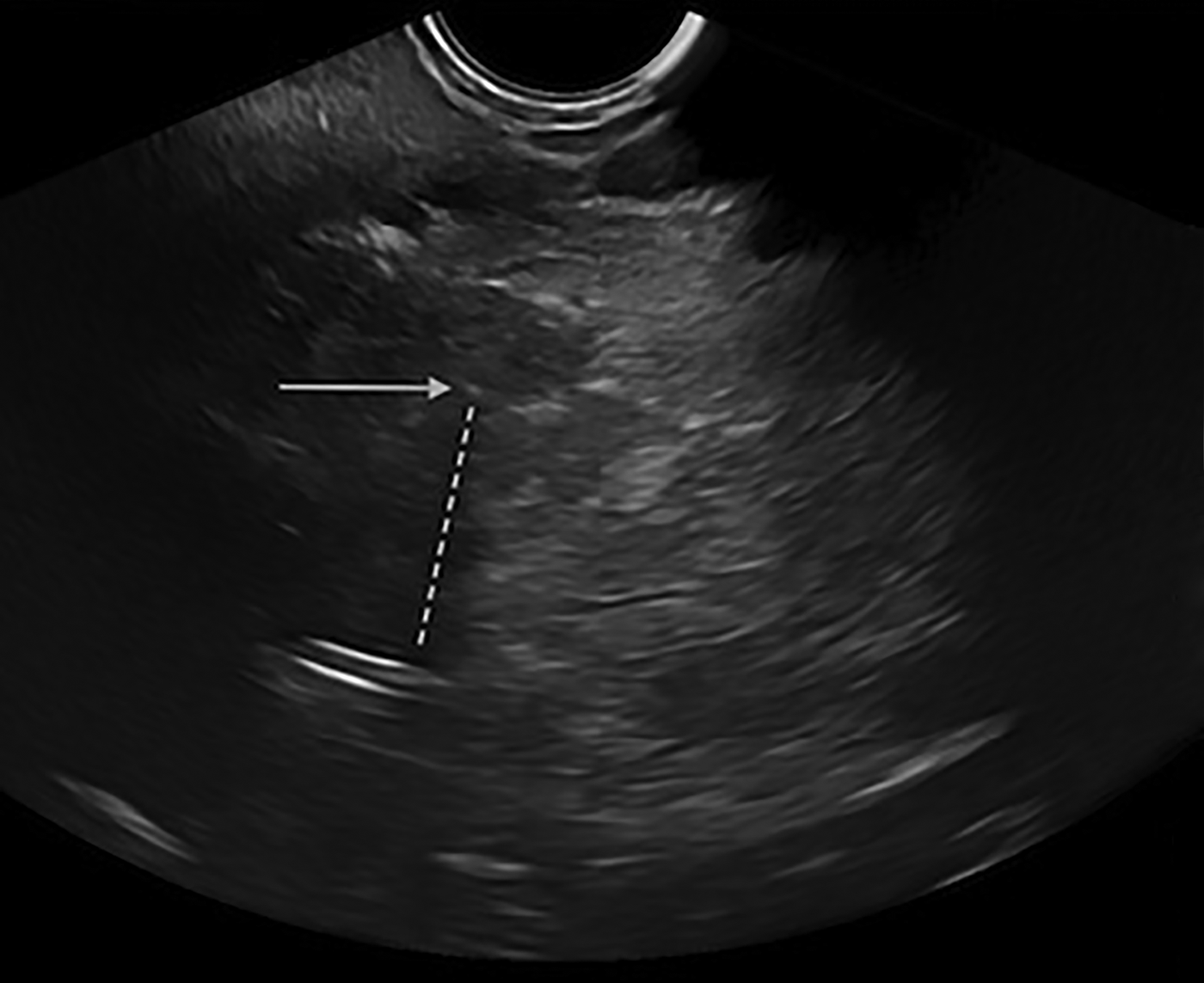
The catheter was observed sonographically to enter the area where the cavity had previously been, confirming that it had been completely emptied (Figure 4). The area was irrigated and the patient was taken to the recovery room. Microbiological analysis of pus samples revealed the presence of Streptococcus epidermidis.
Intra-surgical ultrasound image showing the position of the plastic, non-traumatic catheter (dashed line) and the tip of the catheter within the area formerly occupied by the abscess (arrow). In real-time, instrument movement is seen more clearly and position more accurately than in the static image.
The patient's post-operative course was uneventful. Her symptoms resolved the following day and she was discharged four days later, when laboratory tests had returned to normal.
Discussion
Both external cervical and intra-oral approaches have been used for draining parapharyngeal abscesses, with the latter becoming more popular during the last decade.Reference Amar and Manoukian3 Nevertheless, intra-oral drainage of the parapharyngeal space remains a blind procedure, with dissection through the superior pharyngeal constrictor causing a risk of trauma of the major vessels and failure to locate the cavity. The use of imaging modalities to facilitate an intra-oral approach during surgery has previously been proposed. In a case series of 12 children, Cable et al. used CT image guidance systems to successfully drain parapharyngeal abscesses.Reference Cable, Brenner, Bauman and Mair4
• Intra-oral approach for drainage of a parapharyngeal abscess is considered ideal for draining deep abscesses
• This is the first reported case of intra-oral drainage of a parapharyngeal abscess with the aid of neck ultrasound
• Neck ultrasound may help guide intra-oral instruments to ensure adequate cavity drainage
The use of ultrasound as an intra-operative image guide has been reported only once, by Duque et al. It was used to determine the exact location of parapharyngeal abscesses in a series of three patients.Reference Duque, Guerra and Roy5 However, open (i.e. external) cervical approaches were used. Ultrasound guidance has also been used to access the parapharyngeal space intra-orally for diagnostic purposes, acquire aspirate for cytological examination and facilitate transoral resection of parapharyngeal and retropharyngeal thyroid carcinoma metastases.Reference Andrews, Kwon, Clayman, Edeiken and Kupferman6 To the best of our knowledge, this is the first report of the use of ultrasound as a surgical guide for draining a parapharyngeal space abscess using an intra-oral approach.
In our patient, entry into the parapharyngeal space was facilitated by access through the retropharyngeal cavity. In other cases, the presence of enlarged or inflamed tonsils would necessitate their prior resection or dissection. Entry into the posteromedial parapharyngeal space could then be performed posterior to the tonsils.
The present case suggests that intra-surgical neck ultrasound may assist the surgeon in draining a medial parapharyngeal abscess through an intra-oral approach, which in our case followed drainage of a retropharyngeal abscess. Our case shows that ultrasound performed during dissection may help to navigate surgical instruments within the cavity and confirm adequate drainage. We hope that this paper will encourage more surgeons to follow this method of management in similar cases. This would help clarify the importance and usefulness of ultrasound image-guided intra-oral approaches and contribute to developing a treatment algorithm for children and adults.