Introduction
Delirium is a severe neuropsychiatric syndrome characterized by an acute decline in attention and cognition (American Psychiatric Association, 2000). Although delirium is often viewed as a transient and reversible cognitive disorder, research has shown that it is associated with long-term cognitive impairment and dementia (Jackson et al., Reference Jackson, Gordon, Hart, Hopkins and Ely2004; MacLullich et al., Reference MacLullich, Beaglehole, Hall and Meagher2009; Witlox et al., Reference Witlox, Eurelings, de Jonghe, Kalisvaart, Eikelenboom and van Gool2010).
Neuropsychological test batteries are considered the “gold standard” for investigating cognitive functioning. Yet, most studies that examined the association of delirium with cognitive impairment merely used screening instruments which are inappropriate for fully characterizing and quantifying defects in specific cognitive domains (Jackson et al., Reference Jackson, Gordon, Hart, Hopkins and Ely2004). Therefore, the precise nature of the cognitive impairments and the extent to which particular cognitive domains are affected after delirium remain uncertain (MacLullich et al., Reference MacLullich, Beaglehole, Hall and Meagher2009).
A better understanding of the nature of the cognitive impairments following delirium might help to ascertain the value of different hypotheses that have been proposed to explain the relationship between delirium and cognitive impairment at follow-up. Delirium may persist for months and patients with cognitive impairment at follow-up may in fact experience persistent delirium (Cole et al., Reference Cole, Ciampi, Belzile and Zhong2009). Alternatively, patients with cognitive impairment at follow-up may have depression or have neurodegenerative disease (Lenze et al., Reference Lenze2007; Krogseth et al., Reference Krogseth, Wyller, Engedal and Juliebo2011). The few studies that examined the neuropsychological profiles of elderly patients after an episode of delirium (Katz et al., Reference Katz, Curyto, TenHave, Mossey, Sands and Kallan2001; Benoit et al., Reference Benoit2005; Jankowski et al., Reference Jankowski2011) have not adequately addressed the possibility that the presence of delirium symptoms or depression may have influenced performance on cognitive tests at follow-up.
The aim of the present study was to evaluate cognitive performance at follow-up in elderly hip fracture patients who did or did not experience in-hospital delirium. We used a comprehensive neuropsychological approach and examined to what extent inattention, as an important sign of persistent delirium (Meagher et al., Reference Meagher, Leonard, Donnelly, Conroy, Saunders and Trzepacz2010) and depression (Herrmann et al., Reference Herrmann, Goodwin and Ebmeier2007), is an important feature of the neuropsychological profile of patients who have had delirium three months earlier during their hospital admission.
Methods
Participants
This is a prospective cohort study that was nested within a clinical trial that compared the effectiveness of taurine versus placebo in reducing morbidity and one-year mortality in elderly hip fracture patients aged 75 years and older (Clinicaltrials.gov; registration number NCT00497978). Examining neuropsychological function, three months after hospital discharge, was a pre-specified secondary aim of the trial.
The approval of the regional research ethics committee was obtained. All patients gave signed informed consent. Patients were recruited from a series of consecutively admitted elderly hip fracture patients to a non-academic teaching hospital in Alkmaar, the Netherlands. Eligibility was checked for all patients aged 75 years and older admitted for primary surgical repair of hip fracture. Patients were not eligible if they had no acute trauma, received a total hip prosthesis, had a pathological fracture, were not capable (e.g. dementia in the medical case notes, aphasia, coma) or not willing to provide informed consent or had contraindications regarding the administration of taurine (i.e. renal impairment defined as a creatinine clearing less than 30 mL/min). As these patients are at high risk for developing delirium, all patients received routine care with prophylactic treatment of 0.5 mg haloperidol, three times daily, from the time of admission until postoperative day three, unless contraindications regarding its use were present (Kalisvaart et al., Reference Kalisvaart2005).
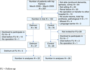
All patients who developed delirium during hospital admission were asked to participate in the follow-up. A subgroup of patients without delirium was selected to participate as controls based on random selection by a computer-generated randomization code. Because we were specifically interested in the effects of in-hospital delirium on neuropsychological test performance three months after hospital discharge, we excluded patients with clinically manifest delirium at follow-up (Figure 1).
Flow chart.

Baseline (preoperative) assessment
Baseline assessment was completed within 12 hours after hospital admission and before surgery. The following demographic variables were documented: age, gender, and educational level. Cognitive functioning was assessed with the Mini-Mental State Examination (MMSE; Folstein et al., Reference Folstein, Folstein and McHugh1975), and pre-existent cognitive decline with the Informant Questionnaire on Cognitive Decline in the Elderly–Short-Form (IQCODE-N; Jorm and Jacomb, Reference Jorm and Jacomb1989; de Jonghe et al., Reference de Jonghe, Schmand, Ooms and Ribbe1997). The Geriatric Depression Scale (GDS) was used as a self-inventory of depressive symptomatology (Sheikh and Yesavage, Reference Sheikh and Yesavage1986; Stek et al., Reference Stek, Gussekloo, Beekman, van Tilburg and Westendorp2004). The number and type of medical co-morbidities and medications at home, the American Society of Anesthesiologists (ASA) physical status classification system (American Society of Anesthesiologists, 2010), and the Acute Physiology and Chronic Health Evaluation II (APACHE II; Knaus et al., Reference Knaus, Draper, Wagner and Zimmerman1985) score were abstracted from the medical record. Visual acuity was assessed with the standardized Snellen test for visual impairment (Hetherington, Reference Hetherington1954). Pre-fracture (instrumental) activities of daily living ((I)ADL) functioning was assessed by a close relative or caregiver with the Barthel Index (BI; Mahoney and Barthel, Reference Mahoney and Barthel1965) and the Lawton IADL scale (Lawton and Brody, Reference Lawton and Brody1969). Delirium was defined according to the criteria of the confusion assessment method (CAM), which consists of an acute onset and fluctuating course of cognitive function, inattention, and either disorganized thinking and/or altered level of consciousness (Inouye et al., Reference Inouye, van Dyck, Alessi, Balkin, Siegal and Horwitz1990). The CAM algorithm was rated on the basis of an interview with the patient and hospital staff, brief cognitive assessment with the MMSE and the expanded digit span test (Lindeboom and Matto, Reference Lindeboom and Matto1994), and screening of the medical and nursing records for signs of delirium. The presence of delirium was assessed daily from the time of hospital admission until at least the fifth postoperative day.
For the IQCODE-N, BI, and Lawton IADL, proxies were asked to describe the patient's condition a week before the fracture as to determine function unbiased by the event of hip fracture itself or any acute or sub-acute event leading to hip fracture.
Three-month follow-up assessment
A neuropsychological test battery was administered three months after hospital discharge by two trained neuropsychologists. The neuropsychological battery was designed to assess multiple cognitive domains and contained standardized and validated instruments. It also included the MMSE, the expanded digit span test, and the GDS, and took approximately one hour to complete. Most patients were examined at home; some patients preferred to visit the hospital.
The CAM was used to screen for delirium at follow-up. CAM positive patients at follow-up were those that demonstrated an acute change or fluctuation in their mental status plus the accompanying inattention and disorganized thinking and/or altered level of consciousness.
The Dutch version of the National Adult Reading Test (DART) was used to measure pre-morbid verbal intelligence (Nelson, Reference Nelson1991; Schmand et al., Reference Schmand, Lindeboom and van Harskamp1992). The DART requires patients to read aloud 50 words with irregular pronunciation.
Episodic memory was examined with the Dutch version of the verbal learning test, i.e. the Groningen Fifteen Words Test (Brand and Jolles, Reference Brand and Jolles1985; van der Elst et al., Reference van der Elst, Van Boxtel, Van Breukelen and Jolles2005). Fifteen words are presented orally and the patient is asked to repeat as many words as possible when the presentation stops. After five trials, a 20-minute delay is interspersed at the end of which the patient is asked to recall and recognize the previously learned words (delayed recall). We analyzed (1) the total number of correctly recalled words summed over five trials; (2) the number of correctly recalled words after the delay; and (3) the number of correctly recognized target and non-target words.
Attention and concentration was first assessed with the expanded digit span test. In this test, the examiner reads a string of digits which the patient has to repeat in either the same (forward) or the reverse (backward) order (Lindeboom and Matto, Reference Lindeboom and Matto1994). The forward and backward tests were analyzed separately with the forward span measuring concentration, and the backward span measuring working memory (Lezak et al., Reference Lezak, Howieson, Loring, Lezak, Howieson and Loring2004). Second, the Expanded Mental Control Task (EMCT), which consists of 12 word lists and arithmetic progressions that the patient has to recite within a certain amount of time, was administered (Lindeboom et al., Reference Lindeboom, Koene and Matto1993). The following measures of the EMCT were analyzed: (1) the combined total score; (2) total time needed to complete all items; and (3) the total number of errors. Third, a computerized simple reaction time (RT) task was used in which the patient had to press the space bar as quickly as possible when the word “YES” appeared on the screen. The time between stimuli presentation varied randomly between 1 and 3 seconds. Forty trials were administered and we analyzed (1) mean reaction time; (2) intra-trial variability of reaction times, i.e. standard deviation of reaction times; and (3) number of premature responses, i.e. response during the inter-stimulus interval.
The Clock Drawing Test measures visuoconstructive function. Patients are asked to draw a clock and set the hands of the clock at ten past eleven. Points are given for (1) drawing a closed circle; (2) correct ordering of the numbers within the clock; (3) correct spatial location of the numbers within the clock; and (4) inserting the hands at the correct time (Pinto and Peters, Reference Pinto and Peters2009).
We administered the Dutch version of the Controlled Oral Word Association Test (COWAT) to examine executive function (Schmand et al., Reference Schmand, Groenink and van den Dungen2008). This verbal fluency task requires patients to name as many words as possible within 60 seconds beginning with a target letter specified by the examiner.
Proxies were asked to complete the IQCODE-N for the second time with reference to the situation three months after hospital discharge.
Outcome
Performance on neuropsychological tests three months after hospital discharge.
Statistical analysis
Statistical calculations were performed using SPSS for Windows, version 14 (SPSS Inc., Chicago, IL). Quantitative variables are presented as mean (standard deviation, SD) or median (inter-quartile range, IQR). The χ2 or Fisher exact tests were used to analyze categorical variables. Continuous variables were analyzed with Student t tests or Mann–Whitney U tests for between-group comparisons and paired t tests or Wilcoxon signed rank tests for within-group comparisons. The assumption of normality was tested with the Kolmogrov–Smirnov test.
To examine whether delirium is associated with neuropsychological test performance independent of important covariates, we fitted a multiple linear regression model for those neuropsychological measures that were associated with delirium in univariate analyses. In the multivariate models, we entered delirium as an independent variable together with age, gender, baseline MMSE, and treatment allocation. These covariates were selected a priori based on their potential to influence neuropsychological test performance. Besides the demographic variables such as age and gender, educational level has also been shown to affect neuropsychological test performance (van der Elst et al., Reference van der Elst, Van Boxtel, Van Breukelen and Jolles2005). However, education was not included as a covariate into our regression models for two reasons: (a) preliminary analyses showed that the results remained unchanged whether or not the variable “level of education” was included in the models; and (b) given our sample size, we aimed to restrict the number of independent variables to a maximum of five. In the regression models that are presented in the results section, we entered the baseline MMSE, and not the baseline IQCODE-N, as a measure of pre-existent cognitive impairment. Compared with the IQCODE-N (which measures intra-individual changes in cognitive functioning), the MMSE provides a score that can more easily be compared between patients. We performed sensitivity analyses to examine whether inclusion of the IQCODE-N (instead of the MMSE) produced similar results. The baseline measures of the GDS and IQCODE-N were entered as covariates, instead of the MMSE, in the multivariate models that examined the association between delirium during hospitalization and the GDS and IQCODE-N at follow-up. The core assumptions of linear regression modeling were tested for each model. Statistical significance was set at p < 0.05.
Results
A total of 53 patients with and without delirium during hospitalization underwent comprehensive neuropsychological testing three months after hospital discharge (Figure 1). Five patients (all of whom also experienced delirium in the hospital) were diagnosed with delirium at follow-up according to CAM criteria. Thus, 48 patients free from clinically manifest delirium at follow-up, of whom 22 had previously experienced delirium, were available for our analyses. Ten of 22 (45%) patients with previous delirium scored one or two points on the CAM at follow-up compared with 3/26 (12%) controls. According to the CAM, 8/22 (36%) patients with in-hospital delirium were inattentive at follow-up compared with 2/26 (8%) controls (p = 0.03). No patient displayed disorganized thinking or an altered level of consciousness at follow-up. The duration between hospital discharge and neuropsychological assessment was on average 81 days and did not differ between both groups (t(46) = 0.05, p = 0.96).
Preoperative comparisons between groups
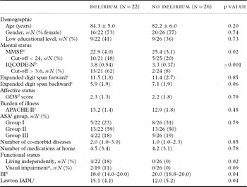
Baseline characteristics of patients with and without in-hospital delirium are presented in Table 1. Baseline cognitive impairment, as evidenced by low MMSE and high IQCODE-N scores, was poorer in patients with delirium than in those without delirium. Also, patients in the delirium group were more dependent in (I)ADL functioning at home. Other baseline characteristics did not differ significantly between both groups.
Baseline characteristics of patients with and without in-hospital delirium

Values are expressed as means ± SD or median (IQR), n/N is number with characteristic/total number, (%) is percentage.
aMMSE is the Mini-Mental State Examination, range 0 (severe cognitive impairment) to 30 (no cognitive impairment), score < 24 indicates cognitive impairment.
bIQCODE-N is the Informant Questionnaire on Cognitive Decline in the Elderly–Short Form, range 16 (cognitive improvement) to 80 (severe cognitive decline), mean score > 3,6 indicates cognitive decline.
cExpanded digit span test, range 0 (poor performance) to 21 (very good performance), forward measures concentration, backward measures working memory.
dGDS is the Geriatric Depression Scale, range 0 (depression not likely) to 15 (depression very likely), score > 4 indicates depression.
eAPACHE II is Acute Physiology and Chronic Health Evaluation II, range 0 (no acute health problems) to 70 (severe acute health problems).
fASA is the American Society of Anesthesiologists physical status classification system, range 1 (normal health patient) to 5 (moribund patient).
gVisual impairment measured with the standardized Snellen test for visual impairment and defined as binocular near vision worse than 20/70 after correction.
hBI is the Barthel Index, range 0 (severe disability) to 20 (no disability).
iLawton IADL is the Lawton Instrumental Activities of Daily Living scale, range 8 (no disability) to 31 (severe disability).
Follow-up comparisons between groups
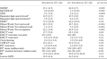
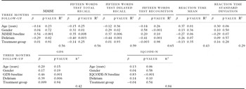
At follow-up, patients with in-hospital delirium performed poorer on tests of global cognitive performance (MMSE), episodic memory, and attention than controls (Table 2). Moreover, patients with in-hospital delirium also showed more signs of depression (GDS) and cognitive decline (IQCODE-N) at follow-up than controls (Table 2). After adjustment for relevant confounders, the association between in-hospital delirium and global cognitive performance (MMSE), episodic memory, and depressive symptoms (GDS) remained significant (Table 3). When sensitivity analyses were performed, in which the baseline MMSE was substituted by the baseline IQCODE-N, the association between in-hospital delirium and the MMSE at follow-up did not remain significant (data not shown).
Global cognitive and affective performance and neuropsychological test scores at three months follow-up of patients with and without in-hospital delirium

Values are expressed as means ± SD or median (IQR).
aMMSE is the Mini-Mental State Examination, range 0 (severe cognitive impairment) to 30 (no cognitive impairment), score < 24 indicates cognitive impairment.
bIQCODE-N is the Informant Questionnaire on Cognitive Decline in the Elderly–Short Form, range 16 (cognitive improvement) to 80 (severe cognitive decline), score >57 (i.e. mean score > 3,6) indicates cognitive decline.
cGDS is the Geriatric Depression Scale, range 0 (depression not likely) to 15 (depression very likely), score > 4 indicates depression.
dExpanded digit span test, range 0 (poor performance) to 21 (very good performance), forward measures concentration, backward measures working memory.
eDART is the Dutch Adult Reading Test, a measure of pre-morbid verbal intelligence, range 0 (no words correctly pronounced) to 100 (all words correctly pronounced).
fEMCT is Expanded Mental Control Task measures attention, range total score 0 (poor performance) to 24 (very good performance).
gCOWAT is the Controlled Oral Word Association Test, a measure of executive function with higher scores indicating a better performance.
hRT is the simple Reaction Time task, a measure of attention.
iBI is the Barthel Index, range 0 (severe disability) to 20 (no disability).
jLawton IADL is the Lawton Instrumental Activities of Daily Living scale, range 8 (no disability) to 31 (severe disability).
Multiple linear regression models of neuropsychological test scores significantly associated with in-hospital delirium in univariate analyses

MMSE = Mini-Mental State Examination; IQCODE-N = Informant Questionnaire on Cognitive Decline in the Elderly–Short Form; GDS = Geriatric Depression Scale.
Preoperative to follow-up comparisons
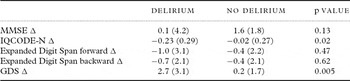
The degree of change in the MMSE over the course of three months between patients with and without in-hospital delirium was not significant (Table 4). However, a comparison within groups revealed that the MMSE of control patients improved over time (t(24) = −4.5, p < 0.001) while no change was observed in the delirium group (t(20) = −0.1, p = 0.92).
Degree of change between baseline and three months follow-up for patients with and without in-hospital delirium

Values are expressed as means ± SD.
MMSE = Mini-Mental State Examination; IQCODE-N = Informant Questionnaire on Cognitive Decline in the Elderly–Short Form; GDS = Geriatric Depression Scale.
In contrast, the degree of change over time in the IQCODE-N between both groups was significant (Table 4) with more deterioration notable in the delirium group (t(15) = −3.2, p = 0.006) but not in the control group (t(25) = −0.46, p = 0.65).
None of the between- and within-group analyses performed on the expanded digit span test showed any significant differences between patients with and without in-hospital delirium (data not shown).
A preoperative to follow-up comparison of GDS between patients with and without in-hospital delirium was significant (Table 4), with patients who experienced in-hospital delirium showing an increase of depressive symptoms (t(17) = −3.7, p = 0.002) while no change was observed among controls (t(24) = −0.6, p = 0.55).
Exploratory subgroup analyses
Among patients who were free of any delirium symptoms at follow-up (i.e. CAM score of null), patients with in-hospital delirium still performed poorer on the total and delayed recall measure of the Fifteen Words Test than non-delirious controls.
Among patients with few to no symptoms of depression at follow-up (i.e. GDS < 4), patients with in-hospital delirium performed poorer on tests of global cognition, episodic memory, attention, and constructional praxis than non-delirious controls.
Finally, patients who previously experienced in-hospital delirium and who did not show any signs of cognitive impairment at baseline (i.e. MMSE > 23 and IQCODE-N < 3.6) performed significantly worse on all measures of the Fifteen Words Test than patients without previous cognitive impairment and delirium.
Discussion
This study evaluated the impact of in-hospital delirium on neuropsychological function three months after hospital discharge in a group of elderly hip fracture patients. In-hospital delirium was found to be independently associated with impairments on tests of global cognition and episodic memory at follow-up.
Strengths of the current study include the systematic and simultaneous assessment of delirium, cognitive impairment, and depression at follow-up using standardized and validated instruments. A limitation of this study is the small patient number. Although this study would ideally have been performed in a larger and purely observational cohort, our multivariate models show highly significant associations between in-hospital delirium and poorer cognitive performance at follow-up. Nevertheless, future studies of larger cohorts would be better able to exclude potential type II errors and provide more robust and definitive conclusions. Such studies would also be better able to take into account the interactions between delirium and numerous other determinants of cognitive impairment.
Interestingly, patients with in-hospital delirium were more often labeled as “inattentive” according to the CAM than controls at follow-up whereas no difference in attention performance was seen on the more objective neuropsychological tests. This discrepancy may be explained by a tendency of raters to label the observed cognitive impairments as having an attentional component when the cognitive deficits are more severe, as they are for patients with previous delirium.
Several hypotheses have been proposed to explain the association between delirium and cognitive impairment. One of these explanations suggests that the high prevalence of cognitive impairment at follow-up among patients with previous delirium may reflect persistence of delirium instead (Cole et al., Reference Cole, Ciampi, Belzile and Zhong2009). Indeed, according to the CAM, elderly hip fracture patients who earlier experienced in-hospital delirium were most likely to show signs of inattention – the cardinal and required symptom of (persistent) delirium (World Health Organization, 1993; American Psychiatric Association, 2000) – at follow-up. However, when important baseline characteristics were taken into account, differences between both groups in attentional performance could not be observed on the more objective neuropsychological tests. This may imply that the poorer cognitive performance at the follow-up of patients who earlier experienced delirium cannot be fully explained by the persistence of a key symptom of delirium. This suggestion is substantiated by our subgroup analyses that showed that patients with in-hospital delirium, but without delirium symptoms at follow-up (i.e. CAM score of null), performed poorer on an episodic memory test than controls who never experienced delirium.
On the contrary, our study also clearly demonstrates that the persistence of delirium can affect cognitive outcome at follow-up in some patients, as almost 20% of our patients with in-hospital delirium were excluded because of persistent (or recurrent) delirium three months after hospital discharge.
When explaining the association between delirium and cognitive impairment at follow-up, the interrelationship between delirium and depression may also be of relevance. Depression is a known risk factor for delirium in elderly hip fracture patients (Olofsson et al., Reference Olofsson, Lundstrom, Borssen, Nyberg and Gustafson2005). In turn, delirium has been associated with higher depressive symptom levels months after hip fracture (Lenze et al., Reference Lenze2007), and importantly, depression can markedly affect cognitive function (Herrmann et al., Reference Herrmann, Goodwin and Ebmeier2007). To date, few, if any, studies have examined the association between delirium and cognitive impairment at follow-up while simultaneously documenting the presence of mood disturbances. Contrary to previous investigations (Olofsson et al., Reference Olofsson, Lundstrom, Borssen, Nyberg and Gustafson2005), depression was not a risk factor for in-hospital delirium in our study. However, patients with in-hospital delirium did show an increase in depressive symptoms at follow-up that may (partially) explain their poorer performance on neuropsychological tests. Yet, the neuropsychological profile of depression in late life is typically characterized by diminished processing speed and impaired executive function (Herrmann et al., Reference Herrmann, Goodwin and Ebmeier2007). In contrast, our delirium patients showed disproportionate memory disturbances. In addition, in patients with few or no depressive symptoms present at follow-up, in-hospital delirium remained associated with poorer performance on a range of neuropsychological tests. These results suggest that an increase of depressive symptoms at follow-up among patients with previous delirium cannot fully explain their poorer cognitive functioning.
As a consequence, studies that include patients with reversible causes of cognitive impairment, such as persistent delirium and depression, may systematically overestimate the strength of the relationship between delirium and newly acquired long-term cognitive impairment or dementia.
If the presence of delirium symptoms or worsening of depressive symptomatology at follow-up cannot fully explain poor cognitive outcome of individuals with previous delirium, what other explanations can be considered? Delirium may unmask early or subclinical dementia or may initiate or accelerate a process of cognitive decline. In most dementias, there is a disproportionate disturbance on tests of global cognition and episodic memory with relatively preserved attentional capacities. Thus, the neuropsychological profile of our patients with previous delirium may seem consistent with the presence or development of a dementia syndrome. However, to test the hypothesis that delirium actually initiates neurodegeneration requires a (seemingly) population without dementia at baseline. Because pre-existent cognitive impairment is an important predisposing risk factor for delirium in elderly individuals (Dasgupta and Dumbrell, Reference Dasgupta and Dumbrell2006), our findings of poor cognitive performance at follow-up among patients with in-hospital delirium may merely reflect the presence (or acceleration) of baseline cognitive impairment. Indeed, many of our patients who did develop delirium showed signs of pre-fracture cognitive decline. Therefore, we adjusted our multivariate models for baseline cognitive impairment and also repeated our analyses in a subgroup of patients that did not show any sign of cognitive impairment at baseline. The results of these analyses suggest that delirium may be more than a marker of pre-existent cognitive decline, although the exact mechanism through which delirium is associated with later cognitive deterioration remains unclear. Factors that precipitate delirium may incite a sequence of events in the brain which may contribute to the development or acceleration of cognitive impairment. Microglia and aberrant stress responses have been suggested to play a role in this detrimental process (MacLullich et al., Reference MacLullich, Ferguson, Miller, de Rooij and Cunningham2008; Cunningham et al., Reference Cunningham2009; van Gool et al., Reference van Gool, van de Beek and Eikelenboom2010).
In conclusion, the current investigation provides evidence of the poor cognitive and affective prognosis of elderly people after delirium and adds to a growing body of evidence that suggests that delirium is associated with various forms of poor long-term outcomes (Witlox et al., Reference Witlox, Eurelings, de Jonghe, Kalisvaart, Eikelenboom and van Gool2010). Future studies with multiple and longer follow-up periods will be needed to draw definitive conclusions as to whether cognitive dysfunction at follow-up after delirium follows a static, fluctuating, gradually resolving or progressive course. Moreover, intervention studies are needed to investigate whether the sequelae associated with delirium can be averted.
Conflict of interest
Funding sources: Dr. van Gool is supported by a ZonMw-TOP grant 40-00812-98-10017.
Description of authors’ roles
Joost Witlox contributed to the conception and design, acquisition of data, analysis and interpretation of data, drafting the paper, final approval for publication. Chantal J. Slor and René W.W.M. Jansen undertook analysis and interpretation of data, critical revision of the manuscript for intellectual content, and final approval for publication. Kees J. Kalisvaart, Piet Eikelenboom, Willem A. van Gool, and Jos F.M. de Jonghe contributed to the conception and design, analysis and interpretation of data, critical revision of the manuscript for intellectual content, and final approval for publication. Mireille F.M. van Stijn and Alexander P.J. Houdijk contributed to the acquisition of data, critical revision of the manuscript for intellectual content, and final approval for publication.
Acknowledgments
We would like to thank Gisela Dekker, RN (data acquisition), Ralph Vreeswijk, RN (data acquisition), Milko van Langen (data acquisition), Tjerk Schoemaker, MSc (data acquisition), Ellen Slijkerman, MSc (data acquisition), and Tjeerd van der Ploeg, MSc (data analysis) for their work on the study. Dr. van Gool is supported by a ZonMw-TOP grant 40-00812-98-10017.
Sponsor's role. None.
Trial name. The Effect of Taurine on Morbidity and Mortality in the Elderly Hip Fracture Patient. Registration number: NCT00497978. URL: http://clinicaltrials.gov/ct2/show/NCT00497978?term=taurine+hip+fracture&rank=1