Introduction
Acute airway compromise secondary to deep neck space abscess contributes to morbidity and mortality associated with the condition. As such, prompt diagnosis and appropriate management is imperative. Infection typically derives from an oropharyngeal or odontogenic source, but other infections resulting from trauma or iatrogenic inoculation can also occur.Reference Osborn, Assael and Bell1
Lymphangiomata are benign lymphovenous malformations that can be primary or secondary in origin. They are most common in the head and neck region, but are only rarely reported to affect the palatine tonsil.Reference Mardekian and Karp2
Airway obstruction secondary to lymphovenous malformations is extremely rare, with only one case reported in the current literature.Reference Harsha, Crawford and Sorensen3 We present a case of an adult with acute airway compromise due to parapharyngeal, parotid and masseteric abscesses that formed in association with lymphangioma of the palatine tonsil.
Case report
Clinical presentation
A 37-year-old, non-smoking male presented to the emergency department with progressive odynophagia, ipsilateral otalgia and fluctuating pyrexia over 48 hours. During his admission, over a 4-hour period, he developed trismus and complete dysphagia. His past medical history included microcytic anaemia of an undetermined cause. He took no regular medications and had no drug allergies.
On examination, the patient was stridulous and acutely dyspnoeic, with objectively increased work and rate of breathing, without signs of decompensation. Assessment of vitals revealed him to be pyrexial, tachycardic and hypotensive (temperature of 37.9°C, heart rate of 106 beats per minute and blood pressure of 92/52 mmHg, respectively). Oral cavity examination was limited by trismus. Copious pooling of oral secretions was noted. Neck examination revealed a diffuse, right cervical mass overlying the parotid gland, becoming confluent with a fluctuant mass in level IIa. Severe torticollis was apparent. Fibre-optic nasendoscopy revealed a right parapharyngeal swelling with pooling of secretions in the valleculae, and secondary erythema and oedema of the supraglottis. The diagnosis of deep neck space collection with impending airway compromise was made.
Acute management was initiated simultaneously with examination. This consisted of high-flow oxygen and intravenous access. Blood was obtained for culture and sensitivity testing. Dexamethasone was administered intravenously with 1.2 g of amoxicillin with clavulanic acid, as stipulated by local microbiology guidance. The patient received two 500 ml crystalloid fluid boluses, with resultant improvement in vital parameters. A urinary catheter was also inserted. Despite this initial resuscitation, the patient remained stridulous. Anaesthetic support was requested and the multidisciplinary decision was made to secure a definitive airway.
The patient was transferred to the operating theatre. An airway was secured via awake fibre-optic nasotracheal intubation. An ENT surgeon was on standby to perform a tracheostomy if required.
The patient subsequently underwent computed tomography of the neck with intravenous contrast. This revealed two distinct deep neck space collections. The first was in the right peritonsillar space, extending laterally into the parapharyngeal space, posterior to medial pterygoid into the masseteric space (Figure 1). The second was within the inferior aspect of the masseter muscle (Figure 2). The decision was made to proceed to surgical drainage.
Axial, contrast-enhanced computed tomography scan demonstrating a loculated, right parapharyngeal abscess (black arrow) extending posterior to the right medial pterygoid muscle into the masseteric space (white arrow). Medialisation of the right palatine tonsil and endotracheal tube is evident.

Axial, contrast-enhanced computed tomography scan demonstrating a loculated abscess in the right masseter muscle (white arrow).

Surgical intervention
Given the location of the abscesses, a combined approach was adopted. Initially, a tracheostomy was sited in a conventional fashion. The right intra-oral mucosa was then opened along the pterygopalatine raphe and a bipolar dissection tonsillectomy completed. The tonsillar tissue was noted to be unusually firm at this stage, and the specimen was sent for both microbiology and histology. The constrictor muscles were bluntly dissected to the parapharyngeal fat and 50 ml of pus was drained.
A modified Blair incision was then made externally and the subplatysmal flap elevated. Dissection continued medial to the sternocleidomastoid. The carotid sheath was opened, and dissection continued superiorly to the parapharyngeal space, releasing a further 30 ml of pus. Blunt dissection was performed between the superficial parotid gland and masseter muscle, where the final abscess was drained. Samples were sent for microbiology.
Washout was performed with saline, and Yeates drains were inserted. A fine-bore nasogastric feeding tube was sited.
Post-operative management
Over the subsequent 48 hours, the patient made good progress physiologically and biochemically, requiring no additional organ support. Gram-positive cocci were seen on the staining of the specimen; antibiotics were rationalised to ceftriaxone, clindamycin and vancomycin.
At 72 hours, there was resolution of both torticollis and trismus. The patient remained apyrexial, with no airway compromise. Oral swallow assessment confirmed no pharyngocutaneous leak.
Feeding resumed after tracheostomy decannulation on day 5 post-operatively. Microbiological specimens confirmed methicillin-sensitive Staphylococcus aureus as the causative organism. The patient was discharged home 7 days post-procedure to complete a course of co-amoxiclav.
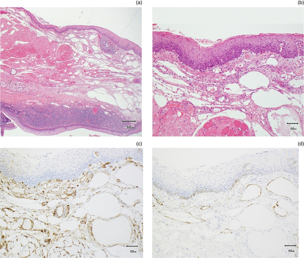
Histopathological analysis confirmed tonsillar tissue, composed of normal overlying squamous epithelium and mucosa-associated lymphoid tissue. However, abutting the striated muscle was a well-developed mass of dilated vascular (clusters of differentiation 31 and 34 positive) and lymphatic (D2-40 positive) spaces without atypia, consistent with a lymphangioma (Figure 3).
Photomicrographs of the right palatine tonsil showing ectatic spaces within peritonsillar submucosa extending into the muscle: (a) low power haematoxylin and eosin stain (×40); (b) high power haematoxylin and eosin stain (×100); (c) high power cluster of differentiation 34 immunohistochemistry, demonstrating that some of the spaces were vascular in nature (×100); and (d) high power D2-40 immunohistochemistry, demonstrating that some of the spaces were lymphatic in nature (×100).

Discussion
The majority of deep neck space infections originate from micro-organisms within the mucosal surfaces of the upper aerodigestive tract or teeth. Other rare causes include oesophageal perforation, thrombophlebitis, cervical osteomyelitis or congenital lymphatic malformations.Reference Nusbaum, Som, Rothschild and Shugar4, Reference Hyo, Fukushima and Harada5 Deep neck space infections relating to congenital lymphatic malformations are usually only discovered when a patient develops recurrent infections and subsequent imaging is used to determine a causative factor.Reference Nusbaum, Som, Rothschild and Shugar4 Our case was characterised by a previously undiagnosed lymphangioma of the palatine tonsil, which presented acutely with multiple deep neck space abscesses causing life-threatening airway compromise.
Lymphangiomata are benign low-flow lymphovascular malformations that typically present in the first two decades of life and seldom present in adulthood. Around 90 per cent occur in the head and neck region.Reference Mardekian and Karp2 They are classified into three morphological types: (1) capillary lymphangioma, comprising thin-walled lymphatic spaces found predominantly under the superficial soft tissues; (2) cavernous lymphangioma, typically consisting of dilated lymphatic tissues found deeper within subcutaneous tissues; and (3) cystic hygroma, characterised by large, dilated cystic lymphatic spaces. These sub-classifications are thought to occur on a spectrum and demonstrate interchangeable features. Sometimes, more than one type can be found within the same lesion.Reference Mardekian and Karp2
Prior to 2013, only 32 cases of lymphangiomata had been described as occurring within the palatine tonsil.Reference Mardekian and Karp2 Although predominantly asymptomatic, recurrent sore throats, foreign body sensations and dysphagia have been reported.Reference Balatsouras, Fassolis, Koukoutsis, Ganelis and Kaberos6 Patients may present with tonsillar asymmetry due to a pedunculated lesion arising from the tonsillar epithelium.Reference Mardekian and Karp2 Cases of airway compromise have been reported secondary to lymphatic malformations within the parapharyngeal region, and may present in a similar fashion to peritonsillar abscesses, but with a dry aspirate.Reference Harsha, Crawford and Sorensen3, Reference Balatsouras, Fassolis, Koukoutsis, Ganelis and Kaberos6, Reference Mra, Emami and Simpson7 To our knowledge, this is the first reported case of a deep neck space infection with airway compromise, secondary to a lymphangiomatous malformation of the palatine tonsil.
The aetiology and pathogenesis of lymphangiomata remains unclear, but their association with recurrent infections is thought to be a result of disrupted lymphatic outflow and stasis.Reference Neri, Montanari, Baraldi, Ricci and Patrizi8 Lymphangiomata also have the propensity to surround and invade local structures, whilst not being overtly malignant.Reference Zadvinskis, Benson, Kerr, Mancuso, Cacciarelli and Madrazo9 We hypothesise that our patient's underlying palatine lymphangioma predisposed them to the development of multiple deep neck space abscesses, because of a disruption in anatomical tissue planes within the parapharyngeal space, combined with lymphatic stasis and the presence of cystic spaces. These factors would allow for rapid propagation of infection from the oropharyngeal submucosa, resulting in multiple, septated collections (specifically peritonsillar, parapharyngeal, masseteric, deep and superficial parotid space abscesses). As is typical, such extensive deep neck space abscesses, with surrounding tissue inflammation, led to rapid airway compromise.
• Lymphovascular malformations of the palatine tonsil are extremely rare; tonsillitis secondary to an underlying lymphangioma is even rarer
• Impaired lymphatic flow within tonsillar lymphangioma can predispose to secondary deep neck space infections, which can culminate in airway compromise
• Such a case has not been previously reported in the literature
• Treatment should be structured around basic surgical principles, including airway management and formal surgical drainage
This report highlights that lymphangiomata of the head and neck can predispose otherwise fit and healthy patients to life-threatening deep neck space infections. Fortunately, prompt management according to basic surgical principles to secure a definitive airway and surgically evacuate the collection(s) resulted in complete recovery. As such, the authors recommend adhering to established treatment protocols in such cases, irrespective of the underlying aetiology. Patients may also be counselled accordingly in the event of confirming the presence of a lymphangioma of the palatine tonsil, particularly in the context of increased risk of deep neck space infections, which the authors stress, is extremely rare.
Acknowledgement
The authors would like to thank Dr K Allan, Consultant Histopathologist, for providing the photomicrographic images.
Competing interests
None declared