Introduction
Human onchocerciasis, or river blindness, remains a major threat in sub-Saharan Africa, Yemen, central and northern South America (Richards et al., Reference Richards, Boatin, Sauerbrey and Seketeli2001a, Reference Richards, Miri, Katabarwa, Eyamba, Sauerbrey, Zea-Flores, Korve, Mathai, Homeida, Mueller, Hilyer and Hopkinsb). The aetiological agent, Ochocerca volvolus, is an obligate parasite of humans and has no known wild animal reservoir (Morales-Hojas et al., Reference Morales-Hojas, Cheke and Post2006). Infections by other Onchocerca species are rare. Since 1965 only a few onchocerciasis cases have been described in humans worldwide, including five from Europe, six from Japan, four from North America and one from the Arabian Peninsula. The species in these cases were identified as Onchocerca gutturosa, Onchocerca cervicalis, Onchocerca reticulata, Onchocerca spp. or Onchocerca dewittei japonica based on morphology, and Onchocerca jakutensis molecularly (Azarova et al., Reference Azarova, Miretskii and Sonin1965; Siegenthaler & Gubler, Reference Siegenthaler and Gubler1965; Beaver et al., 1974, Reference Beaver, Horner and Bilos1989; Ali-Khan, Reference Ali-Khan1977; Hashimoto et al., Reference Hashimoto, Murakami, Fujiwara, Takayasu, Takaoka, Uga, Akao, Kondo and Yoshimura1990; Takaoka et al., Reference Takaoka, Bain, Tajimi, Kashima, Nakayama, Korenaga, Aoki and Otsuka1996, Reference Takaoka, Bain, Uni, Korenaga, Tada, Ichikawa, Otsuka and Eshita2001, Reference Takaoka, Bain, Uni, Korenaga, Kozek, Shirasaka, Aoki, Otsuka, Fukuda, Eshita and Daa2004, Reference Takaoka, Yanagi, Daa, Anzai, Aoki, Fukuda, Uni and Bain2005; Burr et al., Reference Burr, Brown and Eberhard1998; Pampiglione et al., Reference Pampiglione, Vakalis, Lyssimachou, Kouppari and Orihel2001; Wright et al., Reference Wright, Neafie, McLean and Markman2002; Sallo et al., Reference Sallo, Eberhard, Fok, Baska and Hatvani2005; Koehsler et al., Reference Koehsler, Soleiman, Aspock, Auer and Walochnik2007; Hira et al., Reference Hira, Al-Buloushi, Khalid, Iqbal, Bain and Eberhard2008). These species are known filarial parasites of large ungulates, which are commonly found in subcutaneous tissues, and only O. gutturosa and O. cervicalis were tentatively implicated as aetiological agents of ocular onchocerciasis (Otranto & Eberhard, Reference Otranto and Eberhard2011). Recently, the distinct species, Onchocerca lupi, was identified as causing ocular infection in two patients from Turkey and one patient from Tunisia (Otranto et al., Reference Otranto, Sakru, Testini, Gurlu, Yakar, Lia, Dantas-Torres and Bain2011, Reference Otranto, Dantas-Torres, Cebeci, Yeniad, Buyukbabani, Boral, Gustinelli, Mounir, Mutafchiev and Bain2012a). This species was originally described from a Caucasian wolf (Canis lupus) in 1967, although its role in infection of canids was not implicated until 2001. The detailed description of adults and microfilariae of Onchocerca sp. involved in European canine onchocerciasis showed that it only matched O. lupi (Egyed et al., Reference Egyed, Sreter, Szell, Beszteri, Oravecz, Marialigeti and Varga2001), with nucleotide sequences unique within the genus Onchocerca (Egyed et al., Reference Egyed, Sreter, Szell, Beszteri, Oravecz, Marialigeti and Varga2001, Reference Egyed, Sreter, Szell, Nyiro, Marialigeti and Varga2002; Sreter-Lancz et al., Reference Sreter-Lancz, Szell and Sreter2007). In the USA, all cases reported since 1991 were attributed to parasites of cattle, horses or wild angulates (Orihel et al., Reference Orihel, Ash, Holshuh and Santenelli1991; Gardiner et al., Reference Gardiner, Dick, Meininger, Lozano-Alarcon and Jackson1993; Eberhard et al., Reference Eberhard, Ortega, Dial, Schiller, Sears and Greiner2000; Zarfoss et al., Reference Zarfoss, Dubielzig, Eberhard and Schmidt2005). Egyed et al. (Reference Egyed, Sreter, Szell, Beszteri, Oravecz, Marialigeti and Varga2001) argued that the parasites described from the western USA were similar to O. lupi. This conclusion was recently confirmed by molecular analysis of Onchocerca sp. in two cats (Labelle et al., Reference Labelle, Daniels, Dix and Labelle2011). The zoonotic role of canine Onchocerca sp. had already been suspected in Europe, as the clinical signs and histopathology seen in two ocular cases from Albania and the Crimean region were very similar to those of canine ocular onchocerciasis (Sreter et al., Reference Sreter, Szell, Egyed and Varga2002; Sreter & Szell, Reference Sreter and Szell2008). The presence of high numbers of microfilariae in onchocerciasis cases from dogs have also suggested that wolves, dogs and perhaps other canids are the regular definitive hosts of O. lupi (Szell et al., Reference Szell, Sreter, Erdelyi and Varga2001; Sreter & Szell, Reference Sreter and Szell2008).
Here we report an additional case of ocular onchocerciasis involving two, and possibly more, immature worms in a patient from Iran. The involved parasite was found to belong to the genus Onchocerca based on morphological features and the species was confirmed as O. lupi from a partial sequence analysis of 12S ribosomal DNA.
Materials and methods
Diagnosis and treatment
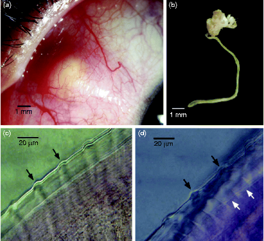
A 20-year-old man was admitted to Farabi Eye Hospital in Tehran with a yellowish conjunctival nodular lesion in his left eye. The lesion had been present for 1 year and caused a mild foreign body sensation with a minor conjunctival hyperaemia over the affected part of the eye. The patient lived in Qom (34.6456°N, 50.8798°E), 130 km south of Tehran, and had no recent history of travel to other cities or abroad. He had uncorrected visual acuity of 20/20 with normal intraocular pressure and eye movement in both eyes. Slit lamp examination showed a yellowish-white subconjunctival nodule of approximately 3 × 3 mm in the superonasal quadrant, 6 mm posterior to the limbus. The nodule was firmly attached to the underlying sclera and had a well-defined border (fig. 1a). Except for a mild conjunctival inflammation, no other abnormality was observed in the anterior or posterior chambers of the eye. The right eye was completely normal. Imaging with ultrasound biomicroscopy revealed a lesion with mixed hypo- and hyper-echogenic signal densities (data not shown). A battery of laboratory tests including complete blood count, erythrocyte sedimentation rate, C-reactive protein, angiotensin converting enzyme and tuberculosis skin test ruled out possible correlated diseases such as sarcoidosis and tuberculosis. Finally, the patient consented to explorative surgery. Initial dissection of the overlying conjunctiva near the lesion revealed a firm, immobile, yellow-white nodule attached to the sclera, tightly involving the superior part of insertion of the medial rectus muscle. Incision of the nodule revealed a long, coiled, white, thread-shaped, moving object that appeared to be a worm. The worm, measuring approximately 10 mm in length, was tightly attached to surrounding tissues. The nodule was promptly extracted and the worm was removed from it prior to photography (fig. 1b). There were two more nodules of almost the same size in the belly of the medial rectus muscle just posterior to the insertion. Fine dissection of one of them exposed another worm similar to the first. Further excision of the nodule was avoided, as there was risk of resection of the medial rectus muscle. Cryotherapy was applied to the remnants of two nodules and the undisturbed one, followed by application of mitomycin C 0.04% for 2 min and then irrigation with 100 ml balanced salt solution. The patient received amniotic membrane transplantation to cover the conjunctival defect. Three hours after surgery, he developed generalized urticaria with severe itching. Intravenous injection of a single dose (8 mg) of dexamethasone resulted in instantaneous resolution of the urticaria. On the first day after surgery, the patient had elevated eosinophil counts and IgE levels. Orbital magnetic resonance imaging with and without contrast revealed no significant abnormalities in the posterior chamber of the eye.
(colour online) Ocular onchocerciasis, showing (a) yellowish subconjunctival nodular lesion (mass) on the superonasal quadrant of the conjunctiva with hyperaemia over the affected part; (b) female Onchocerca lupi with the nodule attached; (c) thick and multilayered cuticle with prominent transverse annular ridges (black arrows) on the external surface of the worm; (d) thick and multilayered cuticle with prominent transverse annular ridges (black arrows) on the external surface and transverse striae on the inner layer (white arrows) of the worm.

Nematode identification
Some worm portions were cleared in lactophenol for 20 min, mounted, morphologically examined under a microscope at 400 × magnification and photographed. To identify the filarial worm molecularly, one worm was used for DNA extraction and the partial 12S ribosomal DNA gene was amplified as described by others (Morales-Hojas et al., Reference Morales-Hojas, Cheke and Post2006). The polymerase chain reaction (PCR) product was sequenced in both directions, using the same primer as that used for amplification, and compared with similar sequences available in the GenBank database using Basic Local Alignment Search Tool (BLAST) analysis. The sequence obtained in this study and similar sequences of other filarioids from the GenBank database were used to construct a phylogenetic tree, using the Jukes–Cantor option of the neighbour-joining method in MEGA 5 software (Tamura et al., Reference Tamura, Peterson, Peterson, Stecher, Nei and Kumar2011). The robustness of the topologies was estimated through 1000 bootstrap replications.
Results and discussion
Microscopic examination of the worm revealed a multilayered cuticle with typical prominent undulated ridges on the external layer (fig. 1c, d) and transverse striae on the internal layer (fig. 1d). Absence of striae (fig. 1c) might be due either to inappropriate preparation of tissue samples or lack of fine contrast adjustment. The distances between annular outer ridges were 38–40 μm on a segment of worm 210 μm wide, with two striae per ridge interval. These features are indicative of female worms of the genus Onchocerca.
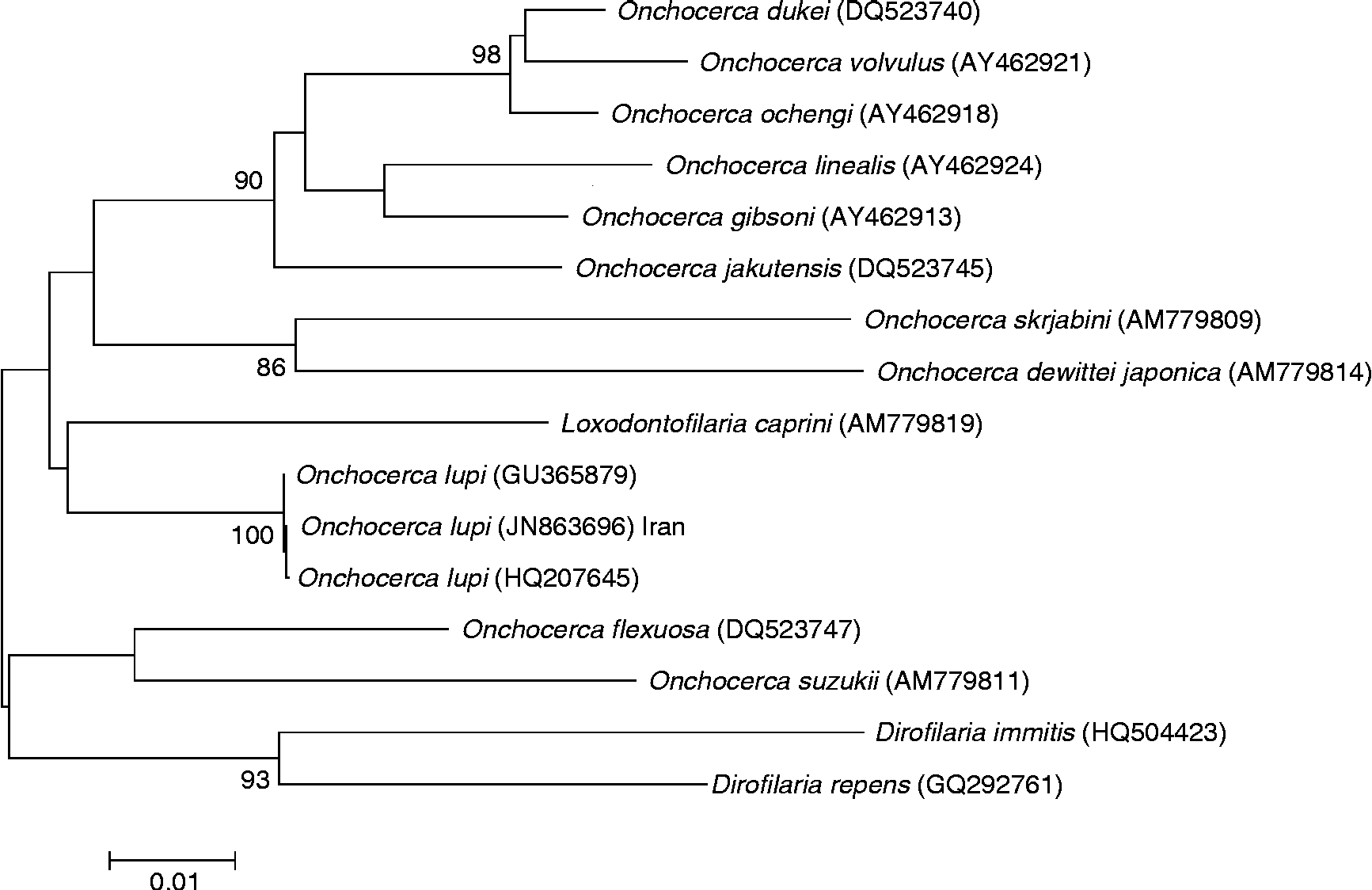
The 12S rRNA gene showed 99% homology with O. lupi of canine onchocerciasis from Portugal (GU365879), and O. lupi involved in human eye infection from Turkey (HQ207645). Phylogenetic analysis grouped the sequence obtained in this study alongside the two above-mentioned sequences, distinct from other species (fig. 2). The sequence data were deposited in GenBank with accession number JN863696.
Phylogenetic tree generated using the 12S rRNA sequence obtained in this study and similar sequences from other filarioids.
In this study we present an additional case of human ocular infection by O. lupi from Iran and for the first time report multiple nodules within the same eye. Multiple nodules in human cases of zoonotic onchocerciasis are very rare. The only documented report was from an O. jakutensis infested patient in Austria who had received long-term immunosuppressive therapy (Koehsler et al., Reference Koehsler, Soleiman, Aspock, Auer and Walochnik2007). To the best of our knowledge, there is no report on the occurrence of human or canine onchocerciasis from Iran. The only documented report available describes the presence of O. cervicalis microfilariae in Equidae from Tehran County (Mirzayans & Maghsoodloo, Reference Mirzayans and Maghsoodloo1977).
So far the information on blackfly vectors of zoonotic onchocerciasis has been very limited. In Japan, home to eight Onchocerca species (Fukuda et al., Reference Fukuda, Otsuka, Uni, Boda, Daisaku, Hasegawa, Takaoka and Bain2011), infective larvae of Onchocerca dewittei japonica and also Onchocerca sp. sensu Fukuda et al. (another parasite of wild boar) were identified molecularly in wild-caught females of Simulium bidentatum (Fukuda et al., Reference Fukuda, Otsuka, Uni, Bain and Takaoka2010a; Takaoka et al., Reference Takaoka, Fukuda, Otsuka, Aoki, Uni and Bain2012). In a separate survey, morphometric studies of infective larvae indicated that S. bidentatum alongside Simulium arakawae, Simulium daisense and Simulium oitanum transmit Onchocerca sp. (another bovine species) and S. arakawae, S. daisense and Simulium kyushuense transmit Onchocerca linealis in Japan (Fukuda et al., Reference Fukuda, Otsuka, Uni, Bain and Takaoka2010b; Takaoka et al., Reference Takaoka, Fukuda, Otsuka, Aoki, Uni and Bain2012). In an attempt to identify potential vectors of O. lupi in Xanthi, Greece, a rural area endemic for this parasite, three blackfly species (Simulium velutinum, Simulium reptans and Simulium pseudequinum) were collected and allowed to feed on two O. lupi infested dogs, but no developing larvae were detected in the dissected blackflies after 21 days (Otranto et al., Reference Otranto, Dantas-Torres, Papadopoulos, Petric, Cupina and Bain2012b). Some 18 blackfly species have been reported from the northern part of Iran; eight of them, including Simulium (Eusimulium) brachyantherum, S. (Montisimulium) assadovi, S. (Nevermannia) brevitarse, S. (Obuchovia) transcaspicum, S. (Simulium) bezzii, S. (Simulium) fontanum and S. (Simulium) variegatum, are from mountainous or hilly areas of Alborz in the north of Tehran Province (Crosskey, Reference Crosskey2002; Yankovsky, Reference Yankovsky2010), the nearest place with abundant potential vectors to Qom. Residents of Qom and Tehran usually commute between these two cities and commonly do not define this as travel. So, there are chances that our patient had contracted the infection in the north of Tehran. No record on blackflies from Qom is available yet. Qom is confined to the desert region of central Iran and swiftly flowing, well-aerated waters are rarely formed in this area. However, substantial marshes, slow-running streams and damp-soil habitats, which are major producers of many Culicoides species, the alternative vectors for Onchocerca spp., are commonly encountered in the suburbs of the city of Qom.
Our patient had lived for the past 2 years in the religious city of Qom where, according to Islamic codes, the proximity of dogs and humans is not common in town. However, stray dogs and sheepdogs are frequently seen in the suburbs of the city and in rural areas. It is unclear when the patient acquired the infection because, unlike the patient from Turkey (Otranto et al., Reference Otranto, Sakru, Testini, Gurlu, Yakar, Lia, Dantas-Torres and Bain2011), he did not recall any fly bites to his face. Interviewing the patient and his family about the time of appearance of the nodule indicated that he might have contracted the infection at least 1 year earlier.
Surgical removal of worms followed by cryotherapy proved to be useful and curative in this case. Generalized urticaria with itching after surgery could have resulted from the triggering of the patient's immune system following incision of nodules during surgery and exposure of in-nodule worm antigens to his systemic circulation.
Recently, human eye infection with Dirofilaria repens and Dirofilaria immitis were reported from Iran (Jamshidi et al., Reference Jamshidi, Jamshidi, Mobedi and Khosroara2008; Fallah Tafti et al., Reference Fallah Tafti, Hajilary, Siatiri, Rokni, Mobedi and Mowlavi2010; Ranjbar-Bahadori et al., Reference Ranjbar-Bahadori, Veshgini, Shirani, Eslami, Mohieddin, Shemshadi and Masooleh2011), and mosquitoes, Culex theileri and Anopheles maculipennis, were found to be naturally infected with third-stage larvae of D. immitis (Azari-Hamidian et al., Reference Azari-Hamidian, Yaghoobi-Ershadi, Javadian, Abai, Mobedi, Linton and Harbach2009). One explanation for the rarity of reports of human ocular infestation with O. lupi might be that mature or immature worms are incorporated in nodules and that their detection is only possible following dissection of the nodules, which may not occur in all cases of resection. Routine dissection of excised nodules or regular histopathology on such nodules should be performed in almost all cases. With reports of four cases of O. lupi induced ocular onchocerciasis from different geographical areas (two from Turkey, one from Tunisia and one from Iran) in less than 2 years, further studies on the zoogeographical distribution of canine onchocerciasis and the potential arthropod vectors in these areas may provide useful information on the epidemiology of this disease and the potential for control, or at least protection against the bites of the appropriate vectors.
Acknowledgements
We acknowledge Dr Elham Razmjoo (Tehran University of Medical Sciences) for her technical assistance in DNA extraction. We also thank Dr Dominico Otranto (University of Bari, Italy) and Dr Mohsen Abolhasani (Pasteur Institute of Iran) for assistance in the revision of the manuscript. This research received no specific grant from any funding agency, commercial or not-for-profit sectors.