Significant outcomes
-
∙ Exposure to an innate aversive stimulus activates transient receptor potential vanilloid type-1 (TRPV1)-positive neurons in the periaqueductal grey (PAG).
-
∙ Fear stimulus seems to recruit TRPV1 neurons mainly in the dorsolateral columns of the PAG.
-
∙ Blockade or TRPV1 channels in the dorsolateral PAG inhibit fear responses.
Limitations
-
∙ Although the present study focussed on the PAG, we did not discard the fact that predator exposure activates TRPV1-positive neurons in other brain regions related to innate defence as well.
-
∙ We did not investigate the effects of TRPV1 blockade on specific models of motor activity. However, a possible motor-impairing action can be discarded, as no effect was observed in capsazepine (CPZ)-treated animals exposed to the dummy cat.
-
∙ The possible endogenous substance responsible for activating TRPV1 channels in the PAG was not investigated.
Introduction
Defensive behaviours are evoked in mammals as a consequence of imminent threat such as the presence of a predator. These behaviours may vary in different species, but they tend to be hierarchically organised accordingly to the distance from the stimulus. As the stimulus approaches, the response may shift from risk assessment and immobility to escape and defensive attack (Reference Blanchard and Blanchard1–Reference Canteras and Blanchard3). In rats, the exposure to an imminent source of fear-provoking situation, such as a live cat, induces unconditioned alertness freezing, fight/flight reactions, analgesia and autonomic changes (Reference Blanchard, Blanchard, Rodgers and Weiss2,Reference Blanchard and Blanchard4). Distinct brain regions comprising the prefrontal cortex, medial amygdala, medial hypothalamic structures and the PAG are proposed to modulate these behaviours (Reference Beijamini and Guimaraes5–Reference Canteras and Goto7). The PAG is proposed as the final pathway of this network that controls active emotional coping strategies, and its chemical or electrical stimulation has been related to panic-like behaviours in animals (Reference Blanchard, Canteras, Markham, Pentkowski and Blanchard8–Reference Graeff10).
Several neurotransmitters have been proposed to modulate defensive behaviours, including glutamate, serotonin, GABA, neuropeptides and nitric oxide (Reference Adamec, Burton, Shallow and Budgell11–Reference Aguiar and Guimaraes15), and more recently the TRPV1 (Reference Di Marzo, Bisogno and De Petrocellis16). This cation-permeable channel was initially identified in primary afferent neurons as a mediator of painful stimulus, sensitive to reduction in pH, temperature and capsaicin (Reference Holzer17). Later on, TRPV1 expression was also detected in the central nervous system including the dorsal horn of the spinal cord (Reference Acs, Palkovits and Blumberg18,Reference Szallasi and Blumberg19), hindbrain, midbrain and forebrain (Reference Toth, Boczan and Kedei20). The expressions of these receptors in regions such as the amygdala, hippocampus and the PAG suggest a role in the control of emotional states. Its putative endogenous agonists are lipid-derived compounds, referred to as endovanilloid, some of which are also cannabinoid CB1 receptor agonists, thus termed endocannabinoids. They include anandamide (N-arachidonoyl ethanolamide, a major endocannabinoid), N-arachidonoyl dopamine, N-oleoyldopamine and some products of lipoxygenase enzymes (Reference Toth, Kedei, Wang and Blumberg21–Reference Hwang, Cho and Kwak24).
Supporting the role of TRPV1 channels in aversive responses, systemic injection of the antagonist CPZ in rats induced anxiolytic-like effects in the elevated plus maze (EPM) (Reference Kasckow, Mulchahey and Geracioti25). Accordingly, reduced anxiety-like behaviour and conditioned fear were observed in TRPV1-knockout mice (Reference Marsch, Foeller and Rammes26). Moreover, antagonists targeting this ion channel induced anxiolytic-like effects when injected into the ventral medial pre-frontal cortex (vmPFC), ventrolateral hippocampus and dorsolateral PAG (dlPAG) (Reference Aguiar, Terzian, Guimaraes and Moreira27–Reference Terzian, Aguiar, Guimaraes and Moreira29), whereas intra-vmPFC injection of the TRPV1 agonist capsaicin induced anxiogenic-like behaviour in rodents submitted to the EPM (Reference Rubino, Realini and Castiglioni30).
Altogether, these results show that TRPV1 blockade favours anxiolytic-like responses in conflict-like situations. The dorsal PAG, however, is also a crucial site in the control of behavioural response to unconditioned fear stimuli under more aversive situations, such as the flight/freezing responses induced by predators (Reference Canteras and Blanchard3,Reference McNaughton and Corr9). Moreover, stimulation of this region has been closely related to panic reactions in humans (Reference Graeff10). Finally, local injection of anandamide inhibits predator-induced panic-like responses in rats, possibly though the CB1 receptor activation (Reference Lisboa, Camargo, Magesto, Resstel and Guimaraes31). The objective of the present study, therefore, was to investigate whether TRPV1, located in the dlPAG, is involved in the defensive responses of rats exposed to a live cat. We performed histological experiments to detect Fos expression in TRPV1-positive neurons and injected a TRPV1 antagonist into this midbrain structure to reverse the behavioural response of rats exposed to a cat.
Material and methods
Subjects
The subjects were adult male Wistar rats (220–240 g) obtained from the colony of pathogen-free rats maintained by the Pharmacy School of Ribeirão Preto, University of Sao Paulo. The animals were housed in groups of four with free access to food and water in a temperature-controlled room (24°C) with a 12 h light/dark cycle. An adult male cat (3 kg), kept at the animal farm of our University Campus with free access to food and water, was used throughout the study. A dummy cat of approximately the same size of the live cat was used as a control. The authors assert that all procedures contributing to this work comply with the ethical standards of the relevant national and institutional committees on human experimentation and with the Helsinki Declaration of 1975, as revised in 2008, and assert that all procedures contributing to this work comply with the ethical standards of the relevant national and institutional guides on the care and use of laboratory animals. The experiments were carried out according to the Brazilian Society of Neuroscience and Behaviour guidelines for care and use of laboratory animals and all efforts were made to minimise animal suffering. The experiment protocol was approved by the local Ethics Committee (083/2008).
Drugs
The TRPV1 antagonist CPZ (60 nmol/0.2 µl; Tocris, Bristol, UK) was dissolved in DMSO. This dose was chosen based on previous results obtained after intra-dlPAG administration (Reference Terzian, Aguiar, Guimaraes and Moreira29,Reference Cassaroto, Terzian and Aguiar32).
Apparatus
The observation box consisted of a rectangular arena (80 cm×22 cm×50 cm) with Plexiglas walls and a metal grid floor. The apparatus was placed in a sound-attenuated 40 lx illuminated room. It was divided into two compartments by a metal grid wall. During the experimental session, each rat was placed in the middle of the compartment opposite to the one containing the live or dummy cat, facing the latter compartment. The box was designed to comfortably contain the cat and to provide space enough for measuring the presence of the rat proximal or distal to the cat compartment. The rat compartment was divided into two equal parts (near and distant to the cat compartment) by an imaginary line. The rational is that when the live cat was not present, the rats have no preference for any part of the box, whereas they escape to the cat distant part and tend to remain there when the predator is present (Reference Moreira and Guimaraes14,Reference Aguiar and Guimaraes15).
Surgery and intra-dlPAG injection
The rats were anaesthetised with 2.5% 2,2,2-tribromoethanol (Sigma–Aldrich, St. Louis, MO, USA, 10 ml/kg, IP), placed in a stereotaxic frame (David Kopf, Tujunga, CA, USA) and unilaterally implanted with a stainless-steel guide cannula (11 mm, length) aimed at the dlPAG (co-ordinates: anterior–posterior (AP)=0 from lambda, L=1.9 mm, D=4.0 mm below the surface of the skull) (Reference Paxinos and Watson33). The cannula was attached to the bones with stainless-steel screws and acrylic cement. An obturator inside the guide cannula prevented obstruction.
Seven days after surgery, intra-dlPAG injections were administered with a thin dental needle (0.3 mm, o.d.) introduced through the guide cannula until its tip was 1 mm below the cannula end. A volume of 0.2 µl was injected for 30 s using an infusion pump (KD Scientific Inc., Holliston, MA, USA). The movement of an air bubble inside the PE 10 polyethylene tubing connecting the pump to the dental needle confirmed drug flow. The drugs were injected 10 min before the exposure to the observation box.
Behavioural procedure
Experiment 1
In this first experiment, we used immunohistochemistry to verify whether threatening stimuli would activate TRPV1-expressing neurons. The animals were divided into three experimental groups as follows: (i) naive, (ii) exposure to the dummy cat and (iii) exposure to the live cat. During 3 consecutive days, the animals were handled daily by the experimenter for 5 min and were habituated to the observation box for 10 min. On the 4th day, the animals were placed in the observation box and exposed to the dummy or the live cat for 10 min. After each trial, the observation box was carefully cleaned with an alcohol solution. To prevent eventual cat smell interference, exposure to the live cat always followed that of the dummy cat. The animals from the naive group remained undisturbed in their home cages until the perfusion for immunofluorescence assay.
Experiment 2
In this experiment, we verified whether TRPV1 antagonism would attenuate the behavioural responses promoted by predator exposure. The animals were divided into the following four experimental groups: (i) vehicle+dummy cat; (ii) CPZ+dummy cat; (iii) vehicle+cat; and (IV) CPZ+cat. The procedure was similar to the first experiment, except that the animals received intra-dlPAG injections of the vehicle or CPZ and were placed into the observation box 10 min later.
All sessions were videotaped and later analysed with the help of ANY-MAZE video-tracking system software (Version 4.73, Stoelting Co., USA). The program detected the position of the animal in the observation box and calculated the time spent and the distance moved in the area near the cat compartment.
Histology
After the behavioural tests, rats from experiment 2 were killed under deep urethane (Sigma–Aldrich, 5 ml/kg, IP) anaesthesia and perfused through the left ventricle of the heart with isotonic saline followed by 10% formalin solution. Subsequently, a dental needle was inserted through the guide cannula and 0.2 μl of fast-green was injected. The brains were removed and, after a minimum period of 3 days, immersed in a 10% formalin solution, and 50-μm sections were obtained in a Cryostat (Cryocut 1800, Reichert-Jung/Leyca, Milton Keynes, UK). The injection sites were identified with the diagrams from the Paxinos and Watson’s atlas (Reference Paxinos and Watson34). Rats that received injections outside the aimed area were excluded from the analysis.
c-Fos and TRPV1 double immunofluorescence
Two hours after exposure to the observation box, the animals from experiment 1 were anaesthetised with an overdose of urethane and perfused transcardially with saline followed by 4% paraformaldehyde in 0.1 M phosphate buffer saline (PBS, pH 7.4). Brains were removed and post-fixed over 2 h in paraformaldehyde and stored for at least 30 h in 30% sucrose for cryoprotection. Coronal sections (25 μm) were obtained in duplicate in a cryostat. Subsequently, the sections were incubated in glycine (0.1 M), washed again and incubated for 1 h in 5% bovine serum albumin (BSA; Sigma–Aldrich) in PBS (0.01 M, pH 7.4) containing 0.4% Triton X-100. Thereafter, the sections were incubated for 2 days at 4°C in a mixture of anti-cFos (1 : 3000; Santa Cruz, Dallas, TX, USA; raised in rabbit) and anti-TRPV1 receptor N-terminus (1 : 100; Santa Cruz raised in goats) diluted in PBS (0.01 M) containing 5% BSA and 0.4% Triton X-100. After incubation in the primary antiserum, the tissue sections were washed in PBS and sequentially incubated in a mixture of Alexa 594 donkey anti-rabbit IgGs (1 : 1000; Molecular Probes, Eugene, OR, USA) and Alexa 488 donkey anti-goat IgGs (1 : 1000; Molecular Probes) for 1 h. Slides were rinsed in PBS and coverslipped with Fluormount-G (Electron Microscopy Sciences, Hatfield, PA, USA). Images were analysed using the Olympus BX50 microscope through a computerised image system (Image Pro-Plus 4.0, Media Cybernetics) and captured using a digital camera DXC-107A (Sony, Tokyo, Japan).
Statistical analysis
The identification method of double-stained cells was similar to that described in previous works (Reference Aguiar and Guimaraes15,Reference Beijamini and Guimaraes35,Reference De Oliveira, Del Bel and Guimaraes36). The number of Fos-like (FLI) and TRPV1 immunoreactive (TRPV1IR) and doubled-stained cells was manually counted with the help of a computerised image analysis system Image J 1.42q (Wayne Rasband, National Institutes of Health, USA). An observer blinded to the group assignment performed the analysis. For each group, one section from each animal was evaluated. All the stained cells in the whole area of each brain region of interest were recorded. Double-stained cells were represented as percentage of TRPV1IR cells. Neuroanatomical sites were identified with the help of the Paxinos and Watson’s atlas (Reference Paxinos and Watson34). The AP localisation from the bregma of the analysed regions were as follows: rostral portion containing the dlPAG, dorsomedial (dmPAG) and ventrolateral (vlPAG) PAG was represented by level −6.96 mm, the intermediate portion containing the dlPAG, dmPAG and vlPAG was represented by level −7.44 mm and the caudal portion containing all the sub-regions was represented by level −7.92 mm.
The behavioural data were analysed by two-way ANOVA, and histological data were analysed by ANOVA. In case of significance, the post-hoc Duncan test was applied. The significance level was set at p<0.05.
Results
Experiment 1
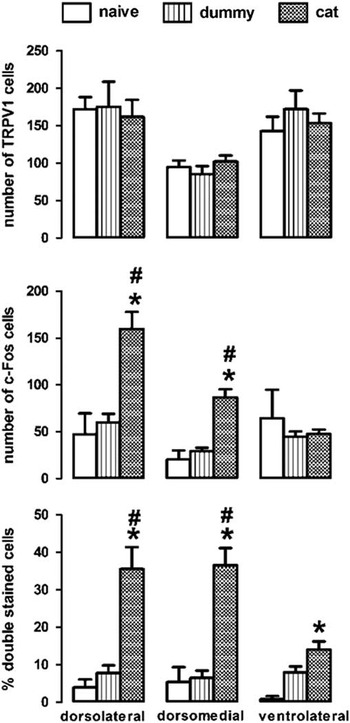
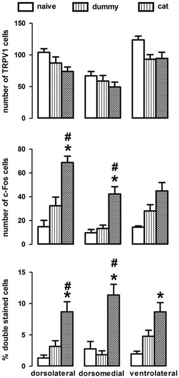
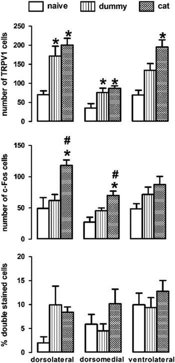
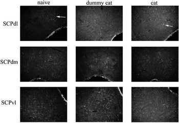
The immunofluorescence results are presented in Figs 1–3 and a representative photomicrography in Fig. 4. Exposure to the live cat induced a significant increase in Fos-like (FLI) cells compared with both naive and dummy cat groups in the following portions of the PAG: (i) rostral: dlPAG: F(2,23)=13.86, p=0.0001; dmPAG: F(2,24)=21.76, p=0.0001 (Fig. 1); (ii) medial: dlPAG: F(2,25)=16.02, p=0.0001; dmPAG: F(2,25)=11.99; p=0.002 (Fig. 2); and (iii) caudal: dlPAG: F(2,23)=12.08, p=0.0001; dmPAG: F(2,24)=8.74; p=0.001 (Fig. 3). In the vlPAG, there was a significant difference compared with naive group only in the medial portion (vlPAG: F(2,24)=4.19; p=0.02; Fig. 4).
Number of transient receptor potential vanilloid type-1 (TRPV1)-positive [TRPV1 immunoreactive (TRPV1IR); a], c-Fos-positive [Fos-like (FLI); b], and percentage of double-stained cells (c) in the rostral portion of the periaqueductal grey (PAG) of rats exposed to a live (n=11–12) or a toy cat (n=10–11). Naive animals (n=4–5) remained undisturbed in their home cages. Data are represented as mean (±SEM). FLI and TRPV1IR are expressed as the number of positive cells in each region. Double-stained cells are expressed as percentage of TRPV1IR cells (*p<0.05 compared with the naive group and #p<0.05 compared with the dummy cat group. ANOVA followed by Duncan test).

Number of transient receptor potential vanilloid type-1 immunoreactive (TRPV1IR; a), Fos-like (FLI; b) and double-stained cells (c) in the medial portion of the periaqueductal grey (PAG) of rats exposed to a live (n=11–12) or a toy cat (n=10–11). Naive animals (n=4–5) remained undisturbed in their home cages. Data are represented as mean (±SEM). FLI and TRPV1IR are expressed as the number of positive cells in each region. Double-stained cells are expressed as percentage of TRPV1IR cells (*p<0.05 compared with the naive group and #p<0.05 compared with the dummy cat group. ANOVA followed by Duncan test).

Number of transient receptor potential vanilloid type-1 immunoreactive (TRPV1IR; a), Fos-like (FLI; b) and double-stained cells (c) in the caudal portion of the periaqueductal grey (PAG) of rats exposed to a live (n=11–12) or a toy cat (n=10–11). Naive animals (n=4–5) remained undisturbed in their home cages. Data are represented as mean (±SEM). FLI and TRPV1IR are expressed as the number of positive cells in each region. Double-stained cells are expressed as percentage of TRPV1IR cells (*p<0.05 compared with the naive group and #p<0.05 compared with the dummy cat group. ANOVA followed by Duncan test).

Photomicrographs showing c-Fos (red arrow, Alexa Fluor 594) and transient receptor potential vanilloid type-1 (TRPV1; white arrow, Alexa Fluor 488) immunofluorescence in the different columns of the medial portion of the periaqueductal grey (PAG) in rats exposed to a dummy or live cat or naive rats. The yellow arrow indicates double-stained neurons. Results obtained in 25-µm-thick sections. Scale bar: 100 µm.

Predator exposure induced a significant increase in the percentage of double-stained cells as compared with naive and dummy cat groups in the in the rostral portion (dlPAG: F(2,23)=12.36, p=0.0001; dmPAG: F(2,24)=21.75, p=0.0001, Fig. 1) and the medial portion (dlPAG: F(2,25)=3.64; p=0.04; dmPAG: F(2,25)=8.02; p=0.002, Fig. 2). In this parameter, the vlPAG had significant differences in the cat-exposed group compared with the naive group in the rostral portion (vlPAG: F(2,25)=7.66; p=0.003, Fig. 3) and the medial portion (vlPAG: F(2,24)=5.43; p=0.01, Fig. 4). No differences for double-stained cells in the caudal portion were found in any columns of the PAG.
In addition, there was no difference in the number of TRPV1-positive cells (TRPV1IR) in the rostral and medial portion of all columns of the PAG. In the caudal portion of the PAG, there was a significant increase in TRPV1IR cells [dlPAG: F(2,24)=5.68, p=0.01; dmPAG: F(2,24)=4.41, p=0.02; vlPAG: F(2,24)=4.97; p=0.016, Fig. 3] in the dummy- and cat-exposed animals compared with naive group.
Experiment 2
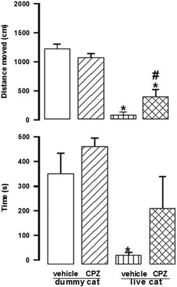
Cat exposure induced a significant fear reaction reflected by a reduction in the distance moved in the compartment near the cat [exposure factor: F(1,14)=16.64, p<0.05; drug factor: F(1,14)=4.86, p<0.05; exposure×drug: F(1,14)=5.76, p<0.05, Fig. 5] and time spent in this compartment [exposure factor: F(1,14)=21.2, p<0.05; drug factor F(1,14)=9.31, p<0.05; exposure×drug: F(1,14)=5.42, p<0.05, Fig. 5], as compared with animals exposed to the dummy cat. Intra-dlPAG administration of CPZ (60 nmol) significantly attenuated the fear behaviour displayed by the presence of the cat, increasing the distance moved and the time spent in the compartment near the cat (Duncan test; p<0.05, as compared with vehicle/cat group).
Effects of capsazepine (CPZ, 60 nmol/0.2 µl) injected into the dorsolateral periaqueductal grey (dlPAG) on the distance moved (a) and time spent (b) in the compartment near the cat after exposure to a live cat for 10 min. Data are represented as means±SEM of 4–5 animals per group (*p<0.05 compared with the dummy cat group and #p<0.05 compared with the live cat group, two-way ANOVA followed by Duncan test).

Discussion
The present study showed that exposure of rats to a live cat promotes an increase in Fos expression in all the subdivisions of the PAG, supporting the proposal that this structure mediates behavioural reactions to proximal threatening stimuli. In addition, exposure to the predator recruited neurons expressing TRPV1 in this region, as revealed by Fos and TRPV1 double staining. This observation is supported by a behavioural experiment, in which local injection of CPZ, a TRPV1 antagonist, inhibited fear reaction.
Fos expression in the PAG was more prominent in the dorsal columns (dlPAG and dmPAG). This region is proposed as the final pathway in the control of active emotional coping strategies (Reference Blanchard, Canteras, Markham, Pentkowski and Blanchard8,Reference McNaughton and Corr9). Interestingly, the vlPAG was activated only in the medial portion, in agreement with other studies showing that this area is preferentially involved in response to more subtle stimuli (Reference Beijamini and Guimaraes5–Reference Blanchard, Canteras, Markham, Pentkowski and Blanchard8,Reference Dielenberg, Hunt and McGregor37). On the contrary, the dorsolateral column (dlPAG) seems to co-ordinate defensive behaviours such as freezing, flight and fight in situations of intense threat such as the presence of a predator (Reference Canteras and Blanchard3,Reference Blanchard and Blanchard4). In line with this view, the dlPAG is activated by different stress stimuli including immobilisation (Reference Cullinan, Herman, Battaglia, Akil and Watson38), exposure to a novel open field (Reference Nagahara and Handa39), foot shocks (Reference Campeau, Falls, Cullinan, Helmreich, Davis and Watson40) and audiogenic stress (Reference Campeau, Akil and Watson41). These results are also in agreement with previous studies describing more intense Fos activation in the dorsal portions of the PAG after exposure to the predator or its odour (Reference Canteras and Goto7,Reference Dielenberg, Hunt and McGregor37) or administration of anxiogenic drugs (Reference Singewald and Sharp42). Cat exposure also induced a significant increase in the percentage of double-stained cells in the rostral and medial regions (dlPAG and dmPAG subdivisions), suggesting that TRPV1-containing receptors are recruited in these regions during situations of extreme fear.
Supporting our initial hypothesis, microinjection of CPZ into the dlPAG reduced defensive behaviours induced by predator exposure. This result indicates that, in addition to conflict-based animal models of anxiety (Reference Terzian, Aguiar, Guimaraes and Moreira29,Reference Casarotto, Terzian and Aguiar43), TRPV1 activation in the dlPAG is also important for regulating aversive states induced by innate fear stimuli. Similar results have been observed after local injection of NMDA antagonist and nitric oxide synthase inhibitors (Reference Aguiar and Guimaraes15). The role of TRPV1 channels in modulating defence responses to a predator is diametrically opposite to that proposed for the cannabinoid CB1 receptor, as local injection of cannabinoids into the PAG also reduced predator-induced fear (Reference Lisboa, Camargo, Magesto, Resstel and Guimaraes31). This is in line with the hypothesis that TRPV1 and CB1 receptors constitute a set-point system that equilibrates defensive responses. They would mediate the opposing actions of a common endogenous agonist, possibly anandamide, to select the more appropriate coping strategy according to the nature and intensity of the stimulus (Reference Moreira, Aguiar, Terzian, Guimaraes and Wotjak44).
The effects obtained with this TRPV1 antagonist also suggest a tonic activation of TRPV1 under basal conditions. This is in agreement with previous studies, which reported decreased anxiety after pharmacological or genetic interruption of TRPV1 signalling (Reference Micale, Di Marzo, Sulcova, Wotjak and Drago45). For instance, systemic injection of CPZ reduced anxiety-like behaviour in rats (Reference Kasckow, Mulchahey and Geracioti25). Moreover, a detailed investigation on the behaviour of TRPV1 knock-out mice unveiled a phenotype of reduced anxiety-like behaviour as well as reduced fear conditioning (Reference Marsch, Foeller and Rammes26). Furthermore, the effect of systemically administered CPZ is mimicked by local injection into brain structures that regulate fear-related responses, namely the ventral hippocampus (Reference Santos, Stern and Bertoglio28), the dlPAG (Reference Terzian, Aguiar, Guimaraes and Moreira29,Reference Casarotto, Terzian and Aguiar43) and the prefrontal cortex (Reference Aguiar, Terzian, Guimaraes and Moreira27,Reference Rubino, Realini and Castiglioni30). This is consonant with neurochemical and electrophysiological data showing that this ion channel stimulates calcium influx and glutamate release in the PAG, increasing local neuronal activity (Reference Li, Mao, Zhang, Yang, Cao and Xu46,Reference Starowicz, Nigam and Di Marzo47). In addition, it has been demonstrated in other brain regions related to anxiety that anandamide, acting through TRPV1, modulates long-term depression of synaptic activity (Reference Chavez, Chiu and Castillo48,Reference Grueter, Brasnjo and Malenka49). Although it remains unclear how these electrophysiological events would reflect behavioural responses, these studies suggest that anandamide could indeed be the endogenous substance activating this ion channel (Reference Chavez, Chiu and Castillo48–Reference Di Marzo50). Other lipids including N-arachidonoyl dopamine, N-oleoyldopamine and some lypo-oxigenase products have also been proposed as main candidates to act as endovanilloids (Reference Di Marzo, Bisogno and De Petrocellis16,Reference Starowicz, Nigam and Di Marzo47).
Conclusion
In conclusion, our results suggest that TRPV1-mediated neurotransmission in the dlPAG plays an important role in defensive responses elicited by predator exposure.
Acknowledgements
D.C.A. performed most of the research, analysed data and wrote the initial version of the paper; A.F.A.S. performed histological experiments and contributed to data analysis; F.A.M. participated in the data analysis and contributed to write the paper; and F.S.G. participated in the design of the study, data analysis and writing the paper. The authors thank J.C. de Aguiar and E.T. Gomes for the excellent technical support.
Financial Support
This research was supported by grants from CNPq, FAPESP (2012/17626-7) and FAPEMIG (APQ-01611-13, PRONEM; APQ-04625-10).
Conflicts of Interest
The authors report no conflicts of interest. The authors alone are responsible for the content and writing of the paper.
Ethical Standards
The authors assert that all procedures contributing to this work comply with the ethical standards of the relevant national and institutional committees on human experimentation and with the Helsinki Declaration of 1975, as revised in 2008, and assert that all procedures contributing to this work comply with the ethical standards of the relevant national and institutional guides on the care and use of laboratory animals.