Implications
The results of this study indicate that selenium source can have a profound effect on the distribution of total selenium and selenium species within the tissues of turkeys. However, higher muscle tissue total selenium contents do not necessarily result in improved meat keeping qualities. This would suggest that selenium sources with superior bioavailability, although not necessarily improving meat quality would provide additional dietary selenium for consumers of animal products.
Introduction
Much of the early work into selenium (Se) was centred on the toxic and detrimental effects that high concentrations of dietary Se had on animal health (Underwood and Suttle, Reference Underwood and Suttle2001). However, during the latter 1950s, it was discovered that Se played an important physiological role in higher animals, as suboptimal levels often resulted in the appearance of myopathies (Schubert et al., Reference Schubert, Muth, Oldfield and Remmert1961). In the early part of the 1970s, a specific biological role for Se became apparent with the discovery of the first selenoprotein glutathione peroxidase (GSH-Px; Rotruck et al., Reference Rotruck, Pope, Ganther, Swanson, Hafeman and Hoekestra1973). GSH-Px catalyses the reduction of lipid and hydrogen peroxides to less harmful hydroxides via the oxidation and subsequent reduction of the selenocysteine (SeCys) active centre of the enzyme (Surai, Reference Surai2006).
The anti-oxidant functions of Se, via the actions of tissue GSH-Px, have been shown to persist post mortem in poultry muscle tissue and to delay the onset of oxidation reactions (DeVore et al., Reference DeVore, Colnago, Jensen and Greene1983). The expression and subsequent activity of all selenoenzymes is dependent upon adequate Se supply. Furthermore, Se within an organism is not evenly distributed as there is a hierarchy of selenoenzymes. Consequently when Se is limiting, there maybe full expression of some selenoenzymes and restricted expression of others (Burke et al., Reference Burke, Hill and Xia2005).
The uptake and assimilation of Se are dependent upon its form; inorganic sources such as sodium selenite (SS) are absorbed via passive diffusion and are either incorporated into selenoenzymes or excreted, whereas organic forms such as selenoyeasts (SY), in which the predominant form of Se is selenomethionine (SeMet), are taken up via methionine transporter mechanisms and can be either utilized for incorporation into selenoenzymes or incorporated non-specifically in place of methionine into general body proteins (Suzuki and Ogra, Reference Suzuki and Ogra2002; Weiss, Reference Weiss2003). Consequently, increases in the Se content of muscle tissue as a result of the increased incorporation of SeMet, may provide additional dietary Se for the consumer of animal products, but may not necessarily result in improvements in meat quality or oxidative stability.
The objective of this study was to determine tissue Se distribution, erythrocyte and tissue GSH-Px activity and post-mortem meat quality in growing female turkeys fed diets containing Se as SS or SY at different doses.
Material and methods
The work was conducted in accordance with the UK Animals (Scientific procedures) Act 1986 (Home Office, 1986) and procedures were undertaken by competent technical staff holding appropriate license authorities under the Act. All birds were housed with sufficient space as prescribed by Home Office (1986) guidelines. Fresh potable water was available at all times.
A total of 250 female BUT8 turkey poults (High Line Turkeys, Salop, UK) were brooded and kept according to breeder specifications (British United Turkeys, 2002). At 6 days of age, all poults were weighed to provide data for experimental blocking. All poults were reweighed at 7 days of age (T0) and a total of 216 poults were selected on the basis of live weight (mean body weight 149.5 ± 1.3 g). Of these 216 selected birds, 24 were randomly selected to form a pre-treatment slaughter group. These birds were euthanized at T0 by cervical dislocation and samples of blood were taken post mortem. In addition, samples of breast muscle (m. pectoralis major [PM]), muscle (m. peroneus longus [PL] and m. gastrocnemius [MG]), heart and liver were retrieved from six randomly selected birds for determination of pre-treatment Se concentrations. All remaining poults were blocked by live weight and randomly allocated to one of four dietary treatments; an unsupplemented control (Con), supplemented with 0.08 or 0.23 mg S/kg dry matter (DM) of SY (SY-L and SY-H, respectively; Sel-Plex, Alltech, Nicholasville, KY, USA) or 0.08 mg/kg DM of an inorganic selenium source (SS-L), to achieve final total Se concentrations of 0.30, 0.45 and 0.30 mg/kg DM, respectively. All diets were formulated to meet the nutritional requirements of growing BUT8 poults (British United Turkeys, 2002)
Poults were housed in groups of eight birds/pen with six pens per treatment. Poults remained within established pen groups throughout the study. Pens were bedded with white wood shavings. Each pen was equipped with one bell type automatic drinker and one plastic poultry tube feeder with a feeding circumference of 60 cm. Birds were offered the experimental starter diet, with respect to treatment designation, for the first 21 days of the study after which a grower-finisher experimental diet, with respect to treatment designation, was introduced and offered for the remainder of the study.
Diets and feed intake
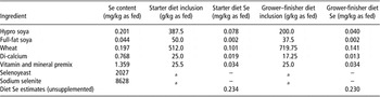
Diets were formulated and manufactured by Dairy Direct (Mill Feed Services, Anglia, UK). Before manufacture, the Se content of ingredients was determined (UT2A laboratories, Pau, France; Table 1). The quantities of either SY or SS supplements required to achieve target doses with respect to treatment were calculated and mineral supplements were subsequently manufactured. Following manufacture, representative samples of each mineral supplement were analysed to confirm the correct level of Se.
Constituent ingredients, inclusion rates, selenium contents and estimated selenium contents of starter and grower–finisher diets

aDetermined by experimental protocol.
Vitamin and mineral premix composition: calcium pantothenate 400 mg/kg, riboflavin (B2) 200 mg/kg, vitamin B12 320 μg/kg, thiamin (B1) 40.0 mg/kg, pyridoxine (B6) 40.0 mg/kg, biotin 2.0 mg/kg, cobalt (cobalt carbonate) 10 mg/kg, iodine (as sulphate) 40 mg/kg, copper (as sulphate) 800 mg/kg, iron (as sulphate) 400 mg/kg, manganese (manganous oxide) 3200 mg/kg, zinc (zinc oxide) 2400 mg/kg, choline chloride 1740 mg/kg, phosphorus 140.6 g/kg, vitamin A 400 000 IU/kg, vitamin D3 120 000 IU/kg, vitamin E 800 mg/kg, vitamin K 80 mg/kg, folic acid 40 mg/kg, niacin 800 mg/kg.
Poults received ad-libitum access to their respective diets throughout the study. Feed offered and refused were weighed and recorded daily for each pen. Weekly samples of each diet were stored at −20°C for subsequent laboratory determination of DM, crude protein (CP), starch, neutral detergent fibre, total sugars, oil ash (Euorofins, Wolverhampton, UK) and Se (UT2A laboratories, Pau, France).
Blood and tissue sampling
Following 42 days of exposure to experimental diets (T42), four poults were randomly selected from each pen (n = 24 birds/treatment) and euthanized by cervical dislocation and samples of blood, breast, thigh, gizzard, kidney and liver were retrieved for determination of total Se concentrations. Following an additional 42 days of exposure (T84) to the experimental diets, all remaining poults (n = 24 birds/treatment) were euthanized by electrical stunning (Wells Poultry, Gwent, UK) followed by exsanguination and samples of breast, thigh, gizzard, kidney and liver were retrieved for determination of total Se concentrations.
At each time point (T42 and T84), blood was collected following exsanguination into three pre-treated lithium heparin tubes/bird (Sarstedt, Leicester, UK). One tube was sent for determination of whole blood GSH-Px activity (Veterinary Laboratories Agency, Shropshire, UK), one for determination of Se content (UT2A, Pau, France) and the third was centrifuged at 1252 × g for 10 min at room temperature in a bench top centrifuge (Weiss-Gallenkamp, Loughborough, UK), after which the plasma fraction was decanted off and sent for determination of Se content (UT2A, Pau, France).
Following slaughter at T42, one bird per pen (six birds/treatment) was randomly selected and dissected. The ventral aspect of the carcass was skinned, and a sample of breast tissue (PM) from the left side of the carcass was retained for Se analysis. The musculature of the left hand thigh was exposed and samples of muscle tissue (PL and MG) were retained for Se analysis. The heart, liver, kidneys and gizzard were removed from the body cavity and retained for Se analysis.
At the T84 slaughter point, the remaining 24 birds/treatment were plucked and dressed and one bird per pen (six birds/treatment) was randomly selected and underwent dissection as previously described. In addition, the right-hand breast and thigh were removed and skinned from 12 birds/treatment, packaged in a zip-lock bag, refrigerated and transported in a cool box on the day of slaughter to the University of Bristol for meat quality assessment (pH, drip loss, thiobarbituric acid reactive substances (TBAR) and tissue GSH-Px activity).
Selenium analyses
Total Se in feeding stuffs, whole blood samples, plasma and tissues was determined according to the method of Mester et al. (Reference Mester, Willie, Sturgeon, Caruso, Fernandez, Fodor, Goldschmidt, Goenaga-Infante, Lobinski and Wolf2006). Briefly, 1 g of each sample was mineralized in 4 ml of 16 M HNO3 and 2 ml of 9.8 M H2O2 within a closed-vessel heating block system. The solution was further diluted with water and Se was subsequently determined using inductively coupled plasma mass spectrometry (ICP-MS; Perkin Elmer Elan 6100 ICPMS, Waltham, MA, USA).
The selenized amino acid (AA) contents of blood were determined by speciation using the method of Bierla et al. (Reference Bierla, Vacchina, Szpunar, Bertin and Lobinski2008a). Samples were initially incubated for 5 h with dl-dithiothreitol and iodoacetamide to reduce and alkylate SeCys. Samples were then spiked with SeMet77 and subsequently incubated for 24 h at 37°C with a mixture of protease and lipase maintained at pH 7.5. Following incubation, the mixture was centrifuged and the supernatant was separated and purified by size-exclusion liquid chromatography. Aliquots of the supernatant were analysed by reversed-phase HPLC using an ICP-MS equipped with a collision cell (Perkin Elmer Elan 6100 ICP-MS, Waltham, MA, USA).
The selenized AA content of tissues (breast, thigh, liver and kidney) was determined by speciation according to the method of Bierla et al. (Reference Bierla, Vacchina, Szpunar, Bertin and Lobinski2008b). Samples were mixed and sieved after which a representative sub-sample was taken. Urea was added to the sub-sample and the sub-sample was sonicated after which it was reduced, alkylated and submitted to proteolysis. The extract was then purified by size-exclusion chromatography and the AAs were quantified by reverse-phase HPLC-ICP-MS.
GSH-Px activity in whole blood was determined using the Olympus AU400 Chemistry Analyzer (Olympus UK, Watford, UK) based on the method of Anderson et al. (Reference Anderson, Berrett and Patterson1978). Results are presented as units per ml.
Meat quality assessment
Skeletal muscle tissue pH was determined in breast tissue at 24 h post mortem by inserting a Testo 230 pH probe (Testo Ltd, Alton, Hants, UK) directly into the muscle tissue. A 20 mm-thick section of tissue was cut from the broadest end of the breast and used for drip loss determination by the method of Honikel (Reference Honikel1998). Briefly, all exterior fat and connective tissues were removed from the tissue section without cutting into the muscle. The tissue section was weighed and suspended covered in plastic netting inside an inflated plastic bag, which was in turn suspended for 48 h at 3°C. The tissue section was reweighed and drip loss was calculated by difference.
The thigh was cut into three 20 mm sections and individually packed into modified atmosphere packs (MAP; O2 : CO2, 75 : 25) and subjected to simulated retail display (3°C, 700 lux for 16 h out of 24 h). Colour (lightness [L*], redness/greenness [a*], yellowness/blueness [b*]) was measured daily on the surface of two of the steaks through the lidding film using a Minolta Chromameter CR200 (Minolta Camera Company, Milton Keynes, UK). Colour chroma (saturation) was calculated from the formula: [a 2 + b 2]0.5. Remaining thigh steak and breast steak were taken for analysis of lipid oxidation as TBARS on the 5th day of display, by the method of Tarladgis et al. (Reference Tarladgis, Watts and Younathan1960), modified by the use of a Büchi 321 distillation unit (Büchi Labortechnik AG, Postfach, Switzerland).
Tissue GSH-Px activity was determined in samples of both breast and thigh tissue taken immediately post-slaughter and from breast and thigh steaks that had undergone 5 days ageing in MAP, using the coupled assay procedure of Paglia and Valentine (Reference Paglia and Valentine1967), modified by DeVore and Greene (Reference DeVore and Greene1982). Results are presented as nmol of NADPH oxidized per mg of protein per min.
Statistical analysis
The study was of a complete block randomized design. Statistically significant differences between treatments for all traits at T42 and T84 were determined by ANOVA using a GLM (Minitab vs. 15, Minitab Inc., State College, PA, USA). Sources of variation within the model included treatment (3 degrees of freedom) and block (5 degrees of freedom). Data pertaining to animal performance (feed intake, live weight gain and feed conversion ratio) were analysed on a group pen basis. Data pertaining to carcass characteristics (carcass weight, dressing out percentage), selenium analysis and meat quality were analysed on an individual bird basis. Results are presented as least square means with the s.e.m. Tukey's simultaneous test was used to establish statistical differences (P < 0.05) among individual treatment means.
Results
Feed analyses
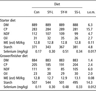
With the exception of Se concentrations, which were dictated by treatment structure, there were no appreciable differences in any determined or calculated feed nutritional parameter (Table 2).
Laboratory-determined nutritive and selenium contents of experimental diets (g/kg as fed, unless otherwise stated)

Con = background selenium (Se) only; SY-L = selenoyeasts 0.3 mg/kg total Se; SY-H = selenoyeasts 0.5 mg/kg total Se; SS-L = sodium selenite 0.3 mg/kg total Se; DM = dry matter; ME = metabolizable energy.
Bird physical performance
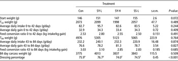
There were no effects of treatment on intake, rates of live weight gain or feed conversion efficiency at either T42 or T84 (Table 3). Similarly, there were no effects of treatment on carcass weight. However, there were differences between treatments on dressing percentage (Table 3), but these followed no discernable treatment pattern and may reflect differences in dressing technique rather than effects of selenium supplementation.
Physical performance of female turkey poults receiving diets containing graded additions of supplementary selenium

Con = background selenium (Se) only; SY-L = selenoyeasts 0.3 mg/kg total Se; SY-H = selenoyeasts 0.5 mg/kg total Se; SS-L = sodium selenite 0.3 mg/kg total Se.
Different superscripts within a row differ significantly (P < 0.05).
Selenium concentration of blood and plasma
There were significant differences between treatments (P < 0.001) to the graded addition of dietary Se on whole blood total Se at both T42 and T84 (Table 4). Whole blood total Se values were 76.04 ± 2.97 ng/g fresh weight at T0 and did not change appreciably between successive time points in those birds receiving treatment Con, which contained no additional Se. Whole blood Se concentrations were higher (P < 0.001) in those birds that had received Se-supplemented diets at both time points. However, whole blood Se values were significantly greater (P < 0.05) in those birds offered SY-supplemented diets when compared with those receiving diets supplemented with a comparable dose of selenite (SY-L v. SS-L), irrespective of time point. Whole blood total Se concentrations were greater (P < 0.001) in treatment SY-H when compared with SY-L at both time points. The same trends were apparent in plasma Se concentrations at T84, in which values were greatest in those treatments that had been supplemented with selenium (P < 0.001), greater in treatment SY-L when compared with SS-L (P = 0.008) and higher in treatment SY-H when compared with SY-L.
Selenium content of whole blood, plasma and whole blood GSH-Px activity of female turkey poults receiving diets containing graded additions of supplementary selenium

GSH-Px = glutathione peroxidase; Con = background selenium (Se) only; SY-L = selenoyeasts 0.3 mg/kg total Se; SY-H = selenoyeasts 0.5 mg/kg total Se; SS-L = sodium selenite 0.3 mg/kg total Se; FW = fresh weight; DM = dry matter.
Basal (T0) whole blood selenium content 76.04 ± 2.97 ng/g FW.
Different superscripts wxyz within a row differ significantly (P < 0.05).
A, unsupplemented diet v. selenium supplemented.
B, selenite v. SY (0.3 mg Se/kg DM).
C, Low-dose SY (0.3 mg Se/kg DM) v. high-dose SY (0.45 mg Se/kg DM).
Selenium concentration in tissues
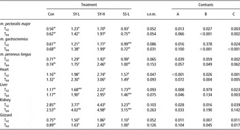
The Se content of tissues at T0 differed among tissue type (Tables 5) with total Se concentrations being greatest in kidney, followed by heart, liver, gizzard and then skeletal muscle. Although there were effects of treatment on the total Se content of each tissue at successive time points (T42 and T84), the hierarchy of tissue Se concentrations seen at T0 was maintained within treatment structure at each time point.
Total Se contents (mg/kg DM) of post-mortem tissues of female turkey poults offered diets containing graded additions of supplementary selenium for either for 42 or 84 days

Con = background selenium (Se) only; SY-L = selenoyeasts 0.3 mg/kg total Se; SY-H = selenoyeasts 0.5 mg/kg total Se; SS-L = sodium selenite 0.3 mg/kg total Se; DM = dry matter.
Basal (T0) m. pectoralis major selenium content 0.46 ± 0.002 mg/kg DM.
Basal (T0) m. gastrocnemius selenium content 0.44 ± 0.007 mg/kg DM.
Basal (T0) m. peroneus longus selenium content 0.33 ± 0.009 mg/kg DM.
Basal (T0) heart selenium content 0.83 ± 0.021 mg/kg DM.
Basal (T0) liver selenium content 1.50 ± 0.023 mg/kg DM.
Basal (T0) kidney selenium content 2.41 ± 0.020 mg/kg DM.
Basal (T0) gizzard selenium content 0.61 ± 0.012 mg/kg DM.
Different superscripts wxyz within a row differ significantly (P < 0.05).
A, unsupplemented diet v. selenium supplemented.
B, selenite v. SY (0.3 mg Se/kg DM).
C, low-dose SY (0.3 mg Se/kg DM) v. high-dose SY (0.45 mg Se/kg DM).
There were effects of treatment (P < 0.05) on the total Se content of all tissues at both time points to the graded addition of SY to the diet. Total Se concentrations increased between T0 and T42 in heart, breast and thigh tissue regardless of treatment, although increases were greater in those treatments that had received some form of Se supplementation (P < 0.05). In addition, total Se concentrations in heart and breast tissue were greater in those birds offered SY-L when compared with SS-L (P < 0.05). At T84, total Se concentrations in heart and skeletal tissue of SS-L poults were similar to those seen at T42 or from treatment group Con at T84. Conversely, total Se concentrations in heart and skeletal tissues of SY-L poults at T84 were greater (P < 0.05) than those of Con and SS-L poults. Similar trends were apparent in the gizzard at both T42 and T84; gizzard total Se concentrations were lowest in Con, greater in SS-L poults than in Con (P < 0.05) and higher in SY-L poults when compared with SS-L poults (P < 0.05).
In both liver and kidney tissue at T42 and T84, there were no appreciable differences between Con and SS-L poults. Although values were greater in SY-L when compared with Con (P < 0.05), no differences were observed between SY-L and SS-L.
Selenized AA content of whole blood
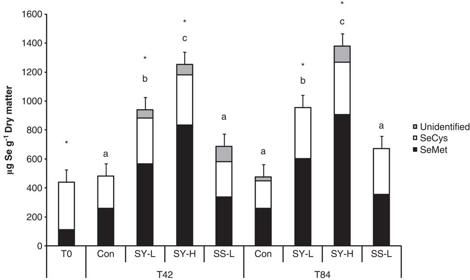
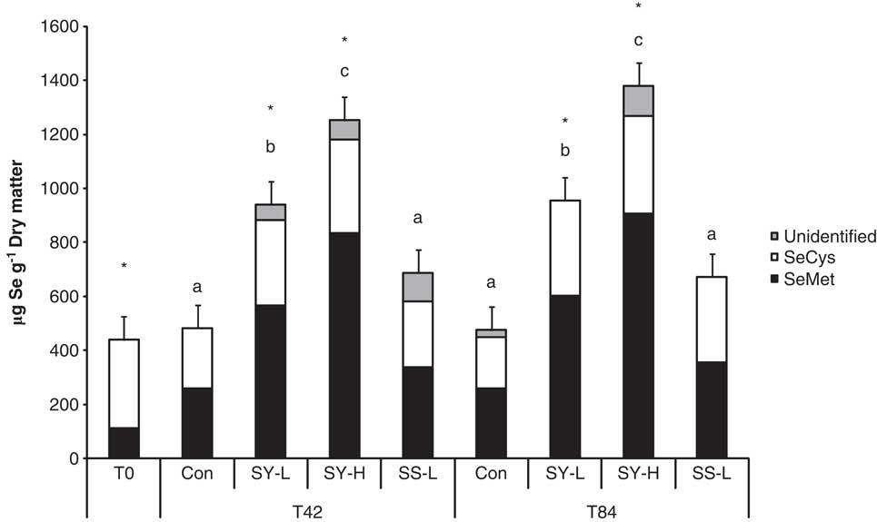
SeCys was the predominant selenized AA within whole blood samples at T0 (Figure 1). Conversely, SeMet values were greatest within whole blood samples of poults fed SY-L and SY-H at both time points (P < 0.05). Despite higher SeMet contents in treatments SY-L and SY-H at T84, SeCys values were numerically higher (P < 0.10) in SY-L and SY-H poults when compared with Con poults. This trend is reflected in whole blood GSH-Px activities (Table 3), which were greater in SY-L and SY-H poults than in Con poults.
Total Se and the proportion of total Se comprised as selenomethionine (SeMet) or selenocysteine (SeCys) in the whole blood of female turkey poults at days 0 (T0), 42 (T42) and 84 (T84). Different lowercase letters within time point denote SeMet differences (P < 0.05) between bars. Asterisks denote differences between SeMet and SeCys within bars.
Selenized AA content of tissues
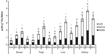
SeCys was the predominant selenized AA in liver and kidney tissue, irrespective of treatment, although values were higher (P < 0.05) in poults fed Se-supplemented diets (Figure 2). SeMet was the predominant selenized AA in breast tissue of birds fed SY supplements (P < 0.05), with SeMet values increasing with ascending dose. SeCys and SeMet comprised almost equal proportions of total Se in the breast tissue of Con and SS-L birds. Furthermore, the differences seen in total Se concentrations in breast tissue among treatments were primarily attributable to apparent increases in the quantity of total Se comprised as SeMet (0.26, 0.84, 0.95 and 0.37 ng SeMet/g DM for treatments Con, SY-L, SY-H and SS-L, respectively). However, this trend was not apparent within thigh tissue as SeMet and SeCys comprised almost equal proportions of total Se in all treatments. However, both the SeCys and SeMet contents of thigh muscle tissue were higher with SY-L and SY-H than with Con and SS-L (P < 0.05).
Total Se and the proportion of total Se comprised as selenomethionine (SeMet) or selenocysteine (SeCys) in the tissues of female turkey poults following 84 days of dietary treatment. Different lowercase letters within tissue denote SeMet differences (P < 0.05) between bars. Different uppercase letters within tissue denote SeCys differences (P < 0.05) between bars. Asterisks denote differences between SeMet and SeCys within bars.

Meat quality
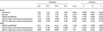
The ultimate pH, drip loss and TBARS of breast tissue were unaffected by the addition of supplementary Se to the diet, irrespective of Se source or dose (Table 6). There were effects of supplementary Se on breast tissue GSH-Px activity at both 0 and 5 days post mortem, activities being greater in the breast tissue of birds receiving diets containing supplementary Se (P < 0.001). However, these responses were predominantly constrained to the breast tissue of birds offered diets supplemented with SY, as GSH-Px activities were similar between Con and SS-L birds, whereas activities were greater in the breast tissue of SY-L birds when compared with SS-L and Con birds (P < 0.05). This is reflected in the positive relationship between breast tissue total Se content and tissue GSH-Px activity (R 2 = 0.437, P = 0.018).
Meat quality of female turkey poults offered diets containing graded additions of supplementary selenium for 84 days

Con = background selenium (Se) only; SY-L = selenoyeasts 0.3 mg/kg total Se; SY-H = selenoyeasts 0.5 mg/kg total Se; SS-L = sodium selenite 0.3 mg/kg total Se; TBARS = thiobarbituric acid reactive substances; MDA = malondialdehyde; GSH-Px = glutathione peroxidase.
Different superscripts xyz within a row differ significantly (P < 0.05).
A, unsupplemented diet v. selenium supplemented.
B, selenite v. SY (0.3 mg Se/kg DM).
C, low-dose SY (0.3 mg Se/kg DM) v. high-dose SY (0.45 mg Se/kg DM).
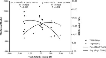
Despite higher tissue total Se concentrations in thigh muscle, TBARS values were approximately four times greater in thigh muscle than in breast muscle, although thigh TBARS values were seen to decline with increasing tissue total Se (R 2 = 0.468, P = 0.012; Figure 3). Thigh tissue GSH-Px activities exhibited the same treatment effects seen in breast tissue such that enzyme activities increased with ascending dietary Se and were seen to persist post mortem. However, as previously mentioned despite the similarities in treatment response between breast and thigh tissue, thigh GSH-Px activities were nearly twice those of breast tissue.
Relationship between thigh skeletal tissue total Se content and thiobarbituric acid reactive substances and thigh tissue total Se content and glutathione peroxidase activity of female turkey poults following 84 days of dietary treatment.

Discussion
Feed analyses
The Se contents of both starter and grower-finisher rations were close to target values.
Selenium content of blood and plasma
The lack of any significant response in blood and plasma Se concentrations of unsupplemented birds would be expected and is consistent with the findings of other authors investigating the effects of supplementary Se on poultry (Echevarria et al., Reference Echevarria, Henry, Ammerman and Miles1988; Pan et al., Reference Pan, Huang, Zhao, Qin, Chen and Hu2007; Yoon et al., Reference Yoon, Werner and Butler2007). The differences seen between comparable doses of the different Se supplements from the current study are indicative of better uptake and incorporation of Se derived from SY and are consistent with findings in sheep (Van Ryssen et al., Reference Van Ryssen, Deagen, Beilstein and Whanger1989; Juniper et al., Reference Juniper, Phipps, Ramos-Morales and Bertin2008a), cattle (Gunter et al., Reference Gunter, Beck and Phillips2003; Juniper et al., Reference Juniper, Phipps, Ramos-Morales and Bertin2008b; Phipps et al., Reference Phipps, Grandison, Jones, Juniper, Ramos-Morales and Bertin2008) and pigs (Kim and Mahan, Reference Kim and Mahan2001). Yoon et al. (Reference Yoon, Werner and Butler2007) reported that there were differences between Se sources in 6-week-old broilers that had received diets containing comparable doses of SS and SY; but differences also existed between comparable doses of different organic sources. Pan et al. (Reference Pan, Huang, Zhao, Qin, Chen and Hu2007) showed that Se responses in the blood of 6-week-old broilers were greater in birds fed SS when compared with birds fed a comparable dose of SY. Pan et al. (Reference Pan, Huang, Zhao, Qin, Chen and Hu2007) postulated that the differences seen between domestic fowl and cattle or pigs could be attributable to animal class differences (avian v. mammalian). However, the results of the current study indicate that this may not be entirely the case. It is likely that differences in Se uptake and assimilation may exist between different Galliformes as differences have been reported in selenium uptake and metabolism between different porcine breeds receiving identical diets (Banoch et al., Reference Banoch, Fajt, Odehnalova, Drabek and Svoboda2009). Furthermore, inconsistencies between the responses reported for different Se sources may be attributable to differences in the composition of the SY under test, as manufacturing processes ultimately affect both the Se content and the proportion of total Se of the SY comprised as SeMet (European Food Safety Authority (EFSA), 2008). As many of the differences observed between the uptake and assimilation of different Se sources are a function of SeMet content, it could be that the differences observed between different studies could be attributable to different SY strains.
Selenium content in tissues
The hierarchy of Se concentrations observed in different tissues tends to reflect the relative metabolic activity of each tissue. Glandular visceral tissues tend to have greater total Se concentrations than muscle, and cardiac muscle has a greater total Se content than skeletal muscle. These data agree with those reported in laying hens (Petrovič et al., Reference Petrovič, Boldižárová, Faix, Mellen, Arpášová and Leng2006; Pan et al., Reference Pan, Huang, Zhao, Qin, Chen and Hu2007) and broilers (Wang and Xu, Reference Wang and Xu2008) and are also similar to those reported in lambs (Juniper et al., Reference Juniper, Phipps, Ramos-Morales and Bertin2008a) and beef cattle (Juniper et al., Reference Juniper, Phipps, Ramos-Morales and Bertin2008b).
The differences seen in cardiac, skeletal and gizzard muscle tissue total Se concentrations, when comparing comparable doses of the two sources of Se, are indicative of improved Se uptake and incorporation within those birds supplemented with SY. However, the same responses were not apparent in liver or kidney tissue as there appeared to be no significant difference in tissue total Se when comparing Se sources at comparable doses. Petrovič et al. (Reference Petrovič, Boldižárová, Faix, Mellen, Arpášová and Leng2006) reported that total Se concentrations were greater in the kidney, liver and pancreatic tissues of SY-supplemented laying hens when compared with those receiving a comparable dose of SS, but there were no differences between Se sources in muscle tissue. Pan et al. (Reference Pan, Huang, Zhao, Qin, Chen and Hu2007) reported that tissue total Se concentrations were similar in liver tissue of laying fowl when comparing sources but greater in the muscle tissue of SY-supplemented birds when compared with SS. Similar effects of Se source have been reported within the cardiac and skeletal muscles of cattle, sheep (Juniper et al., Reference Juniper, Phipps, Ramos-Morales and Bertin2008a and Reference Juniper, Phipps, Ramos-Morales and Bertin2008b) and loin tissue of pigs (Mahan et al., Reference Mahan, Cline and Richert1999; Mateo et al., Reference Mateo, Spallholz, Elder, Yoon and Kim2007), whereby tissue total Se concentrations have been greater in SY-supplemented animals when compared with those receiving a comparable dose of SS.
Selenized AA content of whole blood
The SeCys content of whole blood is usually indicative of selenoenzymes activity, as all selenoenzymes have a functional SeCys core. The results of whole blood speciation of the current study indicate that the bulk of supplementary Se was in the form of SeMet and that the SeCys content of whole blood reflected whole blood GSH-Px activity. In addition, there were no appreciable differences between the two Se sources in terms of both GSH-Px activity and whole blood SeCys, whereas SeMet contents were greater in SY-supplemented birds. This observation in turkey poults is unlike those reported for lambs (Juniper et al., Reference Juniper, Phipps, Ramos-Morales and Bertin2008a), beef cattle (Juniper et al., Reference Juniper, Phipps, Ramos-Morales and Bertin2008b) and dairy cattle (Phipps et al., Reference Phipps, Grandison, Jones, Juniper, Ramos-Morales and Bertin2008) that had received similar sources and doses of supplementary Se, in which SeCys was the predominant selenized AA, irrespective of treatment.
Selenized AA content of tissues
The predominance of SeCys in both liver and kidney tissue has been reported in both lambs (Juniper et al., Reference Juniper, Phipps, Ramos-Morales and Bertin2008a) and beef cattle (Juniper et al., Reference Juniper, Phipps, Ramos-Morales and Bertin2008b) and may reflect the greater metabolic activity of these tissues and the relative associated changes in selenoenzyme activity. However, unlike the current study where SeCys comprised the greater proportion of total Se regardless of treatment, within lamb and beef cattle higher SY doses tended to result in SeMet comprising the greater proportion of total Se in liver tissue. This change in selenized AA partitioning at higher SY doses may reflect saturation of selenoenzymes and subsequent change from incorporation of Se into functional seleno-proteins to the non-specific incorporation of SeMet into liver tissue protein.
The predominance of SeMet in breast tissue is similar to observations that have been observed in the skeletal muscle of beef cattle (Juniper et al., Reference Juniper, Phipps, Ramos-Morales and Bertin2008b) in which SeMet concentrations were seen to increase with ascending SY inclusion, most probably reflecting increases in SeMet intake and subsequent uptake and non-specific incorporation into muscle protein. However, this same trend was not apparent within thigh tissue where SeMet and SeCys comprised almost equal proportions of total Se. In addition, SeCys contents of thigh tissue of SY-L and SY-H poults were greater than those observed in Con and SS-L, probably reflecting improved bioavailability of Se derived from SY and its subsequent incorporation into functional enzymes. It is of interest to note that the SeCys content of SY-L and SY-H thigh tissue was approximately twice that of breast tissue and mirrored the differences seen in thigh and breast tissue GSH-Px activities. This relationship between tissue SeCys content and GSH-Px activity is not surprising as SeCys forms the functional core of all selenoenzymes and is often present in tissues that have greater metabolic activity. The greater SeCys content and GSH-Px activities seen in thigh tissue may therefore reflect increased oxidative activity of thigh skeletal muscle tissue involved in support and locomotion of large framed commercial breeds of poultry.
Meat quality
The greater TBARS values of thigh tissue, when compared with those of breast tissue, may be partially attributable to the lipid content of thigh muscle (red meat) when compared with breast tissue (white meat). The red meat of poultry has nearly twice the lipid content of white meat (Rymer and Givens, Reference Rymer and Givens2006) and it could be likely that given this greater lipid content one would expect greater levels of lipid oxidation and hence greater TBARS values. Furthermore, this increased lipid oxidation potential of thigh tissue may also account for the higher GSH-Px activities seen within thigh when compared with breast. However, the lipid content of tissues was not determined in this study and therefore it is not possible to postulate any further whether TBARS values mirror the lipid content of skeletal tissue. Despite the higher GSH-Px activities in the thighs of SY-L and SY-H birds, it did not result in any commensurate reduction in TBARS values. This overall lack of treatment effect on the oxidative stability of edible muscle tissue is not uncommon. Radmilla et al. (Reference Radmilla, Jovanovic, Baltic, Sefer, Petrujkic and Sinovec2008) and Sevcikova et al. (Reference Sevcikova, Skrivan, Dlouha and Koucky2006) have both reported a lack of effect of Se supplementation on the meat quality attributes of broilers. Mahan et al. (Reference Mahan, Cline and Richert1999) showed little effect of Se source or dose on the drip loss of pig loin tissue and Skřivanová et al. (Reference Skřivanová, Marounek, De Smet and Raes2007) reported a lack of effect of SY supplementation on the oxidative stability of calf loin tissue. Taylor et al. (Reference Taylor, Marchello, Finley, Neville, Combs and Caton2008) and Juniper et al. (Reference Juniper, Phipps, Ramos-Morales and Bertin2008b) both reported that beef steaks from cattle supplemented with Se had similar shelf life attributes to those from unsupplemented animals, despite having higher Se contents. These results tend to suggest that once the Se content of the tissue exceeds the requirements of anti-oxidant enzymes, then further increases in tissue Se do not result in any commensurate improvement in meat keeping quality.
Conclusion
The incorporation of graded additions of SY into the diets of growing turkey poults increased total Se in tissues and blood in a dose-dependent manner. Increased breast tissue total Se appeared to be attributable to greater increases in SeMet, whereas increases in thigh tissue total Se appeared to be attributable to increases in both SeMet and SeCys with changes in thigh tissue SeCys content corresponding to changes in GSH-Px activity, thus indicating that GSH-Px activity and subsequent oxidative stability are more likely to be influenced by SeCys content of tissues rather than total Se. Responses in both blood and tissue were markedly greater in animals offered SY as opposed to SS, reflecting improved uptake and incorporation of SeMet. Despite greater GSH-Px activity in the muscle tissues of turkeys offered diets containing SY, it did not result in any appreciable effect on meat quality. It is therefore likely that increases seen in the total Se content of meat products from SY-supplemented animals, rather than resulting in any benefits regarding product shelf life, may improve the Se status of humans consuming such products in Se-deficient areas.
Acknowledgements
This research was funded by Alltech EU Regulatory Affairs Department. The authors would also like to especially thank Ryszard Lobinski (UT2A laboratories) for performing the selenium analyses and Duncan Marriott for his help with tissue GSH-Px determination.